H+-selective microelectrodes made and calibrated as described by Grandin and Charbonneau (1990a). Each egg was impaled with a potential microelectrode, ...
COMMENTARY Cycling of intracellular free calcium and intracellular pH in Xenopus embryos: a possible role in the control of the cell cycle
NATHALIE GRANDIN and MICHEL CHARBONNEAU* Laboratoire de Biologie et G^nitique du Diveloppement, URA C.N.R.S. 256, University de Rennes I, 35042 Rennes Cedex, France * Author for correspondence
Introduction The scope of this commentary is to propose the incorporation of recent data, obtained from Xenopus eggs and embryos, into the models of cell cycle regulation by MPF, a universal M-phase Promoting Factor operating in most, if not all, mitotic cells, from yeast to human. These new data are: (1) the cycling activity of MPF in Xenopus eggs is temporally and functionally related to the cycling activity of intracellular pH (pHi) (Grandin and Charbonneau, 1990a); (2) cell division in Xenopus embryos is accompanied by oscillations of the intracellular free calcium activity ([Ca2+];) (Grandin and Charbonneau, 1991). There is now definitive evidence that one of the molecular components of the cell cycle, the 'master oscillator' (or cytoplasmic clock), is represented by MPF (Masui and Markert, 1971) and its correlated cdc2 kinase activity and cyclin level (see, for instance, Draetta and Beach, 1989; Murray, 1989; Murray and Kirschner, 1989). In addition, a variety of different systems have revealed a direct implication of [Ca 2+ ]; variations in mitotic events (reviewed by Berridge and Irvine, 1989; Hepler, 1989). Although Xenopus embryonic cells have become, in addition to yeast, one of the most important systems for studying the molecular biology of the cell division cycle, almost no attention has been paid to the possible involvement of ionic messengers, particularly Ca 2+ , in the control of mitosis in Xenopus embryos. This lack of attention to the possible role of [Ca 2+ ]; variations in the cell division cycle of Xenopus embryos was due to their repeatedly noted absence. However, the recent demonstration of Ca 2+ oscillations occurring with a periodicity equal to that of the cell division cycle in Xenopus embryos (Grandin and Charbonneau, 1991) now offers an opportunity of re-evaluating the already proposed models of cell division. MPF: the cdc2 kinase/cyclin complex MPF was initially described as a Maturation-Promoting Factor in frog oocytes (Masui and Markert, 1971; Smith and Ecker, 1971). In amphibians, full-grown immature oocytes are arrested in first prophase of meiosis and, following stimulation with progesterone or microinjection of MPF, are released from this meiotic blockade to attain Journal of Cell Science 99, 5-11 (1991) Printed in Great Britain © The Company of Biologists Limited 1991
the second metaphase of meiosis (reviewed by Masui and Clarke, 1979; Mailer, 1985). Following egg laying, fertilization triggers a number of metabolic reactions (reviewed by Charbonneau and Grandin, 1989), including a second release of the meiotic arrest, which drive the newly fertilized egg into the early embryonic cell cycle, also under the control of MPF. MPF also exists in various cells, always associated with the entry into mitosis (reviewed by Kishimoto and Kanatani, 1977; Meijer and Guerrier, 1984; Kishimoto, 1988). It is now reasonable to conceive that MPF is a universal factor, referred to as M-phase Promoting Factor rather than simply a MaturationPromoting Factor (reviewed by Hunt, 1989; Lohka, 1989; Doree, 1990; Mailer, 1990- Nurse, 1990). MPF consists of the protein kinase p34cdc and cyclin, the two becoming associated to trigger M-phase (see Nurse, 1990). The activity of p S ^ " is partly controlled by the successive synthesis and destruction of cyclin (see Minshull et al. 1989; Murray and Kirschner, 1989; Murray et al. 1989), probably via phosphorylation, since p34cdc2 is activated by dephosphorylation and rephosphorylates upon inactivation (Dor6e et al. 1989). Activation of the cdc2 kinase, leading to a cycling activity, appears to involve the participation of a serine/threonine phosphatase and a tyrosine phosphatase (Dunphy and Newport, 1989; Gautier et al., 1989; Gould and Nurse, 1989; Morla et al. 1989; Felix et al. 1990a). In addition, at the end of mitosis, cyclin degradation is under the control of the cdc2 kinase (Felix et al. 19906). The cdc2 kinase would not simply control mitotic events via the regulation of other enzymes, but also via physiological substrates (histone HI, lamina, nucleolar proteins, vimentin,...) that are readily implicated in the structural events of mitosis (reviewed by Lewin, 1990; Moreno and Nurse, 1990; see also Chou et al. 1990; Peter et al. 1990a,6).
Relations between the cdc2 kinase oscillations and pHi oscillations In Xenopus eggs Oscillations of the intracellular pH (pHi) level in Xenopus embryos were first described by Webb and Nuccitelli (1981). It is interesting to note that Xenopus eggs do not Key words: MPF activity; pHi cycling; Ca2+ oscillations.
possess any of the classical plasma membrane pHiregulating systems (Na + -H + , N a + - H C 0 3 " - C r or H + pumps) existing in most cell types (Grandin and Charbonneau, 19906). We have recently demonstrated that the pHi oscillations in Xenopus eggs represented a component of the basic cell cycle (Grandin and Charbonneau, 1990a). Indeed, pHi cycling, a cytoplasmic activity, was found to be suppressed by treatments that also abolished the cycling activity of MPF, while treatments that blocked cell division without affecting MPF activity cycling did not suppress the pHi oscillations (Grandin and Charbonneau, 1990a). Experiments using another amphibian system, Pleurvdeles waltlii, confirm the view that physiological pHi changes are in tight relation with MPF activity changes. Indeed, the first cell cycle in Pleurodeles eggs is 6h long (versus 1.5 h in Xenopus), followed by 90 min cell cycles (versus 30 min in Xenopus). In Pleurodeles eggs, both the kinetics of the activation-induced increase in pHi and those of MPF inactivation can be superimposed, both activities changing around 45 min after the triggering of egg activation (N. Grandin, J. P. Rolland and M. Charbonneau, unpublished). As in Xenopus eggs, pHi oscillations in Pleurodeles eggs subsequently occur in phase with the embryonic cell cycle, MPF activity cycling and the surface contraction waves (Grandin et al., unpublished). The connection between pHi and other metabolic reactions is still unclear (reviewed by Cohen and lies, 1975; Gevers, 1977; Roos and Boron, 1981; Busa and Nuccitelli, 1984). However, it is well established that general metabolic reactions consume or produce H + , or produce CO2 (reviewed by Gevers, 1977). Indeed, in heart cells, ATP hydrolysis during glycolysis is the principal direct means by which protons are generated in the cytoplasm (Gevers, 1977). Therefore, the cascade of phosphorylations-dephosphorylations occurring during the control of the Xenopus cell cycle by the cdc2 kinase might generate pHi oscillations. A necessary approach to studying the significance of pHi oscillations in Xenopus eggs was to determine the hierarchy of control between the cdc2 kinase and pHi oscillations. Three types of experiments convinced us that pHi cycling was probably a consequence of MPF activity cycling rather than the converse. First, we had noted that treatment of unactivated Xenopus eggs with weak bases (NH4CI or procaine), in a manner that induced a cytoplasmic alkalinization of a similar amplitude to that of the alkalinization induced by sperm during egg activation, did not release the metaphase block and, hence, did not induce the inactivation of MPF normally taking place during egg activation (Grandin and Charbonneau, 1989). In addition, treatments that decrease MPF activity in eggs (unactivated) are activating agents, which, accordingly, always produce the egg activationassociated increase in pHi. In other words, decreasing MPF activity by means of an activating stimulus always results in an increase in pHi. However, increasing pHi in unactivated eggs never produces changes in MPF activity. A second approach consisted in preventing the egg activation-associated increase in pHi in Xenopus embryos using CO2 in the external solution to artificially change pHi, under conditions that were controled with intracellular pH microelectrodes (Grandin and Charbonneau, 19906). CCvinduced suppression of that physiological increase in pHi did not prevent the embryos from reaching the first cell division, suggesting that MPF had correctly N. Grandin and M. Charbonneau
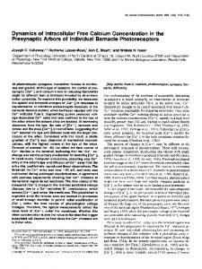
been inactivated following egg activation and been reactivated at the first mitotic metaphase even in the absence of the increase in pHi (Grandin and Charbonneau, 19906). A third line of evidence suggesting that pHi cycling is a consequence of MPF activity cycling is provided by experiments using cycloheximide, an inhibitor of protein synthesis. Cycloheximide prevents the cycling of both MPF activity (Gerhart et al. 1984) and pHi (Grandin and Charbonneau, 1990a) in activated Xenopus eggs. The first cyclic increase in MPF and pHi occurs 1 h after egg activation, the time of the first mitotic metaphase. However, cycloheximide does not affect the egg activation-associated increase in pHi, as shown in Fig. 1, or the pHi level in unactivated Xenopus eggs, but starts having an effect on pHi only at the time corresponding to the synthesis of cyclin, around 50 min after egg activation (Grandin and Charbonneau, 1990a). Thus, the suppression of pHi oscillations, 1 h after egg activation, by cycloheximide (Grandin and Charbonneau, 1990a) cannot result from a direct effect on pHi, but is rather mediated by the suppression of MPF activity cycling resulting from
B
uf
-10
7.3
o.
7.8
Fig. 1. Effects of cycloheximide on the pHi response to egg activation in Xenopus. Intracellular (pHi) was measured with H+-selective microelectrodes made and calibrated as described by Grandin and Charbonneau (1990a). Each egg was impaled with a potential microelectrode, measuring the membrane potential (Em; top trace), and a pH microelectrode, measuring £ m +pHi. Em recorded by the potential microelectrode was subtracted at the pen recorder input from the total value CEm+pHi) recorded by the pH microelectrode, to give the pHi value (pHi; bottom trace). Unactivated eggs were dejellied with 2 % cysteine (in the physiological Fl solution, see Grandin and Charbonneau, 1990a) and immersed in Fl solution with or without 200/igml" 1 cycloheximide. In the example shown here, the egg was incubated in the presence of cycloheximide 40 min before impalement with the microelectrodes (beginning of the trace), always in the presence of cycloheximide, and activated by pricking a few minutes later. Successful egg activation was attested by the occurrence of the activation potential (AP), a Cl~-dependent plasma membrane depolarization, followed 6 min later by a typical increase in pHi, from pH7.34 to pH7.70. The levels of pHi in cycloheximide-treated eggs before egg activation, as well as the kinetics of the egg activationassociated increase in pHi and the elevated pHi level attained 20—30 min after egg activation, were exactly similar to those in untreated eggs. Other criteria of egg activation, observed under a stereomicroscope during electrical recording in cycloheximide-treated eggs, were normal and identical to those in untreated eggs. These morphological criteria were: the elevation of the vitelline envelope, a consequence of cortical granule exocytosis; the cortical contraction; the disappearance of the maturation spot, a consequence of meiosis resumption, reflecting the migration of the egg nucleus deeper in the cytoplasm during the passage from the metaphase Il-arrested stage of meiosis to the pronucleus stage.
inhibition of the synthesis of one of the components of MPF, probably cyclin.
imprecision while measuring the time between the two types of oscillation.
Temporal relationship between pHi oscillations and Ca 2+ oscillations in Xenopus embryos
A model of cell division In Xenopus embryos that integrates the existence of Ca 2+ and pHi oscillations
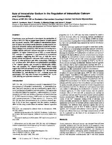
Monitoring the intracellular free calcium level ([Ca2+],) with microelectrodes implanted in Xenopus embryos, we observed Ca 2+ oscillations, which had a period equal to that of the cell division cycle (Grandin and Charbonneau, 1991). These Ca 2+ oscillations did not occur in artificially activated eggs or nocodazole-treated embryos, in both of which the basic cell cycle (for instance, cdc2 kinase activation and inactivation) persists (Grandin and Charbonneau, 1991). This demonstrated that the Ca 2+ oscillations are not required for the basic cell cycle in Xenopus embryos, and suggested that they might rather be linked to the metabolic events occurring only when both nuclear divisions and cleavage take place correctly. Both the opacity of Xenopus embryos and their very rapid period of cell division prevented us, at that time, from establishing a precise relationship between [Ca 2+ ]! and mitotic stages. We have now measured pHi and [Ca2+]i simultaneously in the same embryo, the pHi oscillations serving as a reference mark to determine the corresponding mitotic stages (Fig. 2). Initial attempts, which relied on the observation of nuclei with the light microscope, gave erroneous results because of the asynchrony between blastomeres at advanced stages (256-cell) of development (Grandin and Charbonneau, 1990a). In fact, the acidic peaks of the pHi oscillations correspond to the peaks of MPF activity (metaphase), as seen both in Pleurodeles after direct measurement of MPF activity, and in Xenopus, indirectly, by correlating the cleavage-associated membrane hyperpolarizations (telophase) and the surface contraction waves that occur at the metaphase-anaphase transition, immediately before the onset of cleavage (N. Grandin, J. P. Rolland and M. Charbonneau, unpublished data). Fig. 2 shows that Ca 2+ oscillations occur out of phase with pHi oscillations. Since the period of the cell division cycle, measured as the interval of time between two pHi or Ca 2+ oscillations, varied slightly from one embryo to the other, we will indicate for each of the nine impaled embryos the delay between Ca 2+ and pHi oscillations, as well as the period of the cell cycle. The period between the acidic peak of a pHi oscillation and the peak of the following Ca oscillation, and the period of the cell cycle in the corresponding embryo were, respectively (in min): 9, 21; 13, 23; 16, 24; 10, 22; 9, 21; 6, 25; 9 20; 10, 21; 13, 24; at 26-27 CC. One can see that, in most cases, the peak level of the Ca 2+ oscillation occurs slightly before or slightly after the alkaline peak of the pHi oscillation, that is approximately in the opposite phase with respect to the acidic peak of the pHi oscillation (Fig. 2). Since the acidic peak of the pHi oscillation corresponds to the metaphase stage of mitosis, as seen above, it follows that [Ca 2+ ]| would begin to increase between anaphase or telophase and interphase and would be at its maximal level during interphase or the next prophase. However, we are aware of the fact that the precision of the relation should be improved in the future. For the moment, the two main limitations are: (1) the opacity of the egg, which prevents visual observation of the corresponding mitotic stages; (2) the necessity of recording Ca 2+ and pHi oscillations using a low chart speed, which increases the
As stated above, pHi oscillations always take place in -201-30 kl
-40 _ o.
7.5
E
7.7L -20 -30 -40
6.6
60 min
Fig. 2. Simultaneous measurements of pHi and Ca 2+ oscillations in a single embryo of Xenopus. Since Xenopus embryos display pHi oscillations and Ca 2+ oscillations, both with a period equal to that of the cell cycle (see text), the present experiments were conducted in order to determine the delay, if any, between these two types of oscillations. Xenopus embryos were dejellied and impaled each with two potential microelectrodes, a Ca2+-selective microelectrode (made and calibrated as described by Grandin and Charbonneau, 1991) and a pH microelectrode. Each trace of ion activity measurement has its corresponding membrane potential trace (the one subtracted from the total signal recorded by the ionselective microelectrode) represented above it. In the example shown here, the embryo was impaled at the 8-cell stage. Both [Ca 2+ ], and pHi oscillated around their basal levels, 0.31-0.50/iM (pCa 6.5-6.3) and pH 7.55-7.65, respectively, for 4 or 5 cell cycles, the amplitude of the oscillations being around 50-100 nM [Ca 2+ ]| and 0.04-0.06 pH unit. In this embryo, the duration of the cell cycle was 21-24 min. The peaks of the Ca 2+ oscillations were found to occur 9—12 min after the acidic peaks of the pHi oscillations. That delay increased as the cell cycle lengthened (12 min delay during a 24 min cell cycle), and conversely (9 min delay during a 21 min cell cycle). The great difficulty of recording ion activity changes in dividing embryos (see Grandin and Charbonneau, 1991), was enhanced by the fact that, in the present situation, all four microlectrodes had to remain correctly inserted for several hours. However, such experiments are worthwhile because of the great selectivity and sensitivity allowed by ion-selective microelectrodes. The results of the present experiments provide one more argument (in addition to those developed by Grandin and Charbonneau, 1991) against the existence of artifacts during measurement of intracellular ion activity with microelectrodes. Indeed, the findings that Ca 2+ oscillations did not have the same shape as pHi oscillations, and that they were recorded with a delay between them, argue against the existence of 'mirror-image artifacts' and 'motion artifacts', respectively (see Grandin and Charbonneau, 1991).
pH and Ca2+ cycling in Xenopus embryos
activated eggs, even in the absence of cleavage, whereas Ca 2+ oscillations do not proceed in the absence of cell division. Therefore, we suspect that the existence of these Ca 2+ oscillations in Xenopus embryos might be associated with the presence of dividing nuclei. Accumulation of endoplasmic reticulum (ER) in the perinuclear region of cells might control [Ca 2+ ]; in relation to specific mitotic events. Experimental evidence of this type exists, for instance, in sea-urchin embryos, in which the ER contains a calsequestrin-like protein, which is a Ca2+-binding storage protein (Henson et al. 1989). It is very likely that such organized ER networks, with Ca 2+ storage properties, are present around the nuclei in Xenopus embryos. However, definitive evidence, even in sea-urchin embryos, that the mitosis-associated intracellular Ca 2+ transients originate from the Ca 2+ stored in the ER located in the perinuclear region is still missing. In addition to such possible control of Ca 2+ oscillations in Xenopus embryos by internally stored Ca 2+ , another possibility is control of these oscillations by the entry of extracellular Ca 2+ . However, there is apparently no influx of extracellular Ca 2+ in Xenopus embryos, since immersion of embryos in Ca2+-free medium containing EGTA has no effect on cleavage (Baker and Warner, 1972). This suggests that intracellular Ca 2+ oscillations in Xenopus embryos are not driven by the entry of extracellular Ca 2+ , but would instead depend on internally stored Ca 2+ . Unfortunately, this could not be directly tested on Xenopus embryos impaled with Ca 2+ microelectrodes, since extracellular Ca is needed for healing at the site of microelectrode penetration. In fact, most of our knowledge on the Ca 2+ -binding and -accumulation properties of the ER comes from studies on isolated vesicles from muscle cells. The Ca 2+ binding ability of isolated sarcoplasmic reticulum (SR) vesicles from skeletal and cardiac muscle is markedly pHdependent (Nakamaru and Schwartz, 1970); it decreased as extravesicular pH increased, from 6.3 to 7.5, and Ca 2+ was released from these vesicles as a consequence (Nakamaru and Schwartz, 1970). These experiments were performed in vitro, and, therefore, a smaller alkalinization in vivo might have the same consequence on release of Ca 2+ into the cytoplasm. In addition, dissipation of the pH gradient between the external and internal sides of the SR, following an increase in the extravesicular pH, elicited Ca 2+ release by these vesicles, possibly via a Ca 2+ release channel in the SR membrane (Shoshan etal. 1981). Experiments on skinned muscle fibers and intact muscle fibers led to quite variable, sometimes opposite, results concerning the relationships between [Ca 2+ ]; and pHi (see references quoted by Pressler, 1989; Kaila and Voipio, 1990), perhaps as a result of the diversity of the experimental conditions adopted, as well as of the possibility that the drugs used to change [Ca 2+ ], or pHi may have effects other than those postulated. The experiments on isolated SR vesicles (Nakamaru and Schwartz, 1970; Shoshan et al. 1981) agree nicely with our experiments in Xenopus embryos, in which the peak level of the Ca 2+ oscillations is, roughly, in the opposite phase with respect to the acidic peak of the pHi oscillations (Fig. 2). According to the hypothesis for an active role of pHi oscillations in the generation of Ca 2+ oscillations, the periodical alteration of a pH gradient between the cytoplasm and an internal compartment specifically located around the nuclei would produce a periodical influx of Ca 2+ from that internal compartment into the cytoplasm. As seen above, artificially activated (non8
N. Grandin and M. Charbonneau
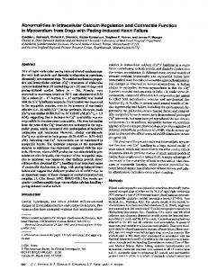
dividing) eggs display pHi oscillations without giving rise to subsequent Ca 2+ oscillations. This is apparently inconsistent with our hypothesis that pHi oscillations are the driving force for Ca 2+ oscillations. However, this contradiction can be explained, considering that pHi oscillations might generate Ca 2+ oscillations only when compartments specifically located around the nuclei and specialized in the storage of Ca 2+ are functional, as is the case in dividing embryos. On the other hand, artificially activated eggs do not possess such functional perinuclear compartments, since they do not divide. In other words, pHi oscillations might generate Ca 2+ oscillations in embryos, but nevertheless exist without generating Ca 2+ oscillations if the structures involved in the generation of the latter are not functional, which is the case in activated non-dividing eggs. Although most of the terms of the proposed cascade of reactions are still hypothetical, none of them is impossible. It is now well established that membranes, particularly the ER and the Golgi apparatus, constitute an important component of the mitotic apparatus, and probably regulate [Ca ]; in order to control the formation and function of spindle fibers and the separation of chromosomes (reviewed by Hepler and Wolniak, 1984). For instance, in sea-urchin embryos, intracellular Ca 2+ transients have been measured in association with events of mitosis, such as the metaphase-anaphase transition and nuclear envelope breakdown (Poenie et al. 1985; Steinhardt and Alderton, 1988). In addition, artificial increases in [Ca 2+ ]; cause premature chromatin condensation and breakdown of the nuclear envelope in sea-urchin embryos (Twigg et al. 1988). Evidence for a close relationship between [Ca 2+ ], variations and specific stages of mitosis also exists in other cell types, such as cultured mammalian cells and plant cells (reviewed by Hepler, 1989; see also Kao et al. 1990; Zhang et al. 1990). In Xenopus embryos, to date there has been no direct evidence for a relationship between [Ca 2+ ]| variations and specific stages of mitosis. However, early experiments, showing that microinjection of Ca-EGTA buffers into Xenopus blastomeres slowed down or even arrested cleavage by specifically lowering the intracellular concentration of Ca 2+ (Baker and Warner, 1972), support the suggestion that Ca 2+ oscillations in Xenopus embryos may play a regulatory role in cell division. We propose an improved model of control of the cell division cycle in Xenopus embryos (Fig. 3), taking into account our recent findings on pHi and Ca 2+ oscillations (Grandin and Charbonneau, 1990a, 1991), the present results and the recently proposed models of control by the cdc2 kinase/cyclin complex. In Fig. 3, we represent the gradual accumulation of cyclin in the egg. At some point in that accumulation, the p S ^ 0 2 kinase is activated following its binding to cyclin. Mitosis is thought to be directly triggered by the activation of the p34 kinase/cyclin complex (MPF). The cdc2 kinase activity, measured in vitro as a histone HI kinase activity or in vivo, in Xenopus oocytes, as a maturation-promoting factor activity, increases at the onset of mitosis and reaches its maximal level during metaphase (indicated as 'high cdc2 kinase activity' in Fig. 3). The association between cyclin and the cdc2 kinase ultimately leads, after a few intermediate steps, to the dephosphorylation of the cdc2 kinase (its active form). Recent studies have demonstrated the existence of a lag period between the addition of cyclin (in vitro) and the appearance of HI kinase activity, even in the presence of a large excess of cyclin (Solomon et al. 1990). The threshold concentration of cyclin required to
activate the cdc2 kinase/cyclin complex and the length of that lag period are regulated by an inhibitor of MPF activation, a type 2A protein phosphatase (not indicated in Fig. 3) (Solomon et al. 1990). It is also suggested that the abrupt transition into mitosis is due to an inhibition (by p34c ) of the initial phosphorylation, on tyrosine, of p34cdc2 by cyclin (during the lag period), which leads to a stimulation of p34cdc2 dephosphorylation (Solomon et al. 1990), one of the last steps prior to the entry into mitosis. At the end of metaphase, cyclin is abruptly degraded, probably via specific proteolysis (Murray and Kirschner, 1989). Recent work shows that, in vitro, cyclin proteolysis is directly triggered by the cdc2 kinase itself, probably via the phosphorylation of an unknown protein that activates the cyclin-specific protease (FeUix et al. 19906). Following cyclin degradation, the cdc2 kinase is rapidly rephosphorylated (its inactive form) or combines with an inhibitor (F61ix et al. 19906). Inactivation of the cdc2 kinase/cyclin complex (indicated as 'low cdc2 kinase activity' in Fig. 3) permits exit from mitosis and entry into interphase. The alternating activated and inactivated forms of the cdc2 kinase/cyclin complex (MPF) represent, together with other activators or inhibitors mentioned above, a sort of autonomous oscillator, which regulates the basic cell cycle. In Xenopus embryos, each of the early cell cycles is 30 min long, with the exception of the first cell cycle, which lasts 90 min. Previous studies (Grandin and Charbonneau, 1990), as well as the present results, strongly suggest that the MPF activity oscillations control - and give rise to pHi oscillations. In addition, we have shown that the
acidic peak of the pHi oscillations corresponds to the metaphase stage of mitosis (Grandin and Charbonneau, 1991; present results), while the alkaline peak corresponds to interphase, when the activity of the cdc2 kinase/cyclin complex is at its lowest level (Fig. 3). However, the molecular mechanisms at the origin of the temporal and functional relationships between MPF activity and pHi remain to be discovered. Taking into account the existence of Ca 2+ oscillations, which are in the opposite phase to the pHi oscillations, and assuming the existence of perinuclear ER that possibly regulates the internal store of Ca 2+ at the origin of the Ca 2+ oscillations, we suggest that Ca 2+ oscillations could be driven by pHi oscillations, as described above. In such a scheme, internal compartments located around the dividing nuclei (perinuclear ER?) would possess in their membrane a pH-dependent Ca 2+ release channel or a C a 2 + - H + exchanger. During the passage between the acidic peak of the pHi oscillation and the alkaline peak of the pHi oscillation, there is a gradual alkalinization of the cytoplasm (pHi is the cytosolic pH). This might lead to the abolition of the pH gradient existing between the cytosol and the internal compartment (perinuclear ER). As explained above, dissipation of that pH gradient might lead to the release of Ca by the ER vesicles through pH-dependent Ca 2+ -release channels contained in the membrane of these vesicles, a situation that exists in muscle cells (see references above). Since pHi continuously cycles during early cell division, the associated cycling of opening and closure of such pH-dependent Ca2+-release channels might result in a cycling of
degradation
.cyclin
. cdc2 kinase activity
alkaline alkalinization
peak
[Ca24} increase high
level
T
mitosis regulation
low
Ca *-H* exchanger or pH-dependent Ca2-release channel
Fig. 3. Improved model of control of the cell cycle in Xenopus embryos, made on the basis of the existence of Ca 2+ and pHi oscillations. All the authors cited in the section MPF: the cdc2 kinase/cyclin complex of the present paper, as well as many of the studies cited by these authors, have contributed by their results to the construction of the present scheme. However, some of these authors have provided general schemes of the control of the cell cycle by the cdc2 kinase and cyclins, so it might be useful for the reader to compare them with the present scheme (Draetta and Beach, 1989; Murray, 1989; F61ix et al. 19906; Minshull et al. 1990; Mailer, 1990; Nurse, 1990). It should noted that the activity of the cdc2 kinase builds up and subsequently drops faster than the parallel changes in H + activity. This is not due to a technical problem, since pH microelectrodes have a response time of a few seconds only. That delay suggests that the coupling between MPF activity oscillations and pHi oscilations involves either slow metabolic reactions or a succession of several coupled reactions.
pH and Ca2+ cycling in Xenopus embryos
cytosolic Ca 2+ (the Ca 2+ oscillations) around the dividing nuclei. It should be noted that a different mechanism, not involving the participation of some intracellular compartment responding to the periodical alteration of a pH gradient between it and the cytosol, might be considered. Indeed, Picard et al. (1990) have recently shown that microinjection of a PSTAIR peptide, which is a conserved sequence of ^M^62, into Xenopus oocytes produced an increase in [Ca2+]i, independently of its histone HI kinase activity. If such a Ca2+-mobilizing activity of the cdc2 kinase was also present in Xenopus embryos, it might explain the generation of Ca 2+ oscillations in response to MPF activity oscillations, directly, using a pathway independent of the pHi oscillations. A major problem with Xenopus embryos is that cells are totally opaque. Therefore, the relations of [Ca 2+ ]! variations to specific mitotic events is much less clear than in other systems, for instance in sea-urchin embryos. However, experiments to try and improve our comprehension of the Xenopus system will be certainly worthwhile. We believe that the two systems, Xenopus and sea-urchin, are different from each other and that new information using the Xenopus system can be complementary to that provided by the study of the sea-urchin system. The pattern of [Ca 2+ ]| variations during embryonic cell division in Xenopus appears to be different from that in sea-urchin embryos, in which intracellular Ca 2+ transients have been recorded in association with pronuclear migration, streak stage, nuclear envelope breakdown, chromatin condensation, onset of anaphase and cytokinesis (Poenie et al. 1985; Steinhardt and Alderton, 1988; Twigg et al. 1988). To date, however, in sea-urchin embryos, nuclear envelope breakdown and chromatin condensation are the only two mitotic events in which there is some substantial evidence for a role for [Ca 2+ ]; (Twigg et al. 1988; Whitaker and Patel, 1990). A major part of our future work will be directed at consolidating our hypothesis, combining [Ca 2+ ], measurements in vivo and the study of the capacity of perinuclear ER to store Ca 2+ by using antibodies directed against ubiquitous Ca2+-binding proteins. An additional requirement will be to uncover some specific targets of the Ca 2+ generated by the Ca 2+ oscillations. For the moment, no such [Ca2+];-regulated target, capable of playing a role in the control of mitosis, has been identified by us or others. Finally, it is important to stress that our scheme (Fig. 3) does not mean that Ca 2+ oscillations represent a solitary endpoint signal for regulation of mitosis. This is particularly evident, considering that to date there has been no mitotic event identified as responding to intracellular Ca 2+ signals in Xenopus embryos. On the contrary, we believe that most of the mitotic targets are under the control of the cdc2 kinase/cyclin complex. Indeed, several mitosis-associated substrates of cdc2 kinase have been recently identified in various systems: histone HI (Arion et al. 1988; Labbe et al. 1988; Langan et al. 1989), major nucleolar proteins (Peter et al. 1990a), lamins (Peter et al. 19906), RNA polymerase H (Cisek and Corden, 1989), elongation factor EF-lr (Bell6 et al. 1989) and vimentin (Chou et al. 1990) (see also the model described by Minshull et al. 1990). We suspect that Ca 2+ oscillations in Xenopus embryos might act in co-operation with the MPF complex to trigger limited and specific mitotic events that have not been identified. We are very grateful to Drs Marie-Anne Felix, Marcel Doree and Beverley Osborne for their comments. This work was 10
N. Grandin and M. Charbonneau
supported by grants from the Ligue contre le Cancer (Comite D6partemental dTlle-et-Vilaine), Association pour la recherche sur le Cancer and Region Bretagne. NG was a recipient of a doctoral fellowship from the Region Bretagne.
References ARION, D., MEIJER, L., BRIZUELA, L. AND BEACH, D. (1988) cdc2 is a
component of the M phase-specific histone HI kinase: evidence for identity with MPF. Cell 55, 371-378. BAKER, P. F. AND WARNER, A. E. (1972). Intracellular calcium and cell cleavage in early embryos of Xenopus laevis. J. Cell Biol. 53, 679-581 BELLE, R., DERANCOURT, J., POULKE, R., CAPONY, J. P., OZON, R. AND
MULNER- LORILLON, O. (1989). A purified complex from Xenopus oocytes contains a p47 protein, an in vivo substrate of MPF, and a p30 protein respectively homologous to elongation factors EF-lr and EF10. FEBS Lett. 255, 101-104. BERRTDGB, M. J. AND IRVINE, R. F. (1989). Inositol phosphates and cell signalling. Nature 341, 197-205 Bu8A, W. B. AND NUCCITKLU, R. (1984). Metabolic regulation via intracellular pH. Am. J Physiol. 246, R409-R438. CHARBONNBAU, M. AND GRANDIN, N. (1989). The egg of Xenopus laevis: a model system for studying cell activation. Cell Differ. Dev. 28, 71-94. CHOU, Y., BISCHOFT, J. R., BEACH, D. AND GOLDMAN, R. D. (1990).
Intermediate filament reorganization during mitosis is mediated by p34cdc3 phosphorylation of vimentin. Cell 62, 1063-1071. CISEK, L. J. AND CORDKN, J. L. (1989). Phosphorylation of RNA
polymerase by the murine homologue of the cell-cycle control protein cdc2 Nature 339, 679-684. COHEN, R. D. AND ILES, R A. (1975). Intracellular pH: measurement, control, and metabolic interrelationships. Crit. Rev. elm. Lab. Sei. 6, 101-143. DOREE, M. (1990). Control of M-phase by maturation-promoting factor. Curr. Opm Cell Biol. 2, 269-273. DOREE, M., LABBE", J. C. AND PICAHD, A. (1989). M phase-promoting
factor its identification as the M phase-specific HI histone kinase and its activation by dephosphorylation. J. Cell Sci. Suppl. 12, 39-51. DRAETTA, G. AND BEACH, D. (1989). The mammalian cdc2 protein kinase: mechanisms of regulation during the cell cycle. J. Cell Sci. Suppl. 12, 21-27. DUNPHY, W. G. AND NEWPORT, J. W. (1989). Fission yeast pl3 blocks mitotic activation and tyrosine dephosphorylation of the Xenopus cdc2 protein kinase. Cell 58, 181-191. FELIX, M. A., COHEN, P. AND KARBENTI, E. (1990a). cdc2 HI kinase is
negatively regulated by a type 2A phosphatase in the Xenopus early embryonic cell cycle: evidence from the effects of okadaic acid. EMBO J. 9, 675-683. FELIX, M. A., LABBS, J. C , DOREE, M. AND KARSBNTI, E. (19906).
Triggering of cyclin degradation in interphase extracts of amphibian eggs by cdc2 kinase. Nature 346, 379-382. GAUTTER, J., MATSUKAWA, T., NURSE, P. AND MALLER, J. (1989).
Dephosphorylation and activation of Xenopus p34cdc2 protein kinase during the cell cycle. Nature 339, 626-629. GBRHART, J. C , Wu, M. AND KIRSCHNBR, M. (1984). Cell cycle dynamics of an M-phase-specific cytoplasmic factor in Xenopus laevis oocytes and eggs. J. Cell Biol. 98, 1247-1255. GEVBRS, W. (1977). Generation of protons by metabolic processes in heart cells J. molec. cell. Cardiol. 9, 867-874. GOULD, K. AND NURSE, P. (1989). Tyrosine phosphorylation of the fission yeast cdc2+ protein kinase regulates entry into mitosis. Nature 342, 39-45. GRANDIN, N. AND CHARBONNEAU, M. (1989). Intracellular pH and the
increase in protein synthesis accompanying activation of Xenopus eggs. Biol. Cell 67, 321-330. GRANDIN, N. AND CHARBONNEAU, M. (1990a). Cycling of intracellular pH during cell division of Xenopus embryos is a cytoplasmic activity depending on protein synthesis and phosphorylation J. Cell Biol. I l l , 523-532 GRANDIN, N. AND CHARBONNBAU, M. (19906). Is the egg activation-
induced intracellular pH increase necessary for the embryonic development of Xenopus laevis (anuran amphibian)? In Mechanism of Fertilization: Plants to Human (ed. B. Dale), pp. 503-507. SpnngerVerlag, Berlin. GRANDIN, N. AND CHARBONNBAU, M. (1991). Intracellular free calcium oscillates during cell division of Xenopus embryos. J. Cell Biol. 112, 711-718. HENSON, J H., BEGG, D. A., BEAULTEU, S. M., FISHKIND, D. J., BONDER, E M., TERASAKI, M., LEBECHE, D. AND KAMINER, B. (1989). A
calaequestrin-like protein in the endoplasmic reticulum of the sea urchin: localization and dynamics in the egg and first cell cycle embryo J. Cell Biol. 109, 149-161.
HEPLBR, P K (1989). Calcium transients during mitosis: observations in flux. J. Cell Biol. 109, 2567-2573. HEPLER, P K. AND WOLNIAK, S. M. (1984). Membranes in the mitotic apparatus: their structure and function. Int. Rev. Cytol. 90, 169-238. HUNT, T. (1989). Maturation promoting factor, cyclin and the control of M-phase. Curr. Opin. Cell Biol. 1, 268-274. KAILA, K. AND VOIPIO, J. (1990). Dependence of intracellular free calcium and tension on membrane potential and intracellular pH in single crayfish muscle fibres. Pflilgers Arch. ges. Phystol. 416, 501-511. KAO, J. P Y., ALDERTON, J. M., TSIEN, R Y AND STEINHARDT, R. A.
(1990). Active involvement of Ca2+ in mitotic progression of SWIBS 3T3 fibroblasts J. Cell Biol. I l l , 183-196. KISHIMOTO, T. (1988). Regulation of metaphaBe by a maturationpromoting factor. Dev. Growth Differ. 30, 105-115. KISHIMOTO, T. AND KANATANI, H (1977). Lack of species specificity of starfish maturation-promoting factor. Gen. comp. Endocrin. 33, 41-44. LABBE, J. C, LEE, M. G., NURSE, P., PICARD, A. AND DOREE, M. (1988).
Activation at M-phase of a protein kinase encoded by a starfish homologue of the cell cycle control gene cdc2+. Nature 335, 251-254. LANGAN, T. A , GAUTIER, J., LOHKA, M., HOLLINGSWORTH, R., MORENO, S., NURSE, P., MAUSER, J. AND SCLAFANI, R. A. (1989). Mammalian +
MURRAY, A. W. (1989). Cychn synthesis and degradation and the embryonic cell cycle. J. Cell Sci. Suppl. 12, 65-76. MURRAY, A. W. AND KIRSCHNER, M. (1989). Cyclin synthesis drives the early embryonic cell cycle. Nature 339, 276-280. MURRAY, A. W., SOLOMON, M. J. AND KIRSCHNER, M. W. (1989). The role
of cyclin synthesis and degradation in the control of MPF activity. Nature 339, 280-286. NAKAMARU, Y. AND SCHWARTZ, A (1970). Possible control of intracellular calcium metabolism by [H] + : sarcoplasmic reticulum of skeletal and cardiac muscle. Bwchem. bwphys. Res. Commun. 41, 830-836. NURSE, P. (1990). Univereal control mechanism regulating onset of Mphase. Nature 344, 503-608. PETER, M., NAKAOAWA, J., DOREE, M., LABBE, J. C. AND NIGG, E. A.
(1990a). Identification of major nucleolar proteins as candidate mitotic substrates of cdc2 kinase. Cell 60, 791-801. PETER, M., NAKAOAWA, J., DOREE, M., LABBE, J. C. AND NIGG, E. A.
(19906). In vitro disassembly of the nuclear lamina and M phasespecific phosphorylation of lamins by cdc2 kinase. Cell 61, 591-602. PICARD, A., CAVADORE, J. C, LORY, P., BKRNENGO, J. C, OJEDA, C. AND
DORBE, M. (1990). Microinjection of a conserved peptide sequence of p34cdc2 induces a Ca a+ transient in oocytes. Science 247, 327-329.
growth-associated HI histone kinase: a homolog of cdc2 /CDC28 protein kinases controlling mitotic entry in yeast and frog cells. Molec. Cell Bwl. 9, 3860-3868. LEWIN, B. (1990). Driving the cell cycle: M phase kinase, its partners, and substrates. Cell 61, 743-752. LOHKA, M. J. (1989). Mitotic control by metaphase promoting factor and cdc proteins J. Cell Set. 92, 131-135. MALLER, J. L. (1986). Regulation of amphibian oocyte maturation. Cell Differ. 16, 211-221. MALLER, J. L. (1990). Xenopus oocytes and the biochemistry of cell division Biochemistry 29, 3157-3166. MASUI, Y. AND CLARKE, H. J. (1979). Oocyte maturation. Int. Rev Cytol 57, 186-223. MASUI, Y. AND MARKBRT, C. L. (1971). Cytoplaamic control of nuclear behavior during meiotic maturation of frog oocytes. J. exp. Zool. 177, 129-146 MBUER, L. AND GUERRIER, P. (1984). Maturation and fertilization in starfish oocytes. Int. Rev. Cytol. 86, 129-196.
PoENffi, M., ALDERTON, J., TSIEN, R, Y. AND STEINHARDT, R. A. (1986).
MINSHULL, J., BLOW, J. J. AND HUNT, T. (1989). Translation of cyclin
TWIGG, J., PATEL, R. AND WHITAKER, M. (1988). Translational control of
mRNA is necessary for extracts of activated Xenopus eggs to enter mitosis. Cell 56, 947-956. and B-type cychn associated cdc2 kinases in Xenopus turn on and off at different times in the cell cycle. EMBO J. 9, 2866-2875. MORENO, S. AND NURSE, P. (1990). Substrates for $34"^: m vivo veritas? Cell 61, 549-551.
InsPs-induced chromatin condensation during the early cell cycles of sea urchin embryos. Nature 332, 366-369. WEBB, D. J. AND NUCCITELLI, R. (1981). Direct measurement of intracellular pH changes in Xenopus at fertilization and cleavage. J. Cell Biol. 91, 662-567. WHITAKER, M. AND PATEL, R. (1990). Calcium and cell cycle control. Development 108, 525-642.
MORLA, A. O., DRAETTA, G., BEACH, D. AND WANG, J. Y. J. (1989).
ZHANG, D. H., CALLAHAM, D. A. AND HBPLER, P K. (1990). Regulation of
MINSHULL, J., GOLOTEYN, R., HILL, C. S. AND HUNT, T. (1990). The A-
Reversible tyrosine phosphorylation of cdc2: dephosphorylation accompanies activation during entry into mitosis. Cell 58, 193-203.
Changes of free calcium levels with stages of the cell division cycle. Nature 315, 147-149. PRESSLER, M. L. (1989). Effects of pCai and pHi on cell-to-cell coupling. Expenentia 43, 1084-1091. Roos, A AND BORON, W. F. (1981). Intracellular pH. Physiol. Rev. 61, 296-434. SHOSHAN, V., MACLENNAN, D H AND WOOD, D. S. (1981). A proton
gradient controls a calcium-release channel in sarcoplasmic reticulum. Proc. natn. Acad. Sci. U.SA. 78, 4828-4832. SMITH, L. D. AND ECKER, R. E. (1971). The interaction of steroids with Rana pipiens oocytes in the induction of maturation. Devi Biol. 25, 232-247. SOLOMON, M. J , GLOTZER, M., LEE, T H., PHILIPPE, M. AND KIRSCHNER,
M. W. (1990). Cyclin activation of p34cdc2. Cell 63, 1013-1024. STEINHARDT, R A. AND ALDERTON, J. (1988). Intracellular free calcium riae triggers nuclear envelope breakdown in the sea urchin embryo. Nature 332, 364-366.
anaphase chromosome motion in Tradescantia stamen hair cells by calcium and related signaling agents. J. Cell Bwl 111, 171-182.
pH and Ca2+ cycling in Xenopus embryos
11