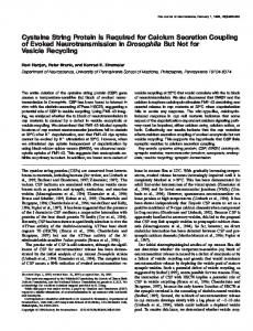
The EMBO Journal Vol.17 No.17 pp.5048–5058, 1998
Cysteine string protein (CSP) is an insulin secretory granule-associated protein regulating β-cell exocytosis
Hilary Brown1,2, Olof Larsson2, Robert Bra¨nstro¨m2, Shao-Nian Yang2, Barbara Leibiger2, Ingo Leibiger2, Gabriel Fried3, Tilo Moede2, Jude T.Deeney2, Graham R.Brown2, Gunilla Jacobsson1, Christopher J.Rhodes4, Janice E.A.Braun5, Richard H.Scheller5, Barbara E.Corkey6, Per-Olof Berggren2 and Bjo¨rn Meister1,7 1Department of Neuroscience, The Berzelius Laboratory, Karolinska Institute, Stockholm, 2The Rolf Luft Center for Diabetes Research, Department of Molecular Medicine, Karolinska Institute, Stockholm, 3Department of Women and Child Health, Karolinska Hospital, Stockholm, Sweden, 4Center for Diabetes Research, Departments of Internal Medicine and Pharmacology, University of Texas South Western Medical Center, Dallas, TX 75235, 5Department of Molecular and Cellular Physiology, Howard Hughes Medical Institute, Beckman Center, Stanford University, Stanford, CA 94305 and 6Diabetes and Metabolism Unit, Evans Department of Medicine, Boston University School of Medicine, Boston, MA 02118, USA 7Corresponding author e-mail:
[email protected]
Cysteine string proteins (CSPs) are novel synaptic vesicle-associated protein components characterized by an N-terminal J-domain and a central palmitoylated string of cysteine residues. The cellular localization and functional role of CSP was studied in pancreatic endocrine cells. In situ hybridization and RT–PCR analysis demonstrated CSP mRNA expression in insulin-producing cells. CSP1 mRNA was present in pancreatic islets; both CSP1 and CSP2 mRNAs were seen in insulin-secreting cell lines. Punctate CSP-like immunoreactivity (CSP-LI) was demonstrated in most islets of Langerhans cells, acinar cells and nerve fibers of the rat pancreas. Ultrastructural analysis showed CSP-LI in close association with membranes of secretory granules of cells in the endo- and exocrine pancreas. Subcellular fractionation of insulinoma cells showed CSP1 (34/36 kDa) in granular fractions; the membrane and cytosol fractions contained predominantly CSP2 (27 kDa). The fractions also contained proteins of 72 and 70 kDa, presumably CSP dimers. CSP1 overexpression in INS-1 cells or intracellular administration of CSP antibodies into mouse ob/ob β-cells did not affect voltage-dependent Ca21-channel activity. Amperometric measurements showed a significant decrease in insulin exocytosis in individual INS-1 cells after CSP1 overexpression. We conclude that CSP is associated with insulin secretory granules and that CSP participates in the molecular regulation of insulin exocytosis by mechanisms not involving changes in the activity of voltage-gated Ca21channels. 5048
Keywords: calcium/chaperone/heat shock protein/ pancreas/secretion
Introduction Cysteine string proteins (CSPs) are highly conserved proteins associated with synaptic vesicles (Zinsmaier et al., 1990; see Braun and Scheller, 1995; Buchner and Gundersen, 1997). The proteins were originally discovered as synapse-associated antigens in the nervous system of Drosophila and, independently, in Torpedo as candidate functional components of presynaptic voltage-sensitive N-type Ca21-channels, expressed ectopically in frog oocytes (Gundersen and Umbach, 1992). CSPs are characterized by a central multiple palmitoylated string of cysteine residues and by an N-terminal J-domain, the latter being a 70 amino acid region of homology shared by bacterial DnaJ, a signature domain present in all members of the DnaJ molecular chaperone/heat shock protein (Hsp) family (Caplan et al., 1993; Braun and Scheller, 1995; see Buchner and Gundersen, 1997). CSPs exist in at least two isoforms, CSP1 and CSP2, generated by alternate RNA splicing (Zinsmaier et al., 1990; Chamberlain and Burgoyne, 1996). CSP2 represents a truncated version of CSP1 lacking the C-terminal 31 amino acid sequence. Several lines of evidence show that CSPs are associated with rat brain synaptic vesicle membranes (Mastrogiacomo and Gundersen, 1995). A general role for CSPs in trafficking events in both neuronal and non-neuronal cells has been implicated since CSP has been identified in membranes of pancreatic zymogen granules (Braun and Scheller, 1995), secretory granules of neurosecretory neurons in the neurohypophysis (Pupier et al., 1997) and in a chromaffin granule-enriched fraction (Kohan et al., 1995; Chamberlain et al., 1996). Detection of CSP mRNA in all human tissues examined suggests a widespread expression of the csp gene (Coppola and Gundersen, 1996). Functionally, CSPs have been demonstrated to be regulatory components of synaptic exocytosis, since deletion of the csp gene in Drosophila generates a phenotype characterized by premature death and temperaturesensitive inhibition of synaptic transmission, resulting in reversible paralysis at elevated temperatures (Zinsmaier et al., 1994). However, the precise role of CSPs in synaptic vesicle exocytosis remains largely unknown. Apart from a suggested functional interaction of CSPs with presynaptic voltage-sensitive N-type Ca21-channels (Gundersen and Umbach, 1992; Mastrogiacomo et al., 1994a), it has been proposed that CSPs operate as molecular chaperones, e.g. assisting in folding or the conformational change of proteins participating in membrane trafficking (Caplan et al., 1993; Zinsmaier et al., 1994; Bohen et al., 1995; © Oxford University Press
CSP in β-cells
see Buchner and Gundersen, 1997). The cysteine-rich string domain in CSPs has led to another hypothetical model for the functional role of CSPs. The cysteine residues of CSPs have been shown to possess a high degree of post-translational fatty acid acylation. This compact acylation conveys a hydrophobic domain to the protein, which also contains highly polar N- and C-termini, and as a consequence CSP has been proposed to be uniquely suited to catalyze membrane fusion (Gundersen et al., 1995). Stimulus–secretion coupling in the pancreatic β-cell involves complex interaction between different signal transduction pathways (see Efendic et al., 1991; Berggren and Larsson, 1994). Glucose metabolism results in an increase in the cellular ATP:ADP ratio, closure of KATPchannels, depolarization of the plasma membrane and opening of voltage-gated L-type Ca21-channels. The subsequent Ca21-influx is a major signal for initiating insulin secretion in the pancreatic β-cell (Berggren and Larsson, 1994). Several proteins that regulate vesicular docking and fusion events, originally characterized in the nervous system, have recently been identified in the endocrine pancreas (Jacobsson et al., 1994), and roles for several of these proteins in the secretion of insulin have been revealed (Jacobsson et al., 1994; Martin et al., 1995, 1996; Regazzi et al., 1995, 1996; Sadoul et al., 1995; Kirali-Bourri et al., 1996; Nagamatsu et al., 1996, 1997; Wheeler et al., 1996; Lang et al., 1997; Mizuta et al., 1997). With the suggestion that CSP may be a modulator of Ca21-channel activity and Ca21-influx being the key event in the initiation of insulin secretion, we have attempted to identify the presence of CSP in pancreatic β-cells and to determine its role in insulin secretion. We have investigated the cellular localization of CSP in the pancreas at the mRNA and protein levels by using in situ hybridization, RT– PCR, RNase protection assay, immunofluorescence histochemistry combined with confocal laser microscopy, immunoelectron microscopy, immunoblotting and subcellular fractionation. The possible functional role of CSP in the β-cell has been evaluated by studies of voltagegated Ca21-channel activity and insulin secretion in individual cells using patch–clamp and amperometric techniques.
Results CSP mRNA in insulin-producing cells In situ hybridization of sections of cultured RINm5F cells as well as rat brain, each with two probes complementary to two different regions of CSP mRNA, showed a strong hybridization signal (data not shown). Hybridization of sections from RINm5F cells with radiolabeled CSP probe in the presence of an excess (1003) of cold probe did not result in any hybridization signal (data not shown). Slides that were emulsion-dipped and hematoxylin-eosin counter-stained, showed silver grains overlying the cytoplasm of individual RINm5F cells (data not shown). Two isoforms of CSP have so far been described, CSP1 and CSP2. The mRNA of the latter isoform has a 72 bp insertion, which contains a stop codon and leads to a shorter 3.3 kDa protein (Chamberlain and Burgoyne, 1996). In order to study which of the isoforms is expressed in the pancreatic islets and in the insulin-producing cell
Fig. 1. (A) Analysis of CSP mRNA isoforms by RT–PCR in rat cerebellum (lane 1), INS-1 cells (lane 2), rat pancreatic islets (lane 3) and ob/ob-mouse pancreatic islets (lane 4). CSP1 isoform corresponds to the 217 bp PCR product and CSP2 isoform to the 289 bp fragment. Lane M shows DNA length marker [pBluescriptII KS(1) digested with HpaII]. (B) Expression of CSP protein detected by immunoblotting using an antiserum to recombinant CSP in rat islets, islets from ob/ob mice, INS-1 cells and rat brain.
line INS-1, we performed RT–PCR analysis employing primers that flank the insertion region. As shown in Figure 1A, INS-1 cells express both isoforms (lane 2) of CSP, whereas pancreatic islets contain only CSP1 (lanes 3 and 4). Therefore, we selected the CSP1 isoform for overexpression studies. CSP-immunoreactivity in rat pancreatic islets, INS-1 and mouse β-cells CSP protein was demonstrated by Western blotting in homogenates from rat pancreatic islets, ob/ob mouse βcells and INS-1 cells (Figure 1B). Incubation of cryostat sections with an antiserum generated to CSP peptide or an antiserum generated to recombinant CSP protein gave identical labeling patterns in both pancreas and brain. In the pancreas, there was strong CSP-like immunoreactivity (CSP-LI) in many cells of the islets of Langerhans and also in nerve fibers and terminals present around blood vessels and extending into the islets (Figure 2A and B). Confocal laser microscopy revealed that CSP-LI was punctate within the cytoplasm of endocrine cells (Figure 2B). The exocrine pancreas demonstrated slightly weaker, punctate CSP-LI in the apical region of acinar cells (Figure 2C). Incubation with non-immune serum gave no fluorescence in the rat pancreas (data not shown). Preabsorption of the CSP peptide antiserum with the corresponding immunogen (10–6 M) gave no immunostaining in exocrine or endocrine cells. Direct double-labeling immunofluorescence histochemistry of rat pancreatic islets demonstrated CSP-LI in insulin- (cf. Figure 3A with B), glucagon- (cf. Figure
5049
H.Brown et al.
3C with D) and somatostatin- (cf. Figure 3E with F) containing cells. Punctate CSP-LI was also demonstrated in cultured RINm5F cells (data not shown), INS-1 cells (Figure 4A) and in cells from ob/ob mouse β-cells (Figure 4B). Doublelabeling of such cells showed extensive, but not complete, co-localization with insulin (Figure 4C). Ultrastructural localization of CSP Immunoelectron microscopy, using an antiserum to recombinant CSP, revealed gold particles localized to the membrane area of secretory vesicles in virtually all endocrine cells of the islets of Langerhans (Figure 5A and B). Insulin was detected in several secretory vesicles. Acinar cells of the exocrine pancreas showed gold particles localized to the membrane area of the zymogen granules (Figure 5C). Localization of gold particles to other organelles of the exocrine pancreas was not apparent. CSP proteins in subcellular fractions and tissue/ cell homogenates The presence and subcellular localization of CSP protein were also examined in pancreas by subcellular fractionation and immunoblotting using antiserum to recombinant CSP. Subcellular fractions of insulinoma cells showed an enrichment of 34 and 36 kDa proteins in the granule fraction with a weak signal at 27 kDa (Figure 6). The membrane fraction showed an enrichment of a 27 kDa protein with a very weak signal at 36 and 34 kDa (Figure 6). The cytosol fraction demonstrated a 27 kDa protein (Figure 6). Homogenates of rat pancreas and liver showed a 36 kDa protein together with a very weakly stained 27 kDa protein (data not shown). Rat brain contained a strong protein band at 34 kDa and a weak band at 27 kDa (Figure 6). Higher molecular weight proteins that reacted with our antiserum to recombinant CSP were also detected. Proteins of 72/70 kDa were identified in all preparations except in brain where the molecular weight was 68 kDa (Figure 6).
Fig. 2. (A–C) Immunofluorescence micrographs of sections of rat pancreas obtained via confocal laser microscopy after incubation with CSP peptide antiserum. Strong CSP-LI is present in the cytoplasm of cells in the islets of Langerhans and also in some nerve fibers and terminals within the islets (solid arrows) (A). At higher magnification (B), it can be seen that the immunofluorescence within the endocrine cells is punctate. Also note weak, punctate CSP-LI in the acinar cells of the exocrine pancreas (open arrow in A). Higher magnification of cells in the exocrine pancreas shows punctate CSP-LI accumulated in the apical region of the acinar cells and in the lumen (C). Bars 5 100 µm.
5050
Effect of CSP overexpression and CSP antibodies on voltage-dependent Ca2F-channels The possible effects of CSP on voltage-dependent Ca21channel activity was tested using the whole-cell configuration of the patch–clamp technique. Overexpression of rat CSP1 in INS-1 cells led to an ~400-fold increase in CSP mRNA amounts (Figure 7A) and a 2.6-fold increase in CSP protein levels (Figure 7B). Immunohistochemical analysis of CSP1 overexpressing cells compared with mock-transfected cells did not reveal any obvious differences in either CSP distribution or co-localization with insulin (Figure 7C). To analyze the effect of CSP1 overexpression on the function of voltage-dependent Ca21-channels at the singlecell level, we co-expressed CSP1 and green fluorescent protein (GFP) in INS-1 cells. Following identification of the positively transfected cells via GFP fluorescence (Figure 8A), the whole-cell configuration was established and membrane currents were recorded during voltage steps to membrane potentials between –60 and 150 mV, from a holding potential of –70 mV. Figure 8B shows the current–voltage (IV) relationship in INS-1 cells following overexpression of CSP1 (CSP1 and GFP, filled circles)
CSP in β-cells
Fig. 3. Immunofluorescence micrographs of sections of rat pancreas obtained via confocal laser microscopy after direct double-labeling combining antiserum to CSP (A, C and E) with antiserum to insulin (B), glucagon (D) and somatostatin (F). Punctate CSP-LI is present in insulin-, glucagonand somatostatin-containing cells (see arrows). Bar 5 100 µm.
or mock-transfection (GFP, open circles), using the lipofectamine transfection technique. No difference in Ca21-channel activity between CSP1 and mock-transfected cells was obtained (Figure 8B). Example current traces from the descending phase of the IV-curve are shown in Figure 8B. In a second approach, we studied the effects of a CSP antiserum on voltage-dependent Ca21-channel currents. Cy3-conjugated secondary antibodies were allowed to perfuse into individual cells via the patch pipette in order to verify accumulation of antibody in the cells (Figure 8C). Mouse β-cells were perfused with CSP antibodies (diluted 1:100) via the recording patch pipette. The cells were then depolarized for 100 ms, from –70 to 0 mV every 20 s. No effect of the CSP antibodies (Figure 8D, bottom traces) on depolarization-induced Ca21-channel currents was obtained during a time period of 15 min, as compared with control cells perfused with mouse IgG (Figure 8D, top traces).
Effect of CSP overexpression on single-cell insulin secretion To investigate the effects of CSP on β-cell secretory capacity, we applied an amperometric technique to study release of serotonin from INS-1 cells preloaded with 5-hydroxy-DL-tryptophan 5-HTP. It is well-established that 5-HTP is converted to serotonin in the β-cell and that serotonin is loaded into the secretory vesicles and cosecreted with insulin by exocytosis (Smith et al., 1995; Aspinwall et al., 1997). Again, CSP1-overexpressing cells were identified by co-expression of GFP. The secretory response to K1-stimulation was significantly decreased in cells overexpressing CSP1 (Figure 9A) as compared with mock-transfected cells (Figure 9B). The number of spikes decreased from 24.8 6 3.6 (n 5 6) in mock-transfected cells to 8.0 6 1.1 (n 5 8) in cells overexpressing CSP1 (P ,0.001; Figure 9C). The area of current spikes (measured in coulombs) can be used to quantitate the moles of detected hormone or transmitter released per
5051
H.Brown et al.
Fig. 4. Immunofluorescence photographs obtained via confocal laser microscopy after incubation of INS-1 cells (A) and ob/ob mouse β-cells (B) with CSP antiserum and after double-labeling with insulin antiserum (C).
vesicle using Faraday’s law (Wightman et al., 1991). No difference in mean spike area between CSP1 and mocktransfected cells could be demonstrated (Figure 9D), indicating that the quality of released serotonin was unaffected by CSP1 overexpression.
Discussion The present study demonstrates that CSP mRNA is expressed in insulin-producing cells and that CSP-LI in these cells is associated with vesicular membranes. Since we could demonstrate the presence of both CSP1 and CSP2 mRNA in insulin-secreting cell lines, we suggest that the 34/36 kDa and the 27 kDa bands represent CSP1 and CSP2, respectively. Furthermore, the granule fraction contains mainly CSP1, whereas CSP2 dominates in the membrane and cytosol fractions of insulinoma cells. Predominance of CSP1 in the granule fraction and the absence of CSP2 in normal islet cells suggests that CSP1 may be 5052
Fig. 5. Immunoelectron microscopy of sections from the endocrine (A and B) and exocrine (C) pancreas after incubation with an antiserum to recombinant CSP. The endocrine pancreas shows gold particles located to the membrane area of secretory vesicles (SV) [arrows in (A)]. (B) Enlargement of rectangle in (A). Insulin (INS) can be seen in some SV. The exocrine pancreas shows gold particles located to the membrane area of zymogen granules (ZG). ER, endoplasmic reticulum; M, mitochondrion; N, nucleus. Bars in (A) and (C) 5 200 nm and bar in (B) 5 100 nm.
involved in catalyzing membrane fusion. There is evidence that CSP is post-translationally palmitoylated in vivo, since treatment with deacylating reagents results in a 7 kDa shift, converting Torpedo CSP from 34 to 27 kDa (Gundersen et al., 1994). Data show that 12 of the 13 cysteine residues of Torpedo CSP are fatty-acylated (Gundersen et al., 1994) and that the cysteine residues are present in a restricted domain, spanning Cys113 to Cys136 (Gundersen et al., 1994). Extensive lipidation has been described for a number of other proteins, including
CSP in β-cells
Fig. 6. Expression of CSP protein detected by immunoblotting using an antiserum to recombinant CSP after subcellular fractionation of rat insulinoma tissue and rat brain. An equal amount of protein has been added to the gel. Proteins corresponding to the expected sizes of 72, 68, 36, 34 and 27 kDa are indicated by arrows.
the proteolipid protein of myelin, GAP-43, SNAP-25, rhodopsin and transferrin receptors (see Gundersen et al., 1994). It has been suggested that this unprecedented degree of acylation in CSP would be well-suited to mediate interactions at membrane interfaces such as those occurring during vesicular membrane fusion events (see Gundersen et al., 1994). It has been shown that the palmitoylated form of CSP2 appears following electrophoretic separation as a 27 kDa protein (Chamberlain and Burgoyne, 1996). Our results show that CSP also occurs in a higher molecular weight (72 kDa) form, which has previously been suggested to represent dimerization of CSP (Braun and Scheller, 1995; Mastrogiacomo and Gundersen, 1995; Chamberlain and Burgoyne, 1996). In this context, it is of interest that two models, the ‘acyl-flip’ and the ‘bilayer collapse’ models, have been proposed to explain how CSP operates as a candidate fusion-promoting agent (Gundersen et al., 1994). Both models suggest that pairs of CSP monomers would be required for fusion to occur (Mastrogiacomo et al., 1994a,b; Gundersen et al., 1995). As shown in the present study, CSP1 overexpression in individual INS-1 cells resulted in a significant decrease in insulin release as compared with mock-transfected cells. The fact that CSP1 overexpression inhibits insulin release may suggest that CSP indeed has a regulatory role on insulin exocytosis under physiological conditions. CSP has earlier been implicated as a regulator of presynaptic Ca21-channels (Gundersen and Umbach, 1992). Using a suppression cloning strategy, whereby N-type Ca21-channels were expressed in Xenopus oocytes, a putative Ca21-channel subunit cDNA was identified (Gundersen and Umbach, 1992). After the injection of CSP antisense RNA, electrophysiological studies demonstrated a complete suppression of Ca21-channel activity (Gundersen and Umbach, 1992). Unexpectedly, sequence analysis revealed that this candidate Ca21-channel subunit was CSP, which has been cloned in Drosophila by Zinsmaier et al. (1990). The inhibitory effect of CSP1 overexpression on insulin secretion, but the absence of effect of CSP1 overexpression or intracellular administra-
Fig. 7. (A) RNase protection analysis of CSP and β-actin mRNA in CSP1-overexpressing INS-1 cells (lane 1) and mock-transfected cells (lane 2). (B) Western blot analysis of CSP protein levels in CSPoverexpressing cells (CSP) and mock-transfected cells (Mock). Equal amounts of protein have been used for gel electrophoresis. (C) Colocalization of CSP- and insulin-immunoreactivity in CSP1overexpressing cells (CSP) [(a) and (c)] and mock-transfected cells (Mock) [(b) and (d)]. Bar 5 10 µm. Overexpression of CSP1 results in a 400-fold increase in CSP1 mRNA and a 2.6-fold increase in CSP protein levels. The distribution of the overexpressed CSP protein and co-localization with insulin is similar to that in mock-transfected cells.
tion of CSP antibodies on voltage-gated Ca21-currents is interesting and suggests that CSP is a regulator of β-cell exocytosis via a mechanism not involving voltage-gated Ca21-channels. Recent results obtained from Drosophila csp mutants which have a temperature-dependent block of synaptic transmission (Umbach et al., 1994) show that CSPs appear to regulate an early step in the Ca21dependent neurotransmitter secretion at the Drosophila neuromuscular junction (Umbach and Gundersen, 1997). The dominant presence of N-type Ca21-channels in neurons and L-type Ca21-channels in β-cells (Berggren and Larsson, 1994) may explain the modulatory effect of CSP on Ca21 fluxes in neurons and absence of effect on Ca21-channel activity in endocrine β-cells. It is of interest that synaptotagmin, but not CSP, synaptophysin or Rab 3A, have been detected in P/Q5053
H.Brown et al.
Fig. 8. (A) CSP1-overexpressing INS-1 cells identified by GFP fluorescence. Confocal image of a cell group reveals a single GFP-expressing cell. Phase-contrast of the cell group having undergone the transfection procedure. Overlay of the confocal and phase-contrast images shows distribution of GFP within the cell cluster. Bar 5 10 µm. (B) Effects of CSP1 overexpression on voltage-dependent Ca21-channel activity in INS-1 cells. Integrated Ca21-currents were recorded during voltage steps (100 ms) to membrane potentials between –60 and 150 mV from a holding potential of –70 mV in INS-1 cells overexpressing CSP1 (open circles; n 5 32) and mock-transfected INS-1 cells (filled circles; n 5 29). Currents are corrected for cell size by dividing the results in each recording by cell capacitance. No difference in cell capacitance between mock (4.18 6 0.27 pF; n 5 18) and CSP1-transfected INS-1 cells (4.18 6 0.25 pF; n 5 34) could be seen. Data are expressed as mean 6 SEM. Examples of whole-cell Ca21-current traces obtained during 100 ms depolarizations from a holding potential of –70 to 110 mV (10 mV steps) in INS-1 cells overexpressing CSP1 (left) and in mock-transfected (right) INS-1 cells. (C) Identification of cells that accumulate Cy3-conjugated secondary antibodies. Cy3-conjugated secondary antibodies (red fluorescence) were perfused into individual mouse β-cells via the patch pipette using the whole-cell configuration and visualized via confocal microscopy. A group of mouse β-cells is seen in transmission microscopy. Overlay of the confocal and transmission images shows a single Cy3-fluorescent (red) cell. Bar 5 10 µm. (D) Effects of CSP-antibodies on integrated Ca21currents in mouse β-cells. Membrane currents were recorded using the whole-cell configuration of the patch–clamp technique. Repetitive depolarizing voltage steps (100 ms) were applied to 0 mV from a holding potential of –70 mV. Test pulses were given every 20 s. The whole-cell configuration was established ~1 min before starting the pulse protocol. Examples of current traces under control conditions and when CSP antibodies (dilution 1:100) were included in the pipette solution. The two top traces are from the same control cell (Ctr) and show the resulting current traces at 1 and 15 min after starting the pulse protocol. Below are corresponding traces in the presence of CSP antibodies (CSPab). This experimental procedure was repeated four times with identical results.
5054
CSP in β-cells
activity can be modulated by protein cofactors, including DnaJ and its homologs. It is known that DnaJ interacts with Hsp70, stimulating its ATPase activity (see Caplan et al., 1993). It has been suggested that DnaJ homologues are regulators of Hsp70 activity (Caplan et al., 1993). It was shown recently that CSP activates the constitutive Hsp70 (Hsc70) in a dose-dependent manner (Braun et al., 1996; Chamberlain and Burgoyne, 1997). Hsc70 is an abundant neural protein with coupled protein binding and ATPase activity, which has been shown to uncoat clathrincoated vesicles in vitro, an obligate step in the fusion of transport vesicles to their target membranes (Schlossman et al., 1984). Hsc70 may function within the biochemical pathways of exo- or endocytosis to promote formation or dissociation of multimeric complexes or to regulate conformational changes (see Braun et al., 1996). Taken together, our results showing reduced insulin secretion after CSP1 overexpression, but absence of regulatory action on voltage-gated Ca21 currents, may be compatible with a chaperone function of CSP in pancreatic β-cells.
Materials and methods
Fig. 9. Amperometric recordings from single, CSP1-transfected or mock-transfected, INS-1 cells preloaded with 5-HTP. (A) and (B) show typical examples of recordings following stimulation of CSP1transfected (A) and mock-transfected (B) INS-1 cells with 25 mM KCl. Current spikes were recorded by a carbon fiber near the cell and each spike represents a single quantum of released catecholamine. The cells were perfused with 3 mM glucose and the arrows above the recordings indicate the application of KCl. Compiled data of number of spikes (C) and mean spike area (D) during 30 s periods under each experimental condition. Note that there is a significant decrease in number of spikes after CSP transfection as compared with mocktransfection. Data are presented as mean 6 SEM. In (C), n 5 4–7; in (D), a total number of 64 spikes in eight different recordings were analyzed in CSP1-transfected cells and 149 spikes in six different recordings following mock-transfection. (E) Example of spikes in CSP1- (top) and mock- (bottom) transfected INS-1 cells. Ten representative spikes from each group were superimposed and aligned at peak current of each spike. P ,0.001.
type Ca21-channel-associated complexes (Martin-Moutout et al., 1996). Trimeric SNARE complexes are implicated in Ca21-dependent exocytosis, which is believed to be partly regulated by synaptotagmin. These observations suggest that CSP is not a part of the SNARE complex. The occurrence of a J-domain indicates that CSP belongs to the DnaJ chaperone/Hsp family. DnaJ proteins have been associated with chaperone-like functions, e.g. to aggregate and disaggregate proteins, facilitate protein transport, facilitate folding or conformational changes in protein, or to regulate protein function (Caplan et al., 1993; see Braun and Scheller, 1995). The common mechanism underlying such diverse functions appears to be the ability of Hsp70 to bind transiently and sequester unfolded regions of substrate proteins, thereby preventing unproductive aggregation (Pelham, 1986). The Hsp70 protein binding and release kinetics are governed by an intrinsic ATPase activity. Both the ATPase activity and the protein-binding
In situ hybridization For in situ hybridization, RINm5F cells were used (Gazdar et al., 1980). RINm5F cells were centrifuged into pellets that were fixed in 4% paraformaldehyde for 1 h, washed with phosphate-buffered saline (PBS) and frozen on dry ice. The pellets were cut at 14 µm section thickness in a cryostat and mounted onto ProbeOn™ microcope Slides (Fisher Scientific, Pittsburgh, PA). Rat brain and newborn mice were frozen and sectioned and used for control purposes. Two oligonucleotide probes (CAGTCCAAGAACATGGTATAACGATTCCCCGGAAGTAGAGAGTGAGCG, probe 1; and GAGTACGAAGTAGGTGTTGACATTCTCCTCCCCAAATTGCTCAGCCAC, probe 2), with an optimal G-C content (50–65%) and homology ,80% with DDBJ/EMBL/GenBankentered nucleotide sequences, were synthesized (Scandinavian Gene Synthesis, Ko¨ping, Sweden), reversed and complementary to rat CSP mRNA (Braun and Scheller, 1995; Mastrogiacomo and Gundersen, 1995). In situ hybridization was performed essentially as previously described (Young, 1990; Dagerlind et al., 1992). Probes were 39end labeled with [α-35S]dATP (NEN, Boston, MA), using terminal deoxynucleotidyl transferase (Amersham Ltd, Amersham, UK) and purified using Nensorb 20 columns (NEN). Sections were air-dried and incubated for 16 h at 42°C with 106 c.p.m. of the labeled probe in a hybridization solution containing 50% deionized formamide, 43 standard saline citrate (SSC) (13 SSC 5 0.15 M NaCl, 0.015 M sodium citrate), 13 Denhardt’s solution [0.02% bovine serum albumin (BSA), 0.02% Ficoll, 0.02% polyvinylpyrolidone], 1% N-lauroylsarcosine, 0.02 M NaPO4 (pH 7.0), 10% dextran sulfate, 500 µg/ml denatured salmon sperm DNA and 200 mM dithiothreitol. Following hybridization, the sections were rinsed in 13 SSC at 55°C for 60 min, rinsed in distilled water, dehydrated in 60 and 95% ethanol and apposed to β-max autoradiography film (Amersham) at –20°C. After two weeks of exposure (probe 1) and one week of exposure (probe 2), the films were developed with Kodak LX 24 and fixed with Kodak AL 4. In addition to film autoradiography, the sections were dipped in Kodak NTB2 autoradiography emulsion in distilled water, exposed for 6 and 3 weeks (probe 1 and probe 2, respectively) at 4°C, developed in Kodak D19 and fixed in Kodak 3000. Sections were rinsed in distilled water and counterstained with hematoxylin-eosin. All sections were examined and photographed under bright- or dark-field illumination, using a Nikon Microphot-SA microscope and Kodak Tmax 100 ASA film. RT–PCR and RNase protection analysis Detection of CSP1 and CSP2 mRNA was performed by RT–PCR using as the upstream primer 59-TTCGTCGTCTGTGGCCTCCT-39 and as the downstream primer 59-TGGTCTCTGTGGCGGATGCT-39. Five micrograms of total RNA obtained from INS-1 cells, rat pancreatic islets, ob/ob mouse islets and rat cerebellum were reverse-transcribed using MMLV revertase. Aliquots of the generated cDNA were used for PCR-mediated amplification using the RT–PCR Kit (Stratagene) and
5055
H.Brown et al. [α-32P]dCTP. PCR was performed in an AutogeneII-thermocycler (Grant, UK) using a linked program (1 cycle of 5 min at 94°C, 5 min at 54°C, 2 min at 72°C; and 30 cycles of 1 min at 94°C, 2 min at 54°C, 2 min at 72°C). PCR products were separated on a 2% agarose gel in TBE. Levels of CSP mRNA were analyzed by RNase-protection analysis. Therefore, the rat CSP cDNA fragment from nucleotides 22 to 467 (according to DDBJ/EMBL/GenBank accession number S81917) was subcloned into pBluescriptII SK(–) (Stratagene, La Jolla, CA). Radiolabeled cRNA was generated on the linearized plasmids by employing the SP6/T7 in vitro-transcription kit (Boehringer Mannheim) and [α-32P]dCTP. Following purification by polyacrylamide gel electrophoresis (6% acrylamide, 7 M urea in 13 TBE), equal c.p.m. of the labeled cRNA probes (53104 c.p.m. final activity) were mixed with the total RNA in hybridization solution, incubated for 5 min at 90°C and hybridized at 45°C overnight. RNase protection was performed using the RPA II kit (Ambion, Austin, TX). Quantification of protected complexes was performed by phosphoimaging. Values obtained for CSP mRNA were normalized by β-actin-mRNA values.
CSP antisera Rabbit polyclonal anti-CSP serum was generated against a CSP/ glutathione S-transferase (GST) fusion protein (anti-CSP). A cDNA encoding the entire rat CSP open reading frame was obtained by PCR from the 2 kb rat CSP clone. The sequence was verified, constructed as a GST-fusion protein and expressed in Escherichia coli. The fusion protein was purified on an agarose–glutathione column before immunization. The characterization of the CSP antipeptide antiserum has been described previously (Braun and Scheller, 1995). Immunofluorescence histochemistry and confocal microscopy Male Sprague-Dawley rats (B & K Universal; 150–200 g) were anesthetized with sodium pentobarbital (Mebumal®; 40 mg/kg i.p.) and perfused via the ascending aorta with Ca21-free Tyrode’s solution (37°C), followed by an ice-cold mixture of formalin–picric acid (4% paraformaldehyde and 0.4% picric acid in 0.16 M phosphate buffer, pH 6.9). The pancreas was rapidly removed and immediately fixed by immersion in the same fixative for 90 min, and rinsed for at least 24 h in a 0.1 M phosphate buffer (pH 7.4) containing 10% sucrose, 0.02% bacitracin and 0.01% sodium azide. Sections were cut at 10 µm thickness in a cryostat and processed for indirect immunofluorescence. INS-1 cells and ob/ob mouse β-cells were prepared and cultured on coverslips. The cells were fixed by immersion in fixative (as above) for 10 min and thereafter washed several times in PBS. The sections and cells were incubated in rabbit antisera to CSP peptide or recombinant CSP protein (diluted 1:500 and 1:800, respectively) for 18–22 h at 4°C. The sections were then washed in PBS for 30 min and incubated with fluoroscein isothiocyanate (FITC)-conjugated donkey anti-rabbit (diluted 1:40; Jackson Immuno Research Laboratories Inc., West Grove, PA) secondary antibody. After a further wash in PBS, the sections were mounted in 0.1% p-phenylendiamine dissolved in PBS and glycerol (1:3). Sections were double-stained by combining rabbit antiserum to CSP peptide with guinea-pig antiserum to insulin (diluted 1:2000; UCB-Bioproducts S.A., Braine-l’Alleud, Belgium) or mouse monoclonal antibodies to glucagon (diluted 1:1000; Novo Nordisk AS, Bagsvaerd, Denmark) or mouse monoclonal antibodies to somatostatin (diluted 1:200; Buchan et al., 1985). The combinations were visualized using a mixture of FITCconjugated donkey anti-rabbit and lissamine-rhodamine (LRSC)conjugated goat anti-guinea-pig (1:40) or goat anti-mouse (1:40; both from Jackson Immuno Research Laboratories Inc.) secondary antibodies. Control sections were stained using non-immune serum (diluted 1:500) as primary antibody and by preabsorption of the antiserum to CSPpeptide with cognate CSP-peptide at 10–6 M. Sections were also examined in a Bio-Rad MRC-600 laser scanning confocal imaging system equipped with a krypton/argon mixed gas laser and a Nikon Optiphot II microscope. The standard K1/K2 dual channel filter sets combined with an excitation filter (488 DF 10 for FITC-induced fluorescence and a 568 DF 10 for LRSC-induced fluorescence) was used to examine the immunoreactivity. The images were produced using a Tektronix Phaser IIsd printer. Electron microscopy Pancreatic tissue was collected from male Sprague-Dawley rats (B & K Universal, Stockholm, Sweden; 150–200 g) and 1 mm3 tissue blocks were fixed at 4°C in a mixture of 4% paraformaldehyde and 0.1% glutaraldehyde in 0.1 M phosphate buffer (pH 7.4) for 4 h. After fixation, the tissue was partially dehydrated in 70% ethanol and embedded in LR White resin (Agar Scientific Ltd, Cambridge, UK). Thin sections were
5056
collected on formvar coated nickel grids. The grids were washed in distilled water and incubated in 5% normal goat serum in PBS containing 0.1% BSA and 0.13% sodium azide (buffer A) for 30 min at room temperature. The sections were placed in primary rabbit anti-recombinant CSP (diluted 1:500) in PBS containing 1% BSA and 0.13% sodium azide for 18–22 h at 4°C. Control sections were incubated in nonimmune serum. The grids were washed thoroughly in buffer A and then transferred to 10 nm gold-conjugated goat anti-rabbit secondary antibodies (diluted 1:50; Amersham) for 30 min at room temperature, followed by a wash in buffer A. The sections were subsequently postfixed in 2% glutaraldehyde in 0.1 M phosphate buffer (pH 7.4) for 15 min and finally stained with uranyl acetate (15 min) and lead citrate (2 min) and examined with a JEOL-1200EX electron microscope. Kodak 4489 film was used for photography.
β-cell granule preparation and tissue/cell homogenization Transplantable rat insulinoma was propagated in New England Deaconess Hospital (NEDH) rats, which were obtained from an in-house breeding colony at the Joslin Diabetes Center, Harvard Medical School, Boston, MA. The subcellular fractionation of the harvested insulinoma by differential and density gradient centrifugation was performed as described previously. Insulinoma tissue was homogenized at 4°C in 0.27 M sucrose, 10 mM HEPES, 1 mM EGTA, pH 6.5 using 8–10 strokes of a Potter homogenizer. The homogenate was then centrifuged at 1700 g for 7 min at 4°C to remove cell debris and nuclei. The supernatant was applied to an iso-osmotic discontinuous density gradient composed of 19.2% (w/v) Nycodenz (Nyegaard Diagnostica AS, Oslo, Norway) mixed with 0.27 M sucrose in the proportions 1:0, 1:1 and 1:3 (5 ml of each) and then centrifuged at 4°C in a Beckman SW 28 rotor at 100 000 g for 60 min. Three fractions consisting of insulin secretory granules, plasma membrane and cytosol were obtained. The band consisting of enriched β-cell granules was removed from the interface between the high and medium density solutions and then washed in the homogenization buffer. The granule fraction was mixed with eight volumes of 27% (v/v) Percoll (Pharmacia, Uppsala, Sweden) in 0.27 M sucrose/10 mM MES, pH 6.5 and centrifuged at 35 000 g at 4°C for 45 min using a Sorvall SS34 rotor. The purified β-cell granule fraction was recovered and washed five or six times in 0.25 M sucrose/10 mM MES, pH 6.5, to remove the gradient material. The fraction was stored in liquid nitrogen. Tissue homogenates were prepared from male Sprague-Dawley rats by homogenization in 50 mM Tris–HCl, 100 mM NaCl and 1 mM EGTA (pH 7.5) containing the following protease inhibitors: 0.2 mM PMSF, 2 µM leupeptin, 10 µM pepstatin, 50 µM benzamidine, 0.5 µM soya trypsin inhibitor and 0.005% DNase (Sigma Chemical Co., St Louis, MO) using a teflon/glass homogenizer. HIT-T15 cells were homogenized in 4 mM HEPES (pH 7.4) and the same protease inhibitors as above. The homogenates were centrifuged at 5000 g for 10 min at 4°C in a Beckman TLA100.2 rotor. RINm5F cell homogenates were prepared by homogenization in 1% SDS using a Branson ultrasonic homogenizer. Protein estimation was performed by protein microassay (Bio-Rad, Hercules, CA). SDS–PAGE and Western blotting Samples were denatured for 5 min at 100°C in SDS–PAGE sample buffer (Hames, 1990). Analysis was performed on a 12% polyacrylamide SDS–PAGE gel (Laemmli, 1970). The proteins were transferred to 0.2 µm nitrocellulose (Schleicher and Schuell, GmbH, Dassel, Germany) in 20% methanol, 20 mM Tris, 150 mM glycine and 0.05% SDS for 12–14 h at 0.35 mA (Towbin et al., 1979). Non-specific binding of the primary antibody was blocked by incubation in 5% milk powder in buffer A (10 mM Tris, 150 mM NaCl, pH 7.4, containing 0.05% Tween-20). The blot was subsequently probed overnight at 4°C with polyclonal rabbit anti-recombinant CSP (diluted 1:10 000; Braun and Scheller, 1995) with buffer A containing 5% milk powder. The blots were washed with buffer A and then incubated for 1 h at room temperature with peroxidase-conjugated goat-anti-rabbit IgG (diluted 1:2000; Organon Teknika Corp., Cappel Research Products, Durham, NC) in buffer A containing 5% milk powder and finally washed with buffer A. Detection was performed using an ECL detection system (Amersham) and the immunoreactive bands visualized with Hyperfilm (Amersham). CSP1 overexpression The rat CSP1 cDNA was subcloned into a pRc/CMV (Invitrogen, Carlsbad, CA) backbone vector, thus generating pCMVrCSP. Plasmid pCMVGFP contains the ‘humanized’ form of the S65T-mutant of the
CSP in β-cells GFP. All expression constructs were purified twice by CsCl-density ultracentrifugation. INS-1 cells were grown in RPMI 1640 medium supplemented with 5.5 mM glucose, 10 mM HEPES (pH 7.4), 1 mM sodium pyruvate, 50 µM β-mercaptoethanol, 100 units/ml penicillin, 100 µg/ml streptomycin, 2 mM glutamine and 10% fetal calf serum at 5% CO2 and 37°C. Transfection was performed overnight by the lipofectamine technique in supplemented RPMI 1640 medium without serum and antibiotics. For co-transfection 1 µg pCMVrCSP or 1 µg pRc/CMV (mock) and 1 µg pCMVGFP was mixed with 6 µl lipofectamine (Gibco-BRL, Paisley, UK) and 1.0 ml serum-free RPMI 1640 medium supplemented as above. Cells were cultured for a further 36 h in Petri dishes for electrophysiological and amperometric studies. For analysis of CSP overexpression at RNA and protein levels, INS-1 cells were co-transfected with pHOOK1 (Invitrogen, Carlsbad, CA) and either pCMVrCSP or pRc/CMV (mock) and transfected cells were collected by the HOOK technique according to the manufacturer’s instruction. One percent SDS extracts of CSP-transfected and mock-transfected INS-1 cells (20 µg) were loaded on 12.5% polyacrylamide gels, and immunoblotted using rabbit CSP antiserum (diluted 1:10 000). Blots were quantified by densitometric analysis using ImageQuant software (Molecular Dynamics Inc., Sunnyvale, CA).
Electrophysiology The activity of the voltage-dependent Ca21-channels was studied in INS-1 cells transfected with CSP as described above and in mouse pancreatic β-cells. For the preparation of single β-cells, mice were starved for 24 h and then killed by decapitation. Pancreatic islets from ob/ob mice were isolated by a collagenase technique (Lacy and Kostianovsky, 1967) and a β-cell suspension was prepared and washed, essentially as previously described (Lernmark, 1974). The cells were resuspended in RPMI 1640 culture medium (Flow Laboratories, Scotland, UK), containing 11 mM glucose, supplemented with 10% fetal bovine serum, 100 IU/ml penicillin, 100 µg/ml streptomycin and 60 µg/ml gentamycin. The cell suspension was seeded into Petri dishes and incubated at 37°C in 5% CO2 for 1–3 days. The culture medium was removed and the cells were washed with a solution composed as follows: 138 mM NaCl; 5.6 mM KCl; 1.2 mM MgCl; 10 mM CaCl2; 10 mM tetraethylammoniumchloride; 5 mM HEPES pH 7.4. Cells were covered with 350 µl of the described solution. The pipette solution contained: 150 mM N-methyl-D-glucamine (NMDG); 110 mM HCl; 1 MgCl2; 2 mM CaCl2; 10 EGTA; 3 mM MgATP; 5 mM HEPES pH 7.15. All experiments were performed at room temperature (22–24°C). The free concentration of Ca21 was calculated to be 60 nM from the binding constants of Martell and Smith (1974). NMDG was substituted for K1 in the pipette solution in order to block outward-directed K1-currents. INS-1 cells co-expressing CSP1 and GFP were detected by GFP fluorescence using a Zeiss Axiovert 35 M fluorescence microscope with the following settings: excitation light from a 75 W xenon lamp with a 475–495 nm excitation filter, dichroic mirror and a 515–565 nm emission filter. Images for documentation were made using fluorescence digital imaging. CSP antibodies were added to the pipette solution at a dilution of 1:100. In order to prove the physiological presence of antibodies applied by the patch pipette inside the mouse β-cells, we used a Cy3-conjugated rabbit IgG secondary antibody and detected it using a confocal microscope (Leica CLSM; Leica Lasertechnik GmbH, Heidelberg, Germany) with the following settings: 403/1.30 oil Leitz fluotar objective lens, excitation wavelength 568 nm (argon/krypton laser) and a long-pass 580 nm emission filter. The whole-cell configuration of the patch–clamp technique (Hamill et al., 1981) was used, utilizing an Axopatch 200 patch–clamp amplifier (Axon Instruments, Inc., Foster City, CA). Voltage-steps were generated, digitized and stored in a computer (IBM AT-clone), using the program pClamp (Axon Instruments) and Labmaster ADC (Scientific Solutions, Inc., Solon, OH). The current responses were filtered at 1 kHz, Bessel filter (–3 dB-point; Frequency Devices, Haverhill, MA). Amperometry Glass-encased carbon fiber microelectrodes were pulled from borosilicate glass capillaries (Hilgenberg, Malsfeld, Germany), containing a single carbon fiber of 9 µm diameter (P-55S; Amoco Corp., Greenville, SC). The carbon fibers were sealed in the tip by dipping them in Sylgard (Dow Corning, Kanagawa, Japan) and then cut at a 35–45° angle. Six to 12 h prior to experiments, cells were pre-incubated in (5-HTP (Sigma Co., St Louis, MO). 5-HTP was prepared as stock solution at a concentration of 25 mM and added to the culture medium giving a final concentration of 1 mM. During experiments, Petri dishes were placed
in a temperature-regulated perfusion chamber (32–36°C), perfused with a standard extracellular (EC) solution consisting of: 138 mM NaCl, 5.6 mM KCl, 1.2 mM MgCl2, 2.6 mM CaCl2, 5 mM HEPES–NaOH, 3 mM glucose at pH 7.40. Electrodes were placed in close contact with the cell surface with a micromanipulator (Narishige PE-2). Cells were stimulated by a transient (10–30 s) pulse of EC with the K1 concentration elevated to 25 mM, using a flow-injector (Transjector, Eppendorf, Hamburg, Germany). Microelectrodes were operated in amperometric mode using an EI-400 potentiostat (Ensman Instrumentation, Bloomington, IN). Records were filtered at 100 Hz (–3 dB value), digitized at 200 Hz (Axon Instrument ADC TL-1) and stored in a computer (Axotape software, Axon Instrument). A holding potential (VC) of 600 mV versus a sodium-saturated calomel electrode was applied to the carbon fiber electrode, sufficient to oxidize 5-HTP (Zhou and Misler, 1996). Amperometric currents were analyzed using in-house software and compared using Student’s t-test. For trace figures, digitized recordings were exported into CorelDraw (Corel Corp., Ottawa, Ontario, Canada) for final layout.
Acknowledgements This research was supported by grants from the Swedish Medical Research Council (04X-10358, 03X-09890, 03X-09891, 03XS-12708, 12P-10169, 19X-00034, 03X-12549), Åke Wibergs Stiftelse, Magnus Bergvalls Stiftelse, United States Public Health Service Grant DK-35914, The Swedish Diabetes Association, The Nordic Insulin Foundation Committee, The Juvenile Diabetes Foundation, Berth von Kantzows Foundation and Funds from the Karolinska Institute.
References Aspinwall,C.A., Brooks,S.A., Kennedy,R.T. and Lakey,J.R.T. (1997) Effects of intravesicular H1 and extracellular H1 and Zn21 on insulin secretion in pancreatic beta cells. J. Biol. Chem., 272, 31308–31314. Berggren,P.-O. and Larsson,O. (1994) Ca21 and pancreatic β-cell function. Biochem. Soc. Trans., 22, 12–18. Bohen,S.P., Kralli,A. and Yamamoto,K.R. (1995) Hold ’em and fold ’em: chaperones and signal transduction. Nature, 268, 1303–1304. Braun,J.E.A. and Scheller,R.H. (1995) Cysteine string protein, a DnaJ family member, is present on diverse secretory vesicles. Neuropharmacology, 34, 1361–1369. Braun,J.E.A., Wilbanks,S.M. and Scheller,R.H. (1996) The cysteine string protein secretory vesicle protein activates Hsc70 ATPase. J. Biol. Chem., 271, 25989–25993. Buchan,A.M.J., Sikora,L.K.J., Levy,J.G., McIntosh,C.H.S., Dyck,J. and Brown,J.C. (1985) An immunocytochemical investigation with monoclonal antibodies to somatostatin. Histochemistry, 83, 175–180. Buchner,E. and Gundersen,C.B. (1997) The DnaJ-like cysteine string protein and exocytotic neurotransmitter release. Trends Neurosci., 20, 223–227. Caplan,A.J., Cyr,D.M. and Douglas,M.G. (1993) Eucaryotic homologues of Escherichia coli dnaJ: a diverse protein family that functions with Hsp70 stress proteins. Mol. Biol. Cell, 4, 555–563. Chamberlain,L.H. and Burgoyne,R.D. (1996) Identification of a novel cysteine string protein variant and expression of cysteine string proteins in non-neuronal cells. J. Biol. Chem., 271, 7320–7323. Chamberlain,L.H. and Burgoyne,R.D. (1997) Activation of the ATPase activity of heat-shock proteins Hsc70 and Hsp70 by cysteine-string protein. Biochem. J., 322, 853–858. Chamberlain,L.H., Henry,J. and Burgoyne,R.D. (1996) Cysteine string proteins are associated with chromaffin granules. J. Biol. Chem., 271, 19514–19517. Coppola,T. and Gundersen,C.B. (1996) Widespread expression of human cysteine string proteins. FEBS Lett., 391, 269–272. Dagerlind,Å., Friberg,K., Bean,A.J. and Ho¨kfelt,T. (1992) Sensitive mRNA detection using unfixed tissue: combined radioactive and nonradioactive in situ hybridization histochemistry. Histochemistry, 98, 39–49. Efendic,S., Kindmark,H. and Berggren,P.-O. (1991) Mechanisms involved in the regulation of the insulin secretory process. J. Intern. Med. Suppl. 2, 229, 9–22. Gazdar,A.F., Chick,W.L., Oie,H.K., Sims,H.L., King,D.L., Weir,G.C. and Lauris,V. (1980) Continuous, clonal, insulin—and somatostatin— secreting cell lines established from a transplantable rat islet cell tumour. Proc. Natl Acad. Sci. USA, 77, 3519–3523. Gundersen,C.B. and Umbach,J.A. (1992) Suppression cloning of the
5057
H.Brown et al. cDNA for a candidate subunit of a presynaptic calcium channel. Neuron, 9, 527–537. Gundersen,C.B., Mastrogiacomo,A., Faull,K. and Umbach,J.A. (1994) Extensive lipidation of a Torpedo cysteine string protein. J. Biol. Chem., 269, 19197–19199. Gundersen,C.B., Mastrogiacomo,A. and Umbach,J.A. (1995) Cysteinestring proteins as templates for membrane fusion: models of synaptic vesicle exocytosis. J. Theor. Biol., 172, 269–277. Hames,B.D. (1990) One-dimensional polyacrylamide gel electrophoresis. In Hames,B.D. and Rickwood,D. (eds), Gel Electrophoresis of Proteins. A Practical Approach. 2nd edn. IRL Press, Oxford, UK, pp. 1–147. Hamill,O.P., Marty,A., Neher,E., Sakman,B. and Sigworth,F.J. (1981) Improved patch–clamp techniques for high-resolution current recording from cells and cell-free membrane patches. Pflu¨gers Arch., 391, 85–100. Jacobsson,G., Bean,A.J., Scheller,R.H., Juntti-Berggren,L., Deeney,J.T., Berggren,P.-O. and Meister,B. (1994) Identification of synaptic proteins and their isoform mRNAs in compartments of pancreatic endocrine cells. Proc. Natl Acad. Sci. USA, 91, 12487–12491. Kirali-Bourri,C., Morgan,A., Burgoyne,R., Weller,U., Wollheim,C.B. and Lang,J. (1996) Soluble N-ethylamide sensitive factor attachment protein and N-ethylamide insensitive factors are required for Ca21stimulated exocytosis of insulin. Biochem. J., 314, 199–203. Kohan,S.A., Pescatori,M., Brecha,N.C., Mastrogiacomo,A., Umbach,J.A. and Gundersen,C.B (1995) Cysteine string protein immunoreactivity in the nervous system and adrenal gland of rat. J. Neurosci., 15, 6230–6238. Lacy,P.E. and Kostianovsky,M. (1967) Method for the isolation of intact islets of Langerhans from the rat pancreas. Diabetes, 16, 35–39. Laemmli,U.K. (1970) Cleavage of structural proteins during the assembly of the head of bacteriophage T4. Nature, 227, 680–685. Lang,J., Fukuda,M., Zhang,H., Mikoshiba,K. and Wollheim,C.B. (1997) The first C2 domain of synaptotagmin is required for exocytosis of insulin from pancreatic β-cells: action of synaptotagmin at low micromolar calcium. EMBO J., 16, 5837–5846. Lernmark,Å. (1974) The preparation of and studies on, free cell suspensions from mouse pancreatic islets. Diabetologia, 10, 431–438. Martell,A.E. and Smith,R.M. (1974) Critical stability constants. Vol I. Amino Acids. Vol II. Amines. Plenum Press, New York, NY. Martin,F., Moya,F., Gutierrez,L.M., Reig,J.A. and Soria,B. (1995) Role of syntaxin in mouse pancreatic beta cells. Diabetologia, 38, 860–863. Martin,F., Salinas,E., Vazquez,J., Soria,B. and Reig,J.A. (1996) Inhibition of insulin release by synthetic peptides shows that the H3 region at the C-terminal domain of syntaxin-1 is crucial for Ca21 but not guanosine-59-[γ-thio]triphosphate-induced secretion. Biochem. J., 320, 201–205. Martin-Moutout,N., Charvin,N., Leveque,C., Sato,K., Nishiki,T., Kozaki,S., Takahashi,M. and Seager,M. (1996) Interaction of SNARE complexes with P/Q-type calcium channels in rat cerebellar synaptosomes. J. Biol. Chem., 271, 6567–6570. Mastrogiacomo,A. and Gundersen,C.B. (1995) The nucleotide and deduced amino acid sequence of a rat cysteine string protein. Mol. Brain Res., 28, 12–18. Mastrogiacomo,A., Parsons,S.M., Zampighi,G.A., Jenden,D.J., Umbach,J.A. and Gundersen,C.B. (1994a) Cysteine string proteins: a potential link between synaptic vesicles and presynaptic calcium channels. Science, 263, 981–982. Mastrogiacomo,A., Evans,C.J. and Gundersen,C.B. (1994b) Antipeptide antibodies against a Torpedo cysteine-string protein. J. Neurochem., 62, 873–880. Mizuta,M., Kurose,T., Miki,T., Shoji-Kasai,Y., Takahashi,M., Seino,S. and Matsukura,S. (1997) Localization and functional role of synaptotagmin III in insulin secretory vesicles in pancreatic β-cells. Diabetes, 46, 2002–2006. Nagamatsu,S., Fujiwara,T., Nakamichi,Y., Watanabe,T., Katahira,H. and Seino,S. (1996) Expression and functional role of syntaxin1/HPC-1 in pancreatic beta cells. Syntaxin 1A, but not 1B, plays a negative role in regulatory insulin release pathway. J. Biol. Chem., 2271, 1160–1165. Nagamatsu,S., Nakamichi,Y., Yamaguchi,K., Sawa,H. and Akagawa,K. (1997) Overexpressed syntaxin1A/HPC-1 inhibits insulin secretion via a regulated pathway, but does not influence glucose metabolsim and intracelllular Ca21 in insulinoma cell line βTC3. Biochem. Biophys. Res. Comm., 231, 89–93. Pelham,H.R. (1986) Speculations on the functions of the major heat shock and glucose-regulated proteins. Cell, 46, 959–961.
5058
Pupier,S., Leveque,C., Marqueze,B., Kataoka,M., Takahashi,M. and Seagar,M.J. (1997) Cysteine string proteins associated with secretory granules of the rat neurohypophysis. J. Neurosci., 17, 2722–2727. Regazzi,C.B. et al. (1995) VAMP-2 and cellubrevin are expressed in pancreatic β-cells and are essential for Ca21 but not for GTPγSinduced insulin secretion. EMBO J., 14, 2723–2730. Regazzi,C.B., Sadoul,K., Meda,P., Kelly,R.B., Halban,P.A. and Wollheim,C.B. (1996) Mutational analysis of VAMP domains implicated in Ca21-induced insulin exocytosis. EMBO J., 15, 6951– 6959. Sadoul,K., Lang,J., Montecucco,C., Weller,U., Regazzi,R., Catsicas,S., Wollheim,C.B. and Halban,P.A. (1995) SNAP-25 is expressed in islets of Langerhans and is involved in insulin release. J. Cell Biol., 128, 1019–1028. Schlossman,D.M., Schmid,S.L., Braell,W.A. and Rothman,J. (1984) An enzyme that removes clathrin coats: purification of an uncoating ATPase. J. Cell Biol., 99, 723–733. Smith,P.A., Duchen,M.R. and Ashcroft,F.M. (1995) A fluorimetric and amperometric study of calcium and secretion in isolated mouse pancreatic beta-cells. Pflu¨gers Arch., 430, 808–818. Towbin,H., Staehelin,T. and Gordon,J. (1979) Electrophoretic transfer of proteins from polyacrylamide gels to nitrocellulose sheets: procedure and some applications. Proc. Natl Acad. Sci. USA, 76, 4350–4354. Umbach,J.A. and Gundersen,C.B. (1997) Evidence that cysteine string proteins regulate an early step in the Ca21-dependent secretion of neurotransmitter at Drosophila neuromuscular junction. J. Neurosci., 17, 7203–7209. Umbach,J.A., Zinsmaier,K.E., Eberle,K.K., Buchner,E., Benzer,S. and Gundersen,C.B. (1994) Presynaptic dysfunction in Drosophila csp mutants. Neuron, 13, 899–907. Wheeler,M.B. et al. (1996) Characterization of the SNARE protein expression in beta-cell lines and pancreatic islets. Endocrinology, 137, 1340–1348. Wightman,R.M., Jankowski,J.A., Kennedy,R.T., Kawagoe,K.T., Schroeder,T.J., Leszczyszyn,D.J., Near,J.A., Diliberto,E.J.,Jr and Viveros,O.H. (1991) Temporally resolved catecholamine spikes correspond to single vesicle release from individual chromaffin cells. Proc. Natl Acad. Sci. USA, 88, 10754–10758. Young,W.S.,III (1990) In situ-hybridization histochemistry. In Bjo¨rklund,A., Ho¨kfelt,T., Wouterlood,F.G. and van den Pol,A.N. (eds), Handbook of Chemical Neuroanatomy: Analysis of Neuronal Microcircuits and Synaptic Interactions. Elsevier, Amsterdam, The Netherlands, pp. 481–512. Zhou,Z. and Misler,S. (1996) Amperometric detection of quantal secretion from patch-clamped rat pancreatic β-cells. J. Biol. Chem., 270, 270–277. Zinsmaier,K.E., Hofbauer,A., Heimbeck,G., Pflugfelder,G.O., Buchner,S. and Buchner,E. (1990) A cysteine-string protein is expressed in retina and brain of Drosophila. J. Neurogenet., 7, 15–29. Zinsmaier,K.E., Eberle,K.K., Buchner,E., Walter,N. and Benzer,S. (1994) Paralysis and early death in cysteine string protein mutants of Drosophila. Science, 263, 977–980. Received February 10, 1998; revised and accepted July 9, 1998