Molecular Human Reproduction Vol.11, No.5 pp. 335–344, 2005 Advance Access publication April 29, 2005
doi:10.1093/molehr/gah171
Cytoplasmic fragmentation in activated eggs occurs in the cytokinetic phase of the cell cycle, in lieu of normal cytokinesis, and in response to cytoskeletal disorder Mina Alikani1,2,3, Tim Schimmel2 and Steen M.Willadsen2 1
Institute of Reproduction and Development, Monash University, Clayton, Australia and 2Tyho-Galileo Research Laboratories, 101 Old Short Hills Road, Suite 501, West Orange, NJ 07052, USA 3
To whom correspondence should be addressed: E-mail:
[email protected]
The timing of cytoplasmic fragmentation in relation to the cell cycle was studied in mature oocytes and early cleavage stages using mouse oocytes and embryos as experimental models. The central approach was to remove the nuclear apparatus, in whole or in part, from non-activated and activated oocytes and early embryos, and follow their response during subsequent culture in vitro. Oocytes arrested in metaphase of the second meiotic division did not fragment following complete removal of the meiotic apparatus, provided they were not subsequently activated. Exposure of spindle-chromosome-complex-depleted oocytes to activation conditions immediately after enucleation led to fragmentation, although not until control embryos entered first mitosis. Delaying activation until 24 h post-enucleation led to earlier fragmentation. Enucleation of normally fertilized or artificially activated oocytes after emission of the second polar body also led to fragmentation coinciding with the first mitosis in nucleated control embryos. However, if artificially activated oocytes were prevented from completing second meiosis, by exposure to cytochalasin, and then enucleated, this universal wave of fragmentation was preceded in some cytoplasts by limited fragmentation after just a few hours in culture, and coinciding with completion of meiosis II in nucleated oocytes. Fragmentation also occurred in the second mitotic cell cycle, but it was limited to blastomeres of fertilized oocytes that were enucleated in late interphase. These results indicate that fragmentation in oocytes and early embryos, though seemingly uncoordinated, is a precisely timed event that occurs only in mitotically active cells, during the cytokinetic phase of the cell cycle, in lieu of normal cytokinesis, and in response to altered cytoskeletal organization. Key words: cytokinesis/cytoplasmic fragmentation/cytoskeleton/enucleation/mitosis
Introduction Both in vitro and in vivo, cleavage stage embryos of practically all mammalian species examined so far have been found often to comprise not only nucleated cells, but also non-nucleated cells, or ‘fragments’, of varying size and number (Edwards et al., 1970; Killeen and Moore, 1971; Enders et al., 1982; Buster et al., 1985). The fragments do not contribute directly to the development of the embryo proper, and therefore the most obvious and immediate result of fragmentation is that the embryo has a smaller than normal cytoplasmic volume and is deprived of any endogenous resources contained in the fragments. This in itself could reduce the developmental potential of the embryo to a point of non-viability, though fragmented embryos generally have a lower viability than can be attributed to the loss of cytoplasm alone. In human embryos, extensive cytoplasmic fragmentation is almost always accompanied by other cytoplasmic and nuclear anomalies, for instance, blastomere multinucleation and chromosomal mosaicism (Pellestor et al., 1994; Kligman et al., 1996; Laverge et al., 1997; Marquez et al., 2000). Collectively, these abnormalities can explain the abnormal pre- and post-implantation development often seen in fragmented embryos (Jackson et al., 1998; Alikani et al., 1999, 2000; Hardy et al., 2003; Racowsky et al., 2003; Van Royen et al., 2003), though it is important to keep in mind that such
embryos are not necessarily non-viable, and that even if non-viable, they may still contain viable cells (Alikani and Willadsen, 2002). It is as yet unclear what causes fragmentation, and what mechanisms are involved. In the mouse, following super-ovulation, fragmentation of unfertilized mature oocytes has been viewed as a manifestation of apoptosis, or programmed cell death (Takase et al., 1995; Morita and Tilly, 1999; Perez et al., 1999; Gordo et al., 2002), and it has been suggested that its occurrence in cleaved embryos is under the control of more than one genetic locus and affected by both parental genotypes (Hawes et al., 2001). Waksmundzka et al. (1984), Ciemerych (1995), and Ciemerych et al. (1998) noted cortical deformations in anucleate halves of bisected (or enucleated) activated mouse oocytes. They pointed out that this ‘autonomous cortical activity’ preceded entry of the nucleated cells into first mitosis but did not identify its cause. Fragmentation of activated oocytes with absent or damaged DNA led Liu et al. (2002) to conclude that this phenomenon represented apoptosis and resulted from activation of a unique checkpoint at first mitosis. In fertilized human oocytes and cleavage stage embryos, cytoplasmic fragmentation is considered by some the final manifestation of a degenerative process, whether apoptotic (Jurisicova et al., 1996, 1998, 2003; Levy et al., 1998; Yang et al., 1998) or triggered
Molecular Human Reproduction q The Author 2005. Published by Oxford University Press on behalf of the European Society of Human Reproduction and Embryology. All rights reserved. For Permissions, please email:
[email protected] 335
M.Alikani, T.Schimmel and S.M.Willadsen otherwise (Van Blerkom et al., 2001). It is unclear, however, how fragmentation as a consequence of cell death could lead to, e.g. the wide range of morphologies and developmental potentials (indeed, viability) seen in fragmented human embryos (Warner et al., 1998; Antczak and Van Blerkom, 1999; Alikani et al., 1999). While it is probable that cytoplasmic fragmentation in mammalian oocytes and embryos has a number of different primary causes, the phenomenon itself, its occurrence and dynamics deserve to be studied in more detail. In the context of in vitro fertilization in humans and animals, and nuclear transplantation in animals, fragmentation seems to take place mainly during the first one or two-cell cycles. Major fragments have not been widely observed to occur in freshly ovulated or freshly matured unfertilized oocytes or in 1-cell embryos prior to the first mitotic division. Indeed, it is doubtful whether fragmentation occurs at all in non-activated mature oocytes. Fragmentation is reminiscent of cell division, at least in the sense that one original cell is partitioned. Normal cell division involves radical reorganization of the cytoskeleton, particularly in the microtubules and their relationship with the cortical microfilaments. It is reasonable to expect, as suggested by early experiments in the sea urchin (described by Rappaport, 1996), that somewhat similar reorganizations are required for fragmentation to take place. This in turn raises the question whether fragmentation takes place at any time during the cell cycle or is restricted to certain phases. The present experiments were undertaken to find an answer to this question, but also, and more importantly, to uncover the causes and nature of the phenomenon and, eventually, explain its association with other abnormalities. Drawing on experience gained in nuclear transplantation, enucleation was chosen as a way of making experimental oocytes and blastomeres ‘fragmentation prone’. The nuclear apparatus was removed from oocytes and embryos during different phases of the meiotic or mitotic cell cycle, and the response of the cells to these manipulations was followed under different culture conditions by light microscopy. The mouse was chosen as the experimental species because, in the mouse, the timing of the second meiotic division and cleavage is well defined and relatively easy to control. Also, in the mouse, the nuclear structures relevant to the particular experimental approach chosen are relatively easy to see in fresh specimens. Laser scanning confocal microscopy was employed to visualize nuclear DNA and the occurrence and distribution of microtubules. In the course of the experiments, cytochalasin B, a destabilizer of microfilaments, and/or colcemid, a microtubule depolymerizer, were used to (1) minimize damage during cell manipulations, and (2) establish culture conditions that temporarily prevented cytokinesis and/or mitosis (e.g. Smith and McLaren, 1977; Surani et al., 1980; Schatten et al., 1985).
Materials and methods Oocyte and embryo collection and culture Eight- to twelve-week-old F1 (C57BL/6 £ BALB/c) female mice were given an intraperitoneal injection of 10 IU of pregnant mare’s serum gonadotrophin (PMSG; Sigma-Aldrich Inc., Saint Louis, MO, USA), followed 48 –49 h later by 10 IU HCG (Sigma-Aldrich Inc., Saint Louis, MO, USA) to induce ovulation. Over the course of past several years, we have found that this dosage assures proper response in the animals in our particular colony without any apparent effect on normal fertilization or development. Ovulated oocytes were collected at 18 h after HCG injection in all experiments but experiment I.1 (parts a and b). In that experiment, the oocytes were collected at 16 h post-HCG to minimize the risk of spontaneous
336
activation (Xu et al., 1997; Abbott et al., 1998). To isolate the oocytes, the ampullae of the oviducts were dissected in modified CZB (MCZB, Chatot et al., 1989), supplemented with 0.1% hyaluronidase and 3% bovine serum albumin (BSA). Zygotes were recovered 20 h after HCG injection, also by dissection of the ampullae. All oocytes and zygotes were cultured in drops of KSOM (Specialty Media, Phillipsburg, NJ, USA) under mineral oil (GenX International, Guilford, CT, USA), at 378C in 5% CO2 in air and 95% humidity.
Ethanol activation of mature oocytes Within 30 min of collection (or enucleation), oocytes to be activated were exposed to 7% ethanol in phosphate-buffered saline (PBS) for 4.5 min (Kaufman, 1982), washed thoroughly in MCZB, and placed in culture. When intact oocytes were ethanol-treated, only activated oocytes showing one polar body and one emerging female pronucleus within 5 h (generally above 85% of those treated) were selected for the experiments. Activation of enucleated oocytes, on the other hand, could not be immediately confirmed. In certain experiments, CCB (10 mg/ml) was included in the culture medium to prevent cytokinesis. In certain other experiments, colcemid (0.02 mg/ml) was included in the culture medium to prevent progression of mitosis beyond metaphase. In these two sets of experiments, experimental material was cultured for 5–24 h, depending on the experiment, in medium containing either CCB or colcemid.
Micromanipulation To minimize damage during micromanipulation, oocytes and embryos were incubated at 378C in 10 mg/ml CCB in MCZB with 3% BSA for 20 min before the start of procedures. Micromanipulation was performed in the same medium. To keep the position of the meiotic spindle well defined, CCB was not used during manipulations in experiment I. Following completion of micromanipulation, the oocytes and embryos were washed through 5–7 drops of culture medium, placed in culture for 10 min and then moved again to a fresh medium drop for further culture. Micromanipulations and examination of live specimens were carried out using differential interference contrast (DIC) microscopy. An Olympus IX70 inverted microscope (Olympus America, Melville, New York, USA), equipped with Hoffman Modulation Optics (Narishige, Tokyo, Japan) and hydraulic miromanipulators (Narishige, Tokyo, Japan) was used. Microtools were manufactured in-house. For enucleation, a pipette with an inner diameter of 25 –30 mm was used. The edges of the pipette tip were rounded with the heated filament of a microforge (model MF-9; Narishige, Tokyo, Japan). When required, the zona pellucida was opened either by laser (Fertilase, ZMS, Vero Beach, FL, USA) or by mechanical dissection using a microneedle (methods summarized by Levron et al., 1995). Zona pellucida dissection needles were closed at the tip and were prepared by pulling pre-pulled pipettes on the microforge. Ten to twenty oocytes or embryos were manipulated at a time and micromanipulation procedures took less than an hour to complete. Enucleations were timed to coincide with different phases of the cell cycle, hence varied in different experiments, as detailed below (for a review of cell cycle timing in the mouse, see Hogan et al., 1994). To slow down heat loss and minimize the risk of microtubule depolymerization while oocytes and embryos were out of the incubator, micromanipulations and examinations were carried out on a heated microscope stage, set to 378C (Thermo Plate, Tokai Hit, Shizuoka-ken, Japan); the work surface of the laminar flow hood used for general handling of oocytes and embryos was also set to 378C.
Enucleation experiments A schematic outline of the study (experiments I –V) is shown in Figure 1.
I. Non-activated metaphase II oocytes The purpose of these experiments (and those under II) was to examine the relationship between fragmentation, metaphase II (MII) arrest and activation. The meiotic spindle-chromosome-complex was removed from these oocytes within 60 min of collection.
Cytoplasmic fragmentation in activated eggs
Figure 1. A schematic presentation of the experiments. EtOH is ethanol; PA is post-activation.
I.1a Removal of the spindle-chromosome-complex and culture of cytoplasts. These oocytes (n ¼ 82) were collected at 16 h post-HCG,
I.2. Immediate activation of spindle-chromosome complexdepleted oocytes. These oocytes (n ¼ 14) were collected at 18 h post-
manipulated to remove the spindle-chromosome-complex, placed in culture and checked every few hours for up to 30 h in culture. Intact MII oocytes (n ¼ 74) collected at the same time and cultured in KSOM served as control in this experiment. I.1b Delayed activation of cytoplasts from I.1a. A proportion of the oocytes from I.1a (n ¼ 69) were exposed to activation conditions (7% ethanol for 4.5 min) after 24 –30 h in culture. They were then washed, placed in culture, and observed every 1–3 h for 6 h and thereupon less frequently, up to 24 h post-activation (PA). A proportion of the intact control oocytes (n ¼ 57) were also activated at the same time.
HCG and were exposed to activation conditions immediately after removal of the spindle-chromosome-complex. They were then washed, placed in culture, and observed every 1 –3 h for 24 h and thereupon less frequently for up to 48 h PA.
II. Enucleation of normally fertilized pronuclear eggs Thirty-one zygotes were enucleated 20–22 h post-HCG (an estimated 7–9 h after fertilization), when both pronuclei could be clearly seen. Intact zygotes cultured in KSOM (n ¼ 25) served as control.
337
M.Alikani, T.Schimmel and S.M.Willadsen
III. Enucleation of ethanol-activated oocytes The purpose of these experiments was to pinpoint the exact timing of fragmentation in relation to oocyte activation and to investigate both the reversibility and preventability of this process. Enucleation was carried out 5 h PA, when the second polar body and the female pronucleus could be clearly observed. The enucleated oocytes were cultured under the following conditions and checked every 1–3 h for 24 h and, thereupon less frequently, up to 48 h: (III.1) culture in KSOM for the duration of the experiment (n ¼ 73), (III.2) culture in KSOM containing: (a) colcemid (n ¼ 72) or (b) CCB (n ¼ 30) for the duration of the experiment, (III.3) Culture in (a) KSOM–colcemid (n ¼ 42) or (b) KSOM–CCB (n ¼ 30) for 14 –22 h, then transfer to KSOM. Intact activated oocytes cultured under the same conditions as above served as controls; respectively, these were nucleated oocytes cultured in KSOM (n ¼ 55), KSOM–colcemid (n ¼ 24) or KSOM–CCB (n ¼ 25) for the duration of the experiment, and nucleated oocytes cultured in KSOM– colcemid (n ¼ 15) or KSOM–CCB (n ¼ 15) and transferred to KSOM at 14 –22 h PA.
Fixation, immunostaining, and laser scanning confocal microscopy (LSM)
The purpose of these experiments was to investigate the possibility of fragmentation during cytokinesis of meiosis II. Ethanol-activated oocytes (n ¼ 51) were cultured in KSOM–CCB for 4–5 h until two emerging pronuclei were observed. They were then subjected to enucleation and afterwards, washed, transferred to and cultured in KSOM. Nucleated oocytes (n ¼ 15), exposed to CCB for 4– 5 h, washed, transferred to, and cultured in KSOM served as control.
For LSM, oocytes and embryos were fixed and extracted for 30 –45 min at 378C in 2% formaldehyde and 0.5% Triton X-100 in PIPES buffer (modified from Allworth and Albertini, 1993). Following fixation and extraction, they were washed and stored (at 48C) in PBS supplemented with 3% BSA and 0.2% azide (PBS/BSA/azide). For indirect immunofluorescence labelling, monoclonal anti-acetylatedtubulin (de Pennart et al., 1988) (T6793, Sigma-Aldrich, Inc., Saint Louis, MO, USA), diluted 1:1200 and monoclonal anti-beta-tubulin (T4026, SigmaAldrich, Inc., Saint Louis, MO, USA) diluted 1:400, alone or in combination were used as primary antibodies. Oocytes and embryos were incubated with primary antibodies for 60 min at 378C; this was followed by two 15-min washes in 0.2% Tween 20 (Sigma-Aldrich Inc., Saint Louis, MO, USA) and a minimum of seven washes in PBS/BSA/azide to remove the Tween. The secondary antibody was an FITC-conjugated goat anti-mouse IgG, whole molecule (F2012, Sigma-Aldrich Inc., Saint Louis, MO, USA). Following 60 min incubation at 378C with secondary antibody, the material was washed through a minimum of seven drops of PBS/BSA/azide. To counter-stain the nuclei/DNA, oocytes/embryos were incubated for 20 min in 0.06 mg/ml propidium iodide after which they were briefly washed. For examination, oocytes and embryos were placed in 2 ml drops of PBS/BSA/azide covered with mineral oil on a glass cover-slip set in a steel chamber (Attofluor cell chamber, Molecular Probes, Eugene, Oregon, USA). Laser scanning confocal microscopy was carried out with a Zeiss LSM 510 laser scanning confocal microscope (Carl Zeiss Inc., Thornwood, NY, USA) equipped with version 3.2 of the LSM software, an argon laser (emitting at wavelength 488 nm) and an He:Ne laser (emitting at wavelength 543 nm).
V. Enucleation of fertilized 2-cell embryos in interphase
Results
The purpose of these experiments was to investigate the possibility of fragmentation during cytokinesis of the second mitotic cycle. Enucleation in these experiments was done either (1) as early as 38 h and up to 45 h postHCG (‘early-mid interphase’ of the second cell cycle), or (2) from 46 to 50 h post-HCG (‘late interphase’ of the second cell cycle), while interphase nuclei were still visible. Under each condition, either (a) one or (b) both blastomeres were enucleated. For early-mid interphase enucleations, the experiments included 28 embryos in which one blastomere was enucleated and 83 embryos in which both blastomeres were enucleated. During late interphase enucleations, 25 embryos had one blastomere enucleated and 75 embryos had both blastomeres enucleated.
The results are summarized in Table I.
IV. Enucleation of ethanol-activated oocytes exposed to CCB to prevent emission of the second polar body
(I) Non-activated MII oocytes I.1a Of the 82 oocytes that were placed in culture following the removal of the spindle-chromosome-complex, none (0/82) showed plasma membrane ruffling, blebbing or fragmentation at any time during a 24 – 30 h culture period. Similarly, none of the intact controls (0/74) divided or fragmented during culture.
Table I. A summary of results in experiments I –V. The key to the experiments is presented in Figure 1 Experiment
No manipulated
No fragmented
Time of fragmentation or period of observation (hours PA)
Status of nucleated controls
I (1a) I (Ib) I (2) II III (1) III (2a) III (2b) III (3a) III (3b) IV
82 69 14 31 73 72 30 42 30 51
V (1b) V (2b)
83 75
0 36 12 21 50 0 0 17 9 27 43 6 56
(17–48)a 2 15 ,20–22b 15 9–48 9–48 16–28c 16–28c 9 16 ,40b ,40b
Metaphase II arrest Fragmented Mitotic Mitotic or cleaved Mitotic Mitotic metaphase arrest Interphase arrest Cleaved Cleaved Meiotic Mitotic Mitotic Mitotic
(0) (52) (86) (68) (68) (0) (0) (40) (30) (53) (84)d (7) (75)
Values in parentheses are percentages Hours post-HCG b Time reperesents an approximation of hours post-fertilization c Fragmentation occured approximately 2–6 h after removal of colcemid or CCB d This proportion reflects the second wave of fragmentation among the same cohort of 51 manipulated oocytes a
338
Cytoplasmic fragmentation in activated eggs
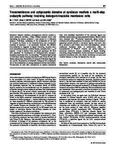
Figure 2. Representative LSM images of non-activated oocytes, collected at 18 h post-HCG (A), and cultured for 30 h (B), or enucleated and cultured for 30 h (C), all in KSOM. The meiotic spindle (ms) is visible in A (front view) and in B; a few asters or microtubule organizing centres (mtoc) are visible in the freshly collected oocyte in A, but their complexity has increased with ageing (B and C). In C, without the spindle-chromosome-complex, many more aster-like microtubule bundles are visible after 30 h of culture. Oocytes have been stained for acetylated and beta tubulin (green) and DNA (red). The oocyte in C is negative for DNA. Images are projections reconstructed from several 3 mm thick optical sections. gc ¼ author please insert. Bar, 20 mm.
I.1b When the spindle-chromosome-complex-depleted oocytes were exposed to activation conditions after 24 – 30 h in culture, many (36/69 or 52%) fragmented within 2 h. A proportion of the control oocytes (9/57 or 16%) also fragmented within that time.
I.2 When the spindle-chromosome-complex-depleted oocytes were exposed to activation conditions, i.e. ethanol-treated, a large proportion (12/14 or 86%) fragmented although not until 15 h after ethanol exposure, at which time the nucleated oocytes subjected to ethanol treatment were found to be in mitosis (see control group for III.1). Figure 2A– C shows LSM images representative of non-activated mature oocytes, collected 18 h post-HCG, mature oocytes collected at 18 h post-HCG after 30 h of culture, and non-activated enucleated oocytes after 30 h of culture. These oocytes were characterized by the absence of a network of microtubules. Intact oocytes were also characterized by more or less exclusive localization of tubulin in the meiotic spindle except for the presence of about 4– 8 cytoplasmic asters (Figure 2A). After in vitro ageing, the spindle showed extensive arrays of astral microtubules pointing away from the spindle poles and toward the cortex; the number of asters did not appear to increase in these oocytes although they became more extensive in structure (Figure 2B). In the spindle-chromosome-complex-depleted oocytes, the number of asters increased following 30 h of culture, as did their size (Figure 2C).
fragmenting completely; at this time, 45% (25/55) of the nucleated controls were in mitosis, that is, they had no visible nuclei. By 24 h PA, a small proportion of the previously fragmenting enucleated oocytes had returned to a smooth spherical single-cell form; the others were still fragmented but the individual fragments had assumed a spherical or ellipsoidal form. Of the nucleated controls, 82% (45/55) had completed division and were in interphase. A pictorial summary of these results is presented in Figure 3A – F.
III.2a None (0/72) of the activated enucleated oocytes cultured in KSOM– colcemid fragmented. Likewise, nucleated control oocytes did not divide or fragment in KSOM– colcemid (0/24). None exhibited pronuclei.
II. Normally fertilized pronuclear oocytes Enucleated zygotes remained round with smooth membranes until 31 h post-HCG or approximately 18 h post-fertilization. By 33 h post-HCG (about 20 h post-fertilization), 29% (9/31) of the enucleated zygotes had fragmented or had ruffled membranes; 2 h later, this proportion had increased to 68% (21/31). At the same time points, i.e. 33 and 35 h post-HCG (20 – 22 h post-fertilization), respectively, 28% (7/25) and 44% (11/25) of the nucleated control zygotes had cleaved into two cells.
III. Ethanol-activated oocytes III.1 Activated enucleated oocytes cultured in KSOM after enucleation remained round with smooth membranes for up to 13 h PA while their nucleated counterparts were in interphase (single female pronucleus clearly visible). By 14 h PA (32 h post-HCG), some cytoplasts showed cortical ruffling, and by 15 h, 68% (50/73) were
Figure 3. Representative DIC images of oocytes in experiment III.1. Activated enucleated (A–C) and activated intact (D –F) oocytes cultured in KSOM are shown at 9, 15, and 24 h PA. Enucleated oocytes remained round with smooth membranes at 9 h PA (A). At 15 h PA, many had fragmented (B) and at 24 h PA, some of the previously fragmented cytoplasts had become ovoid again but in most, fragments remained in the PVS (C). At 9, 15 and 24 h PA, respectively, the nucleated controls were in interphase (one female nucleus and one polar body visible) (D), mitosis (the pronucleus could not be seen) (E) and interphase of the second cell cycle (each of two cells show a prominent nucleus) (F). Author please give an indication of scale
339
M.Alikani, T.Schimmel and S.M.Willadsen
Figure 4. Representative LSM images of activated oocytes following 5 h of culture in KSOM (A) or KSOM–CCB (B), as part of experiment IV. The oocyte in A has extruded a polar body (pb2), and a female pronucleus (fpn) has formed. The meiotic midbody, in the centre (arrow), is visible between the oocyte and the polar body. The inset shows detail of the extensive microtubule network and few microtubule organizing centres in the same oocyte. In the oocyte in B, two pronuclei, both female (fPN), have formed since the polar body is retained in the presence of CCB. The spindle remnant (arrow) is also visible between the two pronuclei. oocytes have been stained for acetylated and beta tubulin (green) and DNA (red). Images are projections reconstructed from several 3 mm thick optical sections. Bar, 20 mm.
Figure 6. Representative LSM (A and B) and DIC (C and D) images of oocytes in experiment IV in which activated oocytes were maintained in KSOM–CCB for 4–5 h immediately PA, were either enucleated or not, washed, and transferred to KSOM. In some enucleated oocytes, the early fragmentation wave culminated in the formation of a polar body-like fragment or bleb (A, arrow). The meiotic spindle remnant is visible in this enucleated oocyte, indicating that removal of the nuclei did not guarantee removal of the meiotic spindle remnant. In B, the arrow points to a similar polar body-like structure in the nucleated control oocyte; the second female pronucleus is present but not visible in this optical section. In other enucleated oocytes, the first wave of fragmentation at 9 h PA led to a more or less even ‘cleavage’ (C). By 16 h PA, both anucleate fragments in the oocyte in C fragmented completely (D). Images in A and B represent a single optical section midway through the oocyte; the oocytes have been stained for acetylated and beta tubulin (shown in grey-scale). Bar, 20 mm.
III.3b Transfer to KSOM following 14 – 22 h of culture in KSOM– CCB led to fragmentation in 30% (9/30) of the enucleated oocytes, again in about 2 – 6 h. When the intact controls were exposed to CCB for the same period and then transferred to KSOM, 33% (5/15) divided to 2-cells within the same period. Division in the control group was often accompanied by fragmentation.
IV. Activated oocytes exposed to CCB
Figure 5. Activated enucleated (A –C) and activated intact (D –F) oocytes that were maintained in KSOM–CCB for 4– 5 h immediately PA then transferred to KSOM. Nine hours PA, some cytoplasts showed cortical ruffling and either produced one or more polar cytoplasts or ‘cleaved’more or less evenly (A). Beginning at 16 h PA and up to 25 h PA, these enucleated oocytes showed more extensive fragmentation affecting the entire oocyte (B, C). At the same time-points, respectively, nucleated control oocytes showed, blebbing and formation of one or more ‘polar bodies’ (D) and entry into mitosis or cleavage (E, F). Bar, 120 mm.
III.2b None (0/30) of the activated enucleated oocytes cultured in KSOM – CCB fragmented during culture. Likewise, nucleated control oocytes did not divide or fragment in KSOM – CCB (0/25), although as expected, a high proportion exhibited two pronuclei.
III.3a Following transfer of activated enucleated oocytes to KSOM after 14 – 22 h of culture in KSOM– colcemid, 40% (17/42) fragmented in about 2– 6 h. Among the nucleated control oocytes, 53% (8/15) divided during the same period. Division in these control groups was often accompanied by fragmentation.
340
A pictorial summary of these results is presented in Figure 5A – F. When emission of the second polar body was prevented by exposing intact activated oocytes to CCB for 4 – 5 h PA, two female pronuclei formed, both placed peripherally in the cytoplasm at some distance from each other (Figure 4). Four hours following removal of these two emerging female pronuclei, at 9 h PA, a fragmentation event, morphologically distinct from that seen in previous experiments was noted in 53% (27/51) of the oocytes: 20% (10/51) showed ruffling and blebbing of the membrane that culminated in the formation of one or several small fragments at or near the location where the second polar body would have formed (Figures 5A and 6A). In the remaining enucleated oocytes, (17/51 or 33%), a cleavage furrow started to form, also in the region where the nuclei had been situated prior to enucleation. The cleavage furrow in some of these divided the oocyte into two equal-sized cytoplasts (Figures 5A and 6C). At or about 16 h PA (continuing up to 25 h PA), a second and universal fragmentation wave occurred in 43/51 or 84% of the oocytes and led to complete fragmentation of the cytoplasts (Figure 5B,C and 6D). Among the nucleated control oocytes, 13 of the 15 showed cortical ruffling and/or formation of ‘polar bodies’ within 9 h of activation (Figures 5D and 6B). At about 16 h PA, coinciding with complete fragmentation of the enucleated oocytes, the majority of these intact oocytes had entered mitosis and some had already
Cytoplasmic fragmentation in activated eggs
Figure 7. Representative LSM images of 2-cell embryos from experiment V in which one of the two cells was enucleated early (A –C) or late (D–E) in interphase. During subsequent culture, the enucleated cells (ec) in A, B, and C are unfragmented and show interphase microtubules. The nucleated cells in A and B are in metaphase and anaphase, of the second cell cycle, respectively. In C, the nucleated cell has divided and the resulting blastomeres are in interphase. The enucleated cell in D shows cortical deformations and that in E has fragmented; three fragments (f) are visible. The nucleated cells in these embryos are in prophase through anaphase of the third cell cycle. Embryos have been stained for acetylated and beta tubulin (green) and DNA (red). The enucleated cells are negative for DNA. Images are projections reconstructed from several 3 mm thick optical sections. Bar, 20 mm.
divided (Figure 5E); by 25 h PA, all had cleaved into two cells (Figure 5F).
V. Fertilized 2-cell embryos V.1a Among the 28 embryos in which only one blastomere was enucleated in early-mid interphase, none (0/28) showed any fragmentation after overnight culture, while their intact sister blastomeres cleaved normally during that period (Figure 7A – C).
V.1b A small proportion (6/83 or 7.2%) of the embryos in which both blastomeres were enucleated in early-mid interphase showed some membrane deformation when checked after overnight culture, but the great majority remained unruffled and ovoid throughout culture (Figure 8A).
V.2a Among the 25 embryos in which one late interphase blastomere was enucleated, 68% (17/25) were found to have fragmented after overnight culture, while the intact sister blastomeres cleaved normally during that period (Figure 7D and E).
V.2b Among the 75 embryos in which both blastomeres were enucleated in late interphase, 74.7% (56/75) were found to be fragmented after overnight culture (Figure 8B). Rounding of the fragmented enucleated cells occurred within the following 24 –48 h of culture, but some fragments remained in the PVS in most instances.
Discussion In the present experiments, cytoplasmic fragmentation did not proceed unless the cells were in division mode, suggesting a general rule that fragmentation does not occur in mitotically inactive cells and therefore is not a predetermined fate of arrested or quiescent cells. The study further suggests that fragmentation in oocytes and embryos occurs during the cytokinetic phase of the cell cycle, in response to the loss of normal interplay between the spindle complex and cortical microfilaments. Whether enucleation immediately followed or preceded artificial activation, the resulting fragmentation in the cytoplasts was a precisely timed event in the cell cycle, rather than a happenstance, and always coincided with entry of nucleated control cells into mitosis. In the cytoplasts, this timing represents the cytokinetic rather than the mitotic phase of the cell cycle, since the removal of the nuclear structure removes the necessity for coordination of nuclear and cytoplasmic divisions (and any delay normally imposed by the former on the latter). When the nucleated control cells had completed cytokinesis and entered interphase, some blebs and fragments receded in the cytoplasts, suggesting further autonomous cell cycling activity in the latter. The dynamic relationship between spindle microtubules and cortical microfilaments is pivotal to normal cytokinesis. Therefore, deliberate depolymerization of microfilaments (by CCB treatment) or depolymerization of microtubules (by colcemid treatment) prevents cytokinesis altogether; here, the same treatments also prevented fragmentation. Drug removal, on the other hand, restored both microtubules and microfilaments, allowing cytokinesis and fragmentation to proceed. It is noteworthy that both following extended exposure to cytoskeletal disruptive drugs, and delayed activation, limited to complete
341
M.Alikani, T.Schimmel and S.M.Willadsen
Figure 8. Representative DIC images of 2-cell embryos from experiment V. Both blastomeres were enucleated in early-mid interphase (A) or later (B) and cultured overnight. Blastomeres in A remained unfragmented after enucleation while blastomeres in B fragmented. Bar, 50 mm.
fragmentation was seen in control embryos. This behaviour, which is generally uncharacteristic of normal mouse development suggests that once induced, cytoskeletal disorder may persist and become overtly expressed in the course of a subsequent cytokinesis, even in the presence of a nuclear apparatus. If our hypothesis that fragmentation is a deviant form of cytokinesis is correct, it should be possible to demonstrate that the phenomenon can also occur during cytokinesis of the second meiosis. We think that this was demonstrated in experiment IV, for in that experiment two distinct waves of fragmentation occurred. The first wave was limited to the region where the nuclei had been situated prior to enucleation and occurred several hours before mitosis 1 in nucleated controls. The second wave was universal and coincided with mitosis 1 in nucleated controls, similar to the events described in experiments I –III. In our opinion, the first of these waves represented cytokinesis of second meiosis, while the second represented cytokinesis of the first mitotic division. We have further investigated meiotic fragmentation in more detail, and the results will form the subject of a separate report (Willadsen and Alikani, in preparation). We also investigated the possibility of inducing fragmentation during cytokinesis of the second cell cycle. In her experiments, Ciemerych (1995) did not observe cortical deformations or any other autonomous activity in the anucleate halves of blastomeres from 2- and 4-cell embryos. This lack of activity was attributed to diminished cytoplasmic autonomy and the low intensity of mitosis
342
promoting factor (MPF) activation during the second (and subsequent) cell cycle(s) (Kubiak and Ciemerych, 2001). In experiment V of the present study, some enucleated blastomeres of fertilized 2-cell embryos did fragment. However, the timing of enucleation was crucial to this outcome: if enucleation was done during early-mid interphase, further activity ceased in the anucleate blastomere; no surface deformations were seen and the microtubules maintained their interphase configuration. However, when enucleation was delayed until late interphase, a large proportion of the enucleated cells either showed cortical deformations or fragmented during subsequent culture. We propose that the failure of the enucleated early blastomeres to undergo fragmentation is reminiscent of the ‘2-cell block’ in the mouse (Goddard and Pratt, 1983) and supports the notion of fragmentation as atypical cytokinesis. The ‘2-cell-block’ takes effect at the late G2/M phase of the second cell cycle when maternal transcripts run out and the zygotic genome must be activated for normal development to continue (Flach et al., 1982). We have assumed that the process of fragmentation in oocytes and embryos is essentially the same whatever the triggering cause(s), i.e. that fragmentation is the diverse expression of a single phenomenon: the failure of the normal dynamic relationship between microtubules and microfilaments during one particular phase of the cell division cycle – cytokinesis. Our results support that assumption. However, we cannot yet be certain that the assumption has more general validity, and for this reason, we can only speculate what the relevance of the results of the present study might be to fragmentation and related phenomena in human oocytes and embryos, not to mention other cell types. In addition, the topography and organization of the cytoskeleton differ between species (Simerly et al., 1995). For instance, the sperm centrosome is thought to play an essential role as the microtubule organizing centre in the human (Sathananthan et al., 1991; Palermo et al., 1994; Van Blerkom, 1996) but not in the mouse (Maro et al., 1985; Schatten et al., 1985). In the human, the genome becomes activated sometime between the 4- and 8-cell stages (Braude et al., 1988), but most fragmentation occurs prior to that. Indeed, it appears that around the time of genomic activation, and particularly after compaction and the establishment of structural junctions (Gualtieri et al., 1992; Nikas et al., 1996), the tendency of human blastomeres to fragment is simply lost. Whether the impetus for this change is the activation of the genome, increased adhesion between the cells, reduced cell size or a combination of these factors remains to be determined. Nevertheless, the basic suggestion of the present experiments is that fragmentation in human embryos is a manifestation of abnormal cytokinesis rather than cell death. This proposal would support a causal relationship between cytoplasmic fragmentation and cytoskeletal changes associated with post-ovulatory ageing (Pickering et al., 1988) or environmental fluctuations during in vitro culture (Almeida and Bolton, 1995; Dale et al., 1998). It would also provide a plausible explanation for the high frequency of fragmentation and its association with nuclear abnormalities, most importantly, post-zygotic chromosome mosaicism among human IVF embryos.
Acknowledgements The authors would like to thank Ms Leona Cohen-Gould, Director of Electron Microscopy Core Facility of Joan and Sanford I. Weill Medical College of Cornell University for her expert assistance with confocal microscopy. Dr Maria Anna Ciemerych of the University of Warsaw is gratefully acknowledged for providing advice on preparation of material for confocal microscopy. We thank professors Alan Trounson, Martin Johnson, and Jacques Cohen for critical reading of the manuscript.
Cytoplasmic fragmentation in activated eggs
References Abbott AL, Xu Z, Kopf GS, Ducibella T and Schultz RM (1998) In vitro culture retards spontaneous activation of cell cycle progression and cortical granule exocytosis that normally occur in in vivo unfertilized mouse eggs. Biol Reprod 59,1515–1521. Alikani M and Willadsen SM (2002) Human blastocysts from aggregated mononucleated cells of two or more non-viable zygote-derived embryos. Reprod Biomed Online 5,56–58. Alikani M, Cohen J, Tomkin G, Garrisi GJ, Mack C and Scott R (1999) Human embryo fragmentation in vitro and its implications for pregnancy and implantation. Fertil Steril 71,836–842. Alikani M, Calderon G, Tomkin G, Garrisi J and Kokot M (2000) Cohen J Cleavage anomalies in early human embryos and survival after prolonged culture in-vitro. Hum Reprod 15,2634–2643. Allworth AE and Albertini DF (1993) Meiotic maturation in cultured bovine oocytes is accompanied by remodeling of the cumulus cell cytoskeleton. Dev Biol 158,101–112. Almeida PA and Bolton VN (1995) The effect of temperature fluctuations on the cytoskeletal organisation and chromosomal constitution of the human oocyte. Zygote 3,357–365. Antczak M and Van Blerkom J (1999) Temporal and spatial aspects of fragmentation in early human embryos: possible effects on developmental competence and association with the differential elimination of regulatory proteins from polarized domains. Hum Reprod 14,429–447. Braude P, Bolton V and Moore S (1988) Human gene expression first occurs between the four- and eight-cell stages of preimplantation development. Nature 332,459–461. Buster JE, Bustillo M, Rodi IA, Cohen SW, Hamilton M, Simon JA, Thorneycroft IH and Marshall JR (1985) Biologic and morphologic development of donated human ova recovered by nonsurgical uterine lavage. Am J Obstet Gynecol 15,211–217. Chatot CL, Ziomek CA, Bavister BD and Torres I (1989) An improved culture medium supports development of random-bred 1-cell mouse embryos in vitro. J Reprod Fertil 86,679–688. Ciemerych MA (1995) Chromatin condensation activity and cortical activity during the first three cell cycles of a mouse embryo. Mol Reprod Dev 41,416–424. Ciemerych MA, Tarkowski AK and Kubiak JZ (1998) Autonomous activation of histone H1 kinase, cortical activity and microtubule organization in one- and two-cell mouse embryos. Biol Cell 90,557– 564. Dale B, Menezo Y, Cohen J, Dimatteo L and Wilding M (1998) Intracellular pH regulation in human oocytes. Hum Reprod 13,964–970. De Pennart H, Houliston E and Maro B (1988) Post translational modifications of tubulin and the dynamics of microtubules in mouse oocytes and zygotes. Biol Cell 64,375– 378. Edwards RG, Steptoe PC and Purdy JM (1970) Fertilization and cleavage in vitro of preovulatory human oocytes. Nature 227,1307– 1309. Enders AC, Hendrickx AG and Binkerd PE (1982) Abnormal development of blastocysts and blastomeres in the Rhesus Monkey. Biol Reprod 26,353–366. Flach G, Johnson MH, Braude PR, Taylor RA and Bolton VN (1982) The transition from maternal to embryonic control in the 2-cell mouse embryo. EMBO J 1,681–686. Goddard MJ and Pratt HP (1983) Control of events during early cleavage of the mouse embryo: an analysis of the ‘2-cell block’. J Embryol Exp Morphol 73,111–133. Gordo AC, Rodrigues P, Kurokawa M, Jellerette T, Exley GE, Warner C and Fissore R (2002) Intracellular calcium oscillations signal apoptosis rather than activation in in vitro aged mouse eggs. Biol Reprod 66,1828–1837. Gualtieri R, Santella L and Dale B (1992) Tight junctions and cavitation in the human pre-embryo. Mol Reprod Dev 32,81–87. Hardy K, Stark J and Winston RM (2003) Maintenance of the inner cell mass in human blastocysts from fragmented embryos. Biol Reprod 68,1165–1169. Hawes SM, Gie Chung Y and Latham KE (2001) Genetic and epigenetic factors affecting blastomere fragmentation in two-cell stage mouse embryos. Biol Reprod 65,1050–1056. Hogan B, Beddington R, Costantini F and Lacy E (1994) Manipulating the Mouse Embryo: A Laboratory Manual. Cold Spring Harbor Laboratory Press, New York, USA. Jackson KV, Ginsberg ES, Hornstein MD, Rein MS and Clarke RN (1998) Multinucleation in normally fertilized embryos is associated with an accelerated ovulation induction response and lower implantation and pregnancy rates in in-vitro fertilization embryo-transfer cycles. Fertil Steril 70,60–66.
Jurisicova A, Varmuza S and Caspar RF (1996) Programmed cell death and human embryo fragmentation. Mol Hum Reprod 2,93–98. Jurisicova A, Rogers I, Fasciani A, Casper RF and Varmuza S (1998) Effect of maternal age and conditions of fertilization on programmed cell death during murine preimplantation embryo development. Mol Hum Reprod 4,139–145. Jurisicova A, Antenos M, Varmuza S, Tilly JL and Casper RF (2003) Expression of apoptosis-related genes during human preimplantation embryo development: potential roles for the Harakiri gene product and Caspase-3 in blastomere fragmentation. Mol Hum Reprod 9,133–141. Kaufman MH (1982) The chromosome complement of single-pronuclear haploid mouse embryos following activation by ethanol treatment. J Embryol Exp Morphol 71,139–154. Killeen ID and Moore NW (1971) The morphological appearance and development of sheep ova fertilized by surgical insemination. J Reprod Fertil 24,63–70. Kligman I, Benadiva C, Alikani M and Munne´ S (1996) The presence of multinucleated blastomeres in human embryos is correlated with chromosomal abnormalities. Hum Reprod 11,1492–1498. Kubiak JZ and Ciemerych MA (2001) Cell cycle regulation in early mouse embryos. Novartis Found Symp 237,79–89. Laverge H, De Sutter P, Verschraegen-Spae MR, De Paepe A and Dhont M (1997) Triple colour fluorescent in situ hybridization for chromosomes X, Y, and 1 on spare human embryos. Hum Reprod 12,809–814. Levron J, Willadsen S, Munne´ S and Cohen J (1995) Formation of male pronuclei in partitioned human oocytes. Biol Reprod 53,209–213. Levy R, Benchaib M, Cordonier H, Couchier C and Guerin JF (1998) Annexin V labelling and terminal transferase-mediated DNA end labelling (TUNEL) assay in human arrested embryos. Mol Hum Reprod 4,775– 783. Liu L, Trimarchi JR, Smith PJ and Keefe DL (2002) Checkpoint for DNA integrity at the first mitosis after oocyte activation. Mol Reprod Dev 62,277–288. Maro B, Howlett SK and Webb M (1985) Non-spindle microtubule organizing centers in metaphase II-arrested mouse oocytes. J Cell Biol 101,1665–1672. Marquez C, Sandalinas M, Bahce M, Alikani M and Munne´ S (2000) Chromosome abnormalities in 1255 cleavage-stage human embryos. Reprod Biomed Online 1,17–26. Morita Y and Tilly JL (1999) Oocyte apoptosis: like sand through an hourglass. Dev Biol 213,1–17. Nikas G, Ao A, Winston R and Handyside AH (1996) Compaction and surface polarity in the human embryo in-vitro. Biol Reprod 55,32–37. Palermo G, Munne´ S and Cohen J (1994) The human zygote inherits its mitotic potential from the male gamete. Hum Reprod 9,1220–1225. Pellestor F, Dufour MC, Arnal F and Humeau C (1994) Direct assessment of the rate of chromosomal abnormalities in grade IV human embryos produced by in-vitro fertilization procedure. Hum Reprod 9,293–302. Perez GI, Tao XJ and Tilly JL (1999) Fragmentation and death (a.k.a. apoptosis) of ovulated oocytes. Mol Hum Reprod 5,414–420. Pickering SJ, Johnson MH, Braude PR and Houliston E (1988) Cytoskeletal organization in fresh, aged and spontaneously activated human oocytes. Hum Reprod 3,978–989. Racowsky C, Combelles CM, Nureddin A, Pan Y, Finn A, Miles L, Gale S, O’Leary T and Jackson KV (2003) Day 3 and day 5 morphological predictors of embryo viability. Reprod Biomed Online 6,323–331. Rappaport R (1996) Cytokinesis in Animal Cells. Cambridge University Press, Cambridge, UK. Sathananthan AH, Kola I, Osborne J, Trounson A, Ng SC, Bongso A and Ratnam SS (1991) Centrioles in the beginning of human development. Proc Natl Acad Sci USA 88,4806–4810. Schatten G, Simerly C and Schatten H (1985) Microtubule configurations during fertilization, mitosis, and early development in the mouse and the requirement for egg microtubule-mediated motility during mammalian fertilization. Proc Natl Acad Sci USA 82,4152–4156. Simerly C, Navara C, Wu GJ and Schatten G (1995) Cytoskeletal organization and dynamics in mammalian oocytes during maturation and fertilization. In Grudzinskas JG and Yovich JL (eds) Cambridge Reviews in Human Reproduction. Gametes: The Oocyte. Cambridge University Press, Cambridge, 5494 pp. Smith R and McLaren A (1977) Factors affecting the time of formation of the mouse blastocoele. J Embryol Exp Morphol 41,79–92. Surani MA, Barton SC and Burling A (1980) Differentiation of 2-cell and 8cell mouse embryos arrested by cytoskeletal inhibitors. Exp Cell Res 125,275–286.
343
M.Alikani, T.Schimmel and S.M.Willadsen Takase K, Ishikawa M and Hoshiai H (1995) Apoptosis in the degeneration process of unfertilized mouse ova. Tohoku J Exp Med 175,69–76. Van Blerkom J (1996) Sperm centrosome dysfunction: a possible new class of male factor infertility in the human. Mol Hum Reprod 2,349–354. Van Blerkom J, Davis P and Alexander S (2001) A microscopic and biochemical study of fragmentation phenotypes in stage-appropriate human embryos. Hum Reprod 16,719–729. Van Royen E, Mangelschots K, Vercruyssen M, De Neubourg D, Valkenburg M, Ryckaert G and Gerris J (2003) Multinucleation in cleavage stage embryos. Hum Reprod 18,1062–1069. Waksmundzka M, Krysiak E, Karasiewicz J, Czolowska R and Tarkowski AK (1984) Autonomous cortical activity in mouse eggs controlled by a cytoplasmic clock. J Embryol Exp Morphol 79,77–96.
344
Warner CM, Cao W, Exley GE, McElhinny AS, Alikani M, Cohen J, Scott RT and Brenner C (1998) Genetic regulation of egg and embryo survival. Hum Reprod 13(suppl),178–190. Yang HW, Hwang KJ, Kwon HC, Kim HS, Choi KW and Oh KS (1998) Detection of reactive oxygen species (ROS) and apoptosis in human fragmented embryos. Hum Reprod 13,998–1002. Xu Z, Abbott A, Kopf GS, Schultz RM and Ducibella T (1997) Spontaneous activation of ovulated mouse eggs: time-dependent effects on M-phase exit, cortical granule exocytosis, maternal messenger ribonucleic acid recruitment, and inositol 1,4,5-trisphosphate sensitivity. Biol Reprod 57, 743–750. Submitted on October 7, 2004; accepted on March 17, 2005