Relative Frequency of Stage-specific CTL Recognizing the 72-kD. Immediate ...... Bangham, C. R. M., P J. M. Oppenshaw, L. A. Ball, A. M. Q; King, G. W. Wertz,.
HUMAN CYTOMEGALOVIRUS-SPECIFIC
CYTOTOXIC T CELLS
Relative Frequency of Stage-specific CTL Recognizing the 72-kD Immediate Early Protein and Glycoprotein B Expressed by Recombinant Vaccinia Viruses BY LESZEK K. BORYSIEWICZ, JULIAN K. HICKLING, SHEK GRAHAM, JOHN SINCLAIR, MARTIN P CRANAGE,' GEOFFREY L. SMITH,' AND J. G. PATRICK SISSONS From the Medical Research Council Clinical Immunology Research Group, Department of Medicine, Royal Postgraduate Medical School, Hammersmith Hospital, London W12 OHS; and the 'Division of Virology, Department of Pathology, University of Cambridge, Cambridge CB2 2QQ,, United Kingdom
Human cytomegalovirus (HCMV)' is a human herpesvirus that establishes lifelong persistent infection, after an often asymptomatic primary infection. Persistent infection may reactivate and become clinically evident after immunosuppression, resulting in significant morbidity and mortality (1). Reactivation in the context of immunosuppression suggests that virus-specific T cells, including HCMVspecific CTL, play a major role in maintaining the normal virus-host equilibrium. We have previously reported that MHC class I-restricted, CD8 + , HCMVspecific CTL precursors (CTLp) are present in PBL of asymptomatic, persistently HCMVinfected individuals (2) . These CTL are present at a precursor frequency of 1/5,000-20,000 PBL, higher than that of varicella zoster virus (VZV)-specific CTL (3). Furthermore, HCMVspecific CTL killed virus-infected cells before viral DNA replication, suggesting that they recognized predominantly nonstructural viral proteins (2-4). However, by limiting dilution analysis a minor population of CTL were identified that only killed HCMVinfected target cells expressing late (structural) viral proteins (3). In murine cytomegalovirus (MCMV) infection, which is similar in some respects to HCMV infection, MCMVspecific CTL may protect against virus infection (5). These protective CTL are specific for viral immediate early (IE) proteins, the first, nonstructural viral proteins expressed in the infected cell (6). After HCMV infection of diploid human fibroblasts, the only cells fully permissive for HCMV replication in vitro, there is a regulated expression of virus gene products (1). Within 4-6 h of infection, viral IE genes are expressed. They code for four to five predominantly intranuclear proteins, the most abundant of which This work was supported by grants from the Medical Research Council and Wellcome Trust. L. K. Borysiewicz is a Lister Research Fellow. 1 Abbreviations used in this paper: CTLp, precursor CTL; E, early; gB, glycoprotein B; HCMV, human cytomegalovirus; IE, immediate early; L, late ; MCMV murine cytomegalovirus; PPF, phosphonoformate ; VZV varicella zoster virus. J. Exp. MED. C The Rockefeller University Press - 0022-1007/88/09/919/13 $2 .00 Volume 168 September 1988 919-931
919
920
HUMAN CYTOMEGALOVIRUS-SPECIFIC CYTOTOXIC T CELLS
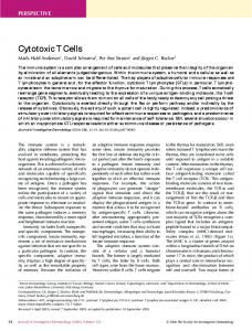
is a 72-kD protein (7, 8). These proteins positively trans-regulate expression of the early (E) viral genes (7-9), and negatively autoregulate their own expression (10). The E genes encode a number of proteins, including the viral DNA polymerase, essential for HCMV DNA replication that occurs N18 h after infection ; only then are the 30-33 late (L) genes, coding mainly for the structural proteins ofthe virion, expressed . The size of the HCMV genome (235 kbp) allows for a large potential coding capacity (1), and thus there are obvious difficulties in determining whether individual viral proteins are immunodominant with respect to the CTL response. The specificity of CTL for particular stages of virus gene expression can be examined using metabolic inhibitors, such as phosphonoformate (PPF) (3, 4), to prevent expression of L genes . However, positive identification of viral determinants recognized by CTL can best be achieved by using expression vectors that permit expression of single HCMV genes . In the case of human persistent virus infections, there is little information on the relative preponderance of CTL specific for individual or stage-specific virus gene products . In these studies we have examined HCMVspecific CTL using recombinant vaccinia viruses that encode the HCMV structural glycoprotein B (vac.gB) (11) and the 72-kD IE protein (vac.IE) . This approach was combined with limiting dilution culture, which permitted both a quantitative and clonal analysis of the CTL response to these viral proteins . Both vac.gB- and vac.IE-infected cells were recognized by HCMVspecific CTL . The frequency of IE protein-specific CTL was between 18 and 58% of HCMVspecific CTLp in the two subjects studied (n = 138/21 and 77, respectively), compared with 0-20/o and 6% for gB. These results suggest that the 72-kD IE protein is recognized by a large proportion of HCMVspecific CTL in the asymptomatic persistently HCMVinfected individual . Materials and Methods Cells and Viruses . HCMV strain AD169 (No . VR538 ; American Type Culture Collection, Rockville, MD) was grown in F5000 (Flow Laboratories, Inc., McLean, VA) diploid human fibroblasts, innoculated at a multiplicity of infection (MOI) of 0.1:1 . Supernatant virus was harvested, plaqued on F5000 cells, and virus stocks with a titer >10' plaqueforming units (PFU)/ml were stored at -70 °C until use . Vaccinia WR strain (vac.WR) was grown in Vero cells and supernatant virus was harvested, plaqued (titer 109), and stored in 1-ml aliquots at -70°C. VZV and HSV were propagated as previously described (3) . Primary human fibroblast lines were established by explant cultures as previously described (2), from individuals of known HLA type. These cells and F5000 cells were maintained in MEM supplemented with 1017o FCS, 2 MM L-glutamine, 100,000 IU/literpenicillin, 10 mg/liter streptomycin, and 1% nonessential amino acids (Gibco Laboratories, Grand Island, NY) . Fibroblasts, free of mycoplasma contamination by DNA staining and culture, and between passage 10-20, were used in these experiments . Recombinant Vaccinia Viruses. Vac .gB encoding HCMV gB was constructed as previously described (11). Vero cells, diploid human fibroblasts, and EBV transformed B cells infected with vac.gB expressed gB, as detected by a mAb (provided by H. Hart, Inveresk Research International, Edinburgh, Scotland [111), in a cytoplasmic distribution, with some weak nuclear membrane fluorescence, again as previously described (11). Vac.IE was constructed from the cDNA clone (pJD083) of the IE gene encoding the 72kD IE protein of HCMV strain AD169, inserted into the Pst I site of pUC9 (provided by J . Oram, Public Health Laboratory Service, Centre for Applied Microbiology & Research, Porton Down, UK) (12) (Fig. 1). A 1.7-kb Eco RI-Hind III fragment containing the com-
BORYSIEWICZ ET AL.
921
EcoRI
H ind 111 17kb EcoRl/Hind III m1E fragment
m
S al
Construction of the vac.1E recombinant from the cDNA ofthe HCMV AD169 72kD IE protein in pJD083 (see text for details) . FIGURE 1 .
plete major IE coding region was gel purified and blunt ended with Klenow fragment . The fragment was ligated into the unique Sma I site of the coinsertion vector pSC11 (13), downstream of the vaccinia early promoter (p7 .5). Correct insertion and orientation ofthe reading frame were confirmed by diagnostic restriction enzyme digestion and agarose gel electrophoresis . Transfection and coinfection with vacWR ofCV1 cells was performed according to established methods (14). TK- progeny virus was selected using BUdR (50 pg/ml) . TK- recombinants were distinguished from spontaneous TK- mutants, by overlaying with 1% agarose containing 0.1% neutral red and 300 wg/ml Xgal (5-bromo-4-chloro-3-indoyl-a-ngalactopyranoside) to screen for a-galactosidase activity (13). Plaques giving an intense blue colour after 4-6h were selected, further plaque purified and recombinant virus stocks grown in CV1 cells (14). The recombinant virus genome was analyzed by restriction endonuclease digestion and Southern blotting (data not shown) which confirmed that the HCMV major IE gene had been inserted into the vaccinia TK gene with no other genomic alterations . Generation of HCMV-speck and gB-specific CTL . PBL from HCMVseropositive subjects were stimulated by cocultivation with autologous fibroblasts (responder/stimulator - 50:1), infected with either HCMV for 48 h or recombinant vaccinia virus for 12 h, then UV inactivated in 24-well plates (Linbro ; Flow Laboratories, Inc.), for 5 d in RPMI supplemented as for MEM but with 10% HCMVseronegative AB serum. The medium was then changed and 2/IU/ml rIL-2 (Boerrhinger Mannheim Biochemicals, Indianapolis, IN) added. Stimulator cells with irradiated autologous PBL (2,500 rad) were added at 7 d, and the cells were allowed to grow for a further 7 d in rIL-2 . Viable cells were harvested by Ficoll/Hypaque density centrifugation and assayed in "Cr-release assays (2) as indicated in the text. Limiting Dilution Cultures. Limiting dilution cultures to estimate the precursor frequency and analyze the clonal specificity of HCMVspecific CTL were performed as described (3) . Briefly, PBL or E' cells isolated from PBL by rosetting with AETtreated SRBC (15) were used. Between 100 and 10,000 lymphocytes were placed in 96-well round-bottomed plates (Linbro ; Flow Laboratories, Inc.) in 30-120 replicate cultures at each input cell concentration. These were divided between several plates to overcome plate-to-plate variation . 2 x 10³ 48-h HCMVinfected autologous fibroblasts, together with autologous irradiated PBL
922
HUMAN CYTOMEGALOVIRUS-SPECIFIC CYTOTOXIC T CELLS
to a final cell concentration of 105 cells/well, were added and the cultures were incubated in RPMI with 10% AB serum supplemented with 2 IU/ml rIL-2 . The cultures were refed at 3-d intervals for 11-14 d. Identical cultures were also established with irradiated (2,500 rad) PBL as feeder cell controls . The cultures were then equally divided and cytotoxicity was determined against four to six different target cells as described in individual experiments . For each target cell, maximum and spontaneous release values between plates were compared for interplate variation by one-way analysis of variance using the Scheffe multiple comparison test (16). Each test well was compared with the "feeder cell only" controls and significant 5'Cr release was determined if the counts were >3 SD above the release observed in the control wells (3) . Each well was scored by the different target cells lysed. Virus-specific, MHC-restricted lysis was observed when the effector cells from an individual well lysed the autologous virus-infected cells and target cells in the presence oflectin, but not HLA-mismatched virus-infected cells, or autologous cells infected with an irrelevant virus. The best straight line fit comparing the logo of the proportion of negative wells against the input cell number was determined by the method of least squares, and the linearity was further examined by regression analysis (3, 16, 17) . From these estimates the frequency of specific CTLp could be estimated as the input cell number that would produce 37 % negative cultures against a particular target(s) (17). All statistical analysis was performed using SPSS/PC + (SPSS Inc ., Chicago, IL) (16). Other Reagents . mAbs L14 (kindly provided byJ . A. Nelson and M. B. A. Oldstone, Scripps Clinic and Research Institute, LaJolla, CA) (18) and Cll (provided by Dr. G. The, University of Gronigen, The Netherlands) against the 72 kD major IE protein of HCMV were used for indirect immunofluorescence offixed and permeabilized infected monolayers of cells (19). Results Expression of 72-kDMajor IE Protein in Cells Infected with vac. IE. Two mAbs specific for the HCMV 72-kD IE protein, L14 and C11, were used to confirm its expression by indirect immunofluorescence. Monolayers of Vero cells and human fibroblasts infected for 12 h at 10 PFU/cell with vac .IE or vac WR, were fixed and permeabilized (19), and then stained with L14 or Cll and (Fab)2, rabbit anti-mouse Ig-FITC (Dako) (Fig. 2). Vac .IE-infected cells had positive nuclear fluorescence, detectable as early as 4 h after infection, increasing up to 12 h. There was no fluorescence in vacWR infected cells . On Western blot analysis a protein of 71-73 kD was detected in HCMV or vac .IE-infected cell lysates, using Cll, although not with L14 (data not shown). HCMV-specific CTL Recognize the 72-kD IE Protein and gB. To determine if the HCMV 72-kD IE protein and gB were recognized by HCMVspecific CTL, PBL from seropositive and seronegative subjects were cocultured with 48-h HCMVinfected autologous fibroblasts under limiting dilution conditions, as described in Materials and Methods . After 11 d individual wells were assayed against HCMVinfected autologous and HLA-mismatched fibroblasts to determine the frequency of all HCMV specific CTL (3). HCMVspecific CTL in subject CH were present at a frequency of 1/11,000 (95% confidence limits: 1/5,000-1/17,000) (Fig. 3). In addition, each individual well was assayed against autologous and HLA-mismatched B cell lines infected with the vaccinia recombinants . HCMV 72-kD IE protein-specific CTL were present at 1/76,000 PBL (1/36,000-116,000) . In the same experiment, HCMV gB-specific CTL were present at the limits of the assay at 1/181,000 PBL . There was no specific lysis of vacWRinfected target cells . No HCMV, HCMV 72-kD IE protein, or HCMV gB-specific CTL were present in seronegative subjects .
923
BORYSIEWICZ ET AL .
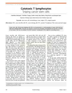
FIGURE 2.
Human primary fibroblasts infected with vac.IE expressed the 72-kD IE protein as detected by mAbs . Fibroblasts were infected for 12 h (MOI 10 :1) with vac.IE (top) (x 200), infected with vac.WR (middle) (x 200), and HCMV for 24 h (bottom) (x 400). The cells were fixed in methanol/acetone, permeabilized, and stained by indirect immunofluorescence using mAb Cll. vac.IE-infected cells expressed the HCMV 72kD IE protein in nuclear distribution .
HUMAN CYTOMEGALOVIRUS-SPECIFIC CYTOTOXIC T CELLS
92 4
3. Frequency analysis of HCMV and vac.IE-specific CTLp. PBL from a seropositive subject (CH) were grown in limiting dilution (120 replicate cultures at each input cell number), with autologous HCMVinfected fibroblasts, irradiated PBL and HL-2 . After 11 d the cultures were assayed against48-h HCMVinfected autologous and HLA-mismatched fibroblasts, to determine the presence of HCMVspecific CTL (/), and against vac.IE- and vac.gB-infected autologous and vac.IEinfected, HLA-mismatched EBVtransformed B cell lines, to determine 72-kD IE-specific CTL (*). Bars indicate 95% confidence limits for each input cell number; hatched area indicates 95% confidence interval for the regression lines. FIGURE
Relative Frequencies of HCMV 72-kD and gB-specific CTL Clones. The relative frequencies of clonally derived (3, 17), vac.IE- and vac.gB-specific CTL were compared, together with the ability of the same clones to mediate lysis of HCMV infected autologous fibroblasts . The data are shown in Table 1 . In three separate experiments in two seropositive subjects (CH and AR), the frequency of vac.IE-specific CTL
TABLE I
Relative Numbers of vac.gB- and vac .IE-speck CTL Clones in Asymptomatic HCMV-seropositive Subjects Exp. 1
2
3
Subject
CH HCMV lysed HCMV not lysed Total CH HCMV HCMV Total AR HCMV HCMV Total
Number of wells (percent of row total) Total vac .IE - -vac . gB Other 25 (18) 4 (2) 29
4 (2) 0 (0) 4
109 (80) 138 (98) 247
138 142 280
lysed not lysed
9 (43) 0 (0) 9
0 (0) o (0) 0
12 (57) 100 (100) 112
21 100 121
lysed not lysed
45 (58) 16 (29) 61
5 (6) 0 (0) 5
27 (36) 40 (71) 67
77 56 133
92 5
BORYSIEWICZ ET AL . 100
7
AUTOLOGOUS
100-1
75
50
PARTIAL MATCH
100 -
MISMATCH HCMV + Uninfected t Vaccinia. WR + Vaccinia . gB
25
0
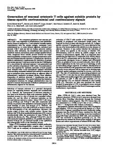
Vac.gB-infected fibroblasts activated HCMV-specific CTL in vitro. PBL from a seropositive subject (AR), were cocultured with vac.gB-infected, UV inactivated, autologous fibroblasts as described in Materials and Methods. After 14 d, viable lymphocytes were assayed in a "Cr-release assay against autologous, partially HLA A and B-matched and HLA-mismatched fibroblasts that were uninfected ( ") or infected with HCMV (L7), vac.gB (O), or vac.WR Bars indicate + 1 SD. FIGURE 4 .
clones was greater than vac.gB-specific clones . In the first and third studies, vac.IEspecific CTL clones derived from the different donors lysed HCMV infected autologous fibroblasts and represented 18.1 and 58 .4%, respectively, of all wells mediating HCMV-specific CTL lysis. However, in these two experiments, although the vac.IEspecific CTL were clonally derived, the cultures lysing HCMVinfected fibroblasts were not by the standard criteria for limiting dilution analysis described by Lefkovitz and Waldeman (17). When the experiment was repeated with cells from donor CH (Table 1, Exp. 2), although fewer clones were studied, both cultures with HCMV-specific and vac.IEspecific CTL activity were clonally derived and 43% of all HCMV-specific CTL clones also lysed autologous vac.IE-infected cells. There was no instance ofan individual well killing both vac.IE- and vac.gB-infected cells. However, a small number ofvac.IE-specific CTL clones did not lyse autologous HCMVinfected fibroblasts. Generation ofHCMV-speck CTL with vac.IE. To determine whether HCMV-specific CTL could be generated using vac.IE, PBL were cocultured with UV-inactivated vac.IE-infected autologous fibroblasts or EBVtransformed B cell lines. In five experiments from three seropositive subjects no HCMV-specific CTL were generated, although in two of the five studies, nonspecific cytotoxicity was observed . Generation of HCMV-specific CTL with vac.gB. To determine whether HCMV memory CTL could be activated in vitro by a single structural HCMV protein (gB) expressed in vaccinia-infected cells, PBL were cocultured with cells infected with vac.gB. Autologous fibroblasts were infected with vac.gB for 12 h, UV treated to inactive residual and released vaccinia virus, and cocultured with PBL from HCMV seropositive and seronegative subjects. The cultures were restimulated with vac.gBinfected cells after 7 d and the lymphocytes were allowed to grow in the presence of rIL-2 for a further week. Viable cells were then assayed against autologous, partially HLA-matched and -mismatched fibroblasts, uninfected or infected with HCMV vacWR, and vac.gB. Only autologous and partially HLA-matched cells infected with the recombinant or HCMV were lysed by the effector cells from a seropositive subject (Fig. 4); there was no significant lysis by PBL from a seronegative subject.
92 6
HUMAN CYTOMEGALOVIRUS-SPECIFIC CYTOTOXIC T CELLS
FIGURE 5.
Limiting dilution analysis of vac .gB-activated, HCMVspecific CTL. 1,000 to 20,000 E*PBL (subject AR) were grown in 30 replicate cultures at each input cell concentration, with 2,000 vac.gB-infected, UVinactivated autologous fibroblasts, irradiated (2,500 rad) PBL and IL-2 for 12 d. The individual wells were assayed against HCMVinfected (48 h) ("), vac.gBinfected (12 h) ( ") autologous fibroblasts, and K562 cells with PHA (/). No significant lysis of uninfected or VZVinfected autologous fibroblasts was detected (data not shown) . Precursor frequencies from limiting dilution plot : HCMVspecific CTL, 1/20,000; vac.gB-specific CTL, 1/16,000 .
To analyze the frequency of such CTLp the experiment was repeated under limiting dilution conditions (Fig. 5). A similar frequency of CTL lysing vac.gB- and HCMV infected cells would be expected under these conditions, as only a single HCMV protein was used to stimulate the response. 1/20,000 E+ PBL lysed HCMVinfected autologous fibroblasts and 1/16,000 lysed vac.gB-infected fibroblasts. Furthermore, there was almost complete concordance when the lysis of HCMV and vac.gB-infected cells by individual wells containing the progeny of a single cell, as predicted from the limiting dilution plots, was compared (data not shown) . In parallel experiments the HCMVspecific CTLp frequency, using autologous HCMV infected fibroblasts as stimulator cells in limiting dilution cultures, was 1/5000 E+PBL (3). Discussion In this study we have shown, using vaccinia recombinants encoding the HCMV 72-kD IE protein and gB, that HCMVspecific CTL from individuals persistently infected with HCMV recognized these viral proteins . The relative number ofHCMV 72-kD IE protein-specific CTL in PBL was much greater than gB-specific CTL in the subjects studied. HCMVspecific memory CTL were activated by coculture with vac.gB-, but not vac.IE-infected fibroblasts. These results suggest that whereas some CTL specific for HCMV structural proteins, such as gB, are present in PBL, a considerably greater proportion ofHCMVspecific CTL recognize a nonstructural protein, the 72-kD IE protein. We have previously shown that HCMVspecific CTLp were present in PBL of normal clinically asymptomatic HCMV seropositive individuals (2) and at a relatively high frequency (3). In addition, the majority of HCMVspecific CTL clones
BORYSIEWICZ ET AL .
92 7
recognized target cells that expressed only the nonstructural IE and E proteins . However, these results also showed that a small number of CTL clones lysed HCMV infected target cells expressing L proteins only (3). To further identify which HCMV proteins were recognized by CTL, we decided to express individual HCMV genes in target cells for use in cytotoxicity assays . Initial attempts were made to achieve this by direct transfection with a plasmid (pES) encoding the HCMV AD169 72-kD IE protein (20), but