DEVELOPMENTAL DYNAMICS 240:979–989, 2011
a
SPECIAL ISSUE REVIEWS–A PEER REVIEWED FORUM
Dedifferentiation and the Role of Sall4 in Reprogramming and Patterning During Amphibian Limb Regeneration
Developmental Dynamics
Anton W. Neff,1* Michael W. King,2 and Anthony L. Mescher1
A central feature of epimorphic regeneration during amphibian limb regeneration is cellular dedifferentiation. Two questions are discussed. First, what is the origin and nature of the soluble factors involved in triggering local cellular and tissue dedifferentiation? Secondly, what role does the key stem cell transcription factor Sall4 play in reprogramming gene expression during dedifferentiation? The pattern of Sall4 expression during Xenopus hindlimb regeneration is consistent with the hypothesis that Sall4 plays a role in dedifferentiation (reprogramming) and in maintaining limb blastema cells in an undifferentiated state. Sall4 is involved in maintenance of ESC pluripotency, is a major repressor of differentiation, plays a major role in reprogramming differentiated cells into iPSCs, and is a component of the stemness regulatory circuit of pluripotent ESCs and iPSCs. These functions suggest Sall4 as an excellent candidate to regulate reprogramming events that produce and maintain dedifferentiated blastema cells required for epimorphic regeneration. Developmental Dynamics 240:979–989, 2011. V 2011 Wiley-Liss, Inc. C
Key words: Xenopus; limb; regeneration; dedifferentiation; plastic; patterning; reprogramming; Sall4 Accepted 29 December 2010
INTRODUCTION The importance of cell and tissue dedifferentiation for organ regeneration was clear from the earliest histological studies of limb, tail, and lens regeneration in amphibia (Morgan, 1901). That cells of salamander limb regeneration blastema could be derived solely from local tissues of the amputated limb stump was demonstrated early in the 20th century (Stocum, 1995; Carlson, 2007). In urodeles, extensive histolysis of internal limb tissues after epidermal closure of the wound releases proliferating mesenchymal cells to form a distal popu-
lation of undifferentiated cells called the blastema, which undergoes patterning and produces new tissues integrated with those more proximal in the limb stump. Myogenic cells within the blastema originate both from reserve satellite cells of dedifferentiating muscle (Morrison et al., 2006, 2010) and by fragmentation of the multinucleated muscle fibers with cell cycle re-entry of individual mononucleated cells (Lo et al., 1993; Kumar et al., 2000). Other limb tissues in urodeles may also contain similar reserve cell populations (Young, 2004). Complete regeneration in limbs of anuran amphibians only occurs, fol-
lowing amputation in hindlimbs, at early developmental stages in premetamorphic larvae, before significant cytological differentiation of mesenchymal cells of the limb buds. As studied in larval Xenopus laevis, once differentiation begins in cell of developing hindlimbs, regeneration after amputation is normally incomplete and the ontogenetic decline in limb regenerative capacity correlates with the degree of differentiation in limb tissues at the time of amputation (Dent, 1962; Korneluk and Liversage, 1984; Khan and Liversage, 1990). Histological analysis of postmetamorphic Xenopus limbs indicates that amputation produces
1 Indiana University School of Medicine-Bloomington, Indiana University Center for Regenerative Biology and Medicine, Bloomington, Indiana 2 Indiana University School of Medicine-Terre Haute, Indiana University Center for Regenerative Biology and Medicine, Terre Haute, Indiana Grant sponsor: National Science Foundation; Grant number: IOS-0814399. *Correspondence to: Anton W. Neff, IUSM-Bloomington, Jordan Hall, Bloomington, IN 47405. E-mail:
[email protected]
DOI 10.1002/dvdy.22554 Published online 7 February 2011 in Wiley Online Library (wileyonlinelibrary.com).
C 2011 Wiley-Liss, Inc. V
Developmental Dynamics
980 NEFF ET AL.
very limited histolysis locally and negligible dedifferentiation of stump tissues, with most distal muscle fibers remaining intact (Korneluk and Liversage, 1984). Dedifferentiation involves reprogramming of differentiated or committed cells of the limb or limb bud into less differentiated, multipotent, proliferating mesenchymal cells, which accumulate to form the blastema from which the amputated portion of the limb is reformed. The mechanisms that allow dedifferentiation after tissue injury and the plasticity or reprogramming potential of dedifferentiated limb cells are of key importance in the field of vertebrate regeneration and recent work in this field has been reviewed by several investigators (Brockes and Kumar, 2002; Odelberg, 2005; Straube and Tanaka, 2006; Stocum and Zupanc, 2008; Tsonis, 2008; Christen et al., 2010; Tamura et al., 2010). The present review addresses two questions about dedifferentiation in regenerating amphibian limbs, which are discussed in light of new studies in other models of regeneration or cell reprogramming. First, what is the origin and nature of the soluble factors involved in triggering local cellular and tissue dedifferentiation? Secondly, what role does the key stem cell transcription factor Sall4 play in reprogramming gene expression during dedifferentiation?
STIMULI FOR DEDIFFERENTIATION The term ‘‘dedifferentiation’’ was used historically in studies of limb regeneration to refer to the demolition of the differentiated distal tissues of the limb stump and the release of cells that will form the regeneration blastema. In this sense, dedifferentiation of tissues in amputated urodele limbs includes the simultaneous enzymatic degradation of most existing extracellular matrix components, formation and release from tissues of mesenchymal cells resembling those of the embryonic limb bud, and concomitant re-entry of those cells into the cell cycle. In the blastema of an amputated early Xenopus limb bud, the ‘‘dedifferentiated’’ cells are likely derived from the population of mesenchymal cells already present at the
time of amputation. As a descriptor of this key early regeneration phase, the concept of dedifferentiation was based mainly on light and electron microscopic observations. Except for studies of certain major hydrolytic enzymes that act on the extracellular matrix (ECM), the molecular basis of the events that trigger dedifferentiation in amphibian regeneration remains poorly understood (Straube and Tanaka, 2006; Carlson, 2007). It has been known for some time, however, that the initial stimuli for dedifferentiation clearly emerge in the aftermath of traumatic injury to the limb and are not dependent on the other tissue interactions required for blastema formation and regeneration (Straube and Tanaka, 2006). A denervated salamander limb, in which there is little cell proliferation and no formation of a blastema, nevertheless undergoes tissue dedifferentiation and entry of the mesenchymal cells into the cell cycle (Mescher and Tassava, 1975; Barger and Tassava, 1985). Dedifferentiation and cell cycling are also seen in an amputated limb that is unable to form distal wound epithelium, the major signaling center required for blastema formation (Mescher, 1976; Loyd and Tassava, 1980). The dedifferentiated histological nature of limb stump tissues becomes more apparent in the presence of nerve axons and the wound epithelium as cells continue proliferating and give rise to a blastema under the mitogenic influence of factors released specifically from these two sources. However, initial dedifferentiative events such as histolysis by matrix metalloproteinases and cell cycle reentry are triggered by the tissue injury alone (Tassava and Mescher, 1975; Yang and Bryant, 1994). The injury-dependent stimuli for dedifferentiation in the amputated amphibian limb are likely to be similar to those for cell activation and ECM breakdown during the initial, inflammatory phase of wound healing and tissue repair in other animals. Study of inflammation in many different mammalian systems has revealed various major sources of matrix hydrolases, mitogens, and other signaling factors that stimulate the coordinated events of cell proliferation, migration, and ECM remodeling, which culminate
in repair of the injured tissues (Gurtner et al., 2008; Medzhitov, 2008). Three such sources will be discussed briefly here, along with relevant studies of epimorphic regeneration.
Serum A highly complex array of proteases and bioactive molecules begins to appear immediately at the time of injury from extravasated blood platelets and plasma proteins. Contact of platelets with collagen causes their immediate exocytosis of various hydrolytic enzymes, specific protease inhibitors, growth factors, thrombospondin, and many vasoactive compounds such as histamine and serotonin. Tissue factor, a membrane-bound glycoprotein present in platelets, on leukocyte surfaces, and in subendothelial vascular tissues, interacts with specific polypeptide factors present in plasma to catalyze the cleavage of plasma prothrombin to thrombin, which in turn cleaves plasma fibrinogen into the clotforming protein fibrin. The extravascular fibrin clot not only serves to prevent excessive blood loss and as a substrate for leukocytes entering the damaged tissue from the blood, it also functions as a depot for the slow release of numerous bioactive peptides formed during the many proteolytic steps of the coagulation and complement cascades (Markiewski and Lambris, 2007). Serum, produced in vivo only at sites of injury, contains the myriad mitogens, enzymes, and other regulatory substances released from platelets, leukocytes, and the proteolytic cascades. Tanaka and colleagues have clearly shown that proteins activated by thrombin and plasmin proteolysis induce dedifferentiation and cell cycle re-entry in vitro of newt myogenic cells that differentiated as multinucleated myotubes when levels of fetal bovine serum were lowered to 0.5% (Tanaka et al., 1999). Straube and Tanaka (2006) have reviewed the evidence that the still unidentified S-phase re-entry factor assayed in this cultured myotube system is also produced by serum treatment with plasmin, but not with various more specific proteases; is not abolished by delipidation or dialysis of other small compounds from serum; and acts via phosphorylation of the retinoblastoma
Developmental Dynamics
DEDIFFERENTIATION, SALL4, AND LIMB REGENERATION 981
protein. Moreover, additional factors are required for nuclei of the myotubes to undergo mitotic activity and form mononucleated myoblasts. During lens regeneration in newt eyes, thrombin activity is also required for dedifferentiation of epithelial cells of the dorsal iris (Imokawa and Brockes, 2003). Also in this system the thrombin activity is generated by localized expression of tissue factor in the dorsal iris (Godwin et al., 2010). Other active serum factors that have been implicated in dedifferentiation, reprogramming, and cell activation during regeneration are two key components of the complement activation cascade, C3 and C5. Besides being generated in serum, both of these complement components are newly expressed by cells in both regenerating limbs and lens of newts. Although neither factor is found or expressed in normal limb or lens cells, regenerating limbs express C3 in mesenchymal blastema cells and C5 in the wound epithelium (Del Rio-Tsonis et al., 1998; Kimura et al., 2003). During newt lens regeneration, C3 is expressed in the stroma and dedifferentiating pigmented epithelial cells of the iris and C5 in new lens vesicle (Kimura et al., 2003). C3, as well as other components of the complement pathway, is seen to be differentially expressed when comparing Xenopus regeneration-complete and regeneration-incomplete limb blastemas and pseudoblastemas (Grow et al., 2006). C3 and C5 mediate a wide spectrum of functions during inflammation and their expression in these regenerating systems suggests their involvement in sustaining important aspects of regeneration after the initial inflammatory response is resolved.
Leukocytes Neutrophils, macrophages, lymphocytes, and other white blood cells enter tissues of the amputated limb immediately from the cut blood vessels and continue to cross the endothelium of intact vasculature by diapedesis during the period of dedifferentiation. Activated leukocytes release a large number of growth factors, cytokines, and other regulatory factors, as well as matrix metalloproteinases and other proteases specific for various
ECM components. Expression studies of the collagenase Mmp-9 during dedifferentiation of amputated amphibian limbs suggest that neutrophils and macrophages, whose major roles are removal of microorganisms and debris, respectively, may also be important sources of this protease. In situ hybridization of larval Xenopus (Carinato et al., 2000), adult newt (Vinarsky et al., 2005), and axolotl limbs (Yang et al., 1999), especially with sectioned material, reveals Mmp-9 -expressing cells as scattered cells located throughout the forming mesenchymal cells and the wound epithelium. In larval Xenopus limb stumps explanted to organ culture immediately after amputation, expression of various markers of dedifferentiation occurs normally, but not that of Mmp-9 (our unpublished observations). In newt limbs, Mmp-9 and related proteases are also highly expressed in bone marrow cells of the humerus (Vinarsky et al., 2005). Whatever the source of the MMPs during regeneration, their activity is required for normal growth and patterning of the new limb (Vinarsky et al., 2005) and work in many systems indicates that this activity includes many important effects related to cellular plasticity besides remodeling of the ECM (Odelberg, 2005; Heissig et al., 2010). For example, besides degrading collagen, MMP-9 also specifically cleaves and activates precursors for the regulatory factors TGF-b, Interleukin-1, and TNF-a, as well as several chemokines important for cell migration during regeneration and repair (Stamenkovic, 2003). The importance of leukocytes has been examined in various regenerating systems after interference with expression of the gene PU.1, which is required for myelopoiesis. Despite the absence of neutrophils and macrophages, regeneration occurred normally in tails of PU.1 morphant zebrafish larvae (Mathew et al., 2007) and skin wounds in adult PU.1 null mice healed in a scar-free manner like that normally found only with fetal mouse skin (Martin et al., 2003). Tail regeneration in Xenopus larvae was also found to be potentiated during its refractory period (stages 45–47) in animals injected with PU.1 morpholinos at the two-cell stage (Fukazawa et al., 2009). These studies not only indicate that
most proteolytic enzymes and mitogenic factors released from neutrophils and macrophages in wounds and amputated appendages can be replaced with similar activities from other sources, they also emphasize the possibility that these cells may mediate processes inhibitory to regeneration or scarless healing (Mori et al., 2008).
Damaged Cells and ECM The ECM of vertebrate tissues sequester large amounts of polypeptide growth factors, including the fibroblast growth factors, bound mainly to heparan sulfate and other components of proteoglycans. Upon tissue injury, these mitogens are released and activated by the hydrolases acting throughout the ECM locally (Stamenkovic, 2003; Mott and Werb, 2004). Moreover, fragments released from various collagens themselves also directly regulate cellular activities, for example fine-tuning the rate of new capillary outgrowth (Mott and Werb, 2004). Immunological studies indicate that several ECM component fragments and substances released from damaged cells, such as ATP and the chromatin protein HMGB1, act as endogenous ‘‘danger signals’’ causing macrophages and other leukocytes to release their stores of mitogens and hydrolases and activating dendritic cells and other antigen-presenting cells to initiate site-specific T- and Bcell responses within the site of injury (Matzinger, 2007; Medzhitov, 2008; Manfredi et al., 2009). Together with signals triggered by microbes introduced with the injury, the endogenous danger signals released specifically during cell necrosis (but not apoptosis) not only elicit a local immune reaction appropriate for the situation, but also help produce a microenvironment favorable for tissue repair.
CELLULAR REPROGRAMMING AND PLASTICITY DURING DEDIFFERENTIATION Classic descriptive studies of regenerating amphibian limbs strongly suggested that each tissue of the limb stump contributes most of the cells for the corresponding tissue in the
Developmental Dynamics
982 NEFF ET AL.
regenerate and that the dedifferentiated mesenchymal cells of the early blastema normally remain lineagerestricted (Goss, 1969; Mescher, 1996; Brockes and Kumar, 2005). Proliferating blastema cells appear as a homogeneous population although they emerge from diverse tissues and make up a heterogeneous population of progenitor cells, heterogeneity that has been demonstrated in newt limbs by clonal analysis and monoclonal antibodies (Brockes and Kumar, 2005). Recent work using axolotl limb tissues labeled with an integrated green fluorescent protein (GFP) transgene have again shown that blastemal cell types are largely restricted to their own tissue identity during the dedifferentiative phase of regeneration, without extensive metaplasia or transdifferentiation (Kragl et al., 2009). Like previous studies, that of Kragl et al. (2009) found evidence of plasticity only among ‘‘fibroblasts,’’ with labeled cells of dermis contributing to other connective tissue types in the regenerate, including tendons and cartilage. Overall, the evidence from regeneration studies in amphibians indicates that blastema cells are not pluripotent but at best only multipotent (Christen et al., 2010). Dedifferentiation of tissue in the amputated amphibian limb involves reprogramming of differentiated cells into a less differentiated, multipotent mesenchymal state. In recent years, many of the molecular regulatory pathways involved in reprogramming differentiated mammalian cells into a more undifferentiated state and the stemness genes responsible for maintaining this state have been uncovered. The observation that forced expression in differentiated cells of a limited set of transcription factors associated with pluripotency in embryonic stem cells (ESC) resulted in induced pluripotent stem cells (iPSC) (Takahashi and Yamanaka, 2006; Takahashi et al., 2007; Yu et al., 2007) provides insights into the dedifferentiation process during amphibian limb regeneration (Christen et al, 2010). Reversion of differentiated cells to a stem cell–like state (reprogramming) involves epigenetic phenomena such as changing chromatin structure (Shafa et al., 2010), express-
ing new specific miRNAs (Mallanna and Rizzino, 2010; Melton et al., 2010; Nakamura et al., 2010), and establishing defined transcriptional networks with a limited number of pluripotency factors, including Oct4, Sox2, c-Myc, Klf4, Nanog, Lin28, Tbx3, and Sall4 (Yang et al., 2008, 2010; Yu et al., 2008; Han et al., 2010; Li et al., 2010; van den Berg et al., 2010). These reprogramming factors are involved in both positive autoregulatory loops and repressive activities that prevent differentiation (Yang et al., 2008). Expression of such factors, including Sall4 (Neff et al., 2005), Tbx3 (Grow et al., 2006), Sox2 and c-Myc (Maki et al., 2009; Christen et al., 2010), has been shown in cells of regenerating amphibian limb blastemas. Unlike many genes related to limb patterning, such as Shh and Lmx-1, which are not expressed uniformly throughout the blastema, stemness genes such as Sall4 and cMyc show pan-blastema expression (Neff et al., 2005; Christen et al., 2010). Consistent with the conclusions from the cell lineage studies of regenerating limbs, the proteomics profile of dedifferentiated cells in the early blastema of regenerating larval Xenopus limbs is more similar to that of multipotent ‘‘adult’’ tissue stem cells rather than embryonic stem cells (ESC) (King et al., 2009). Takahashi (2010) states that in ESC, Sall4 acts as a ‘‘tower of pluripotency’’ in association with Oct4, Sox2, and Nanog. Other recent work in cell reprogramming emphasizes that Sall4 is one of the few genes, if not the only one, that is not only involved in the stem cell properties of ESC, but also those of adult tissue stem cells (Yang et al., 2010). We will discuss here evidence suggesting that Sall4 may play a central role in cell dedifferentiation and reprogramming during amphibian limb regeneration. Sall4 genes are present in Ambystoma (Putta et al., 2004; Smith et al., 2005), but its expression during urodele limb regeneration has not yet been investigated. Our discussion will focus on studies with the anuran Xenopus laevis where more molecular analyses and data from studies of limb regeneration are available.
EXPRESSION PATTERN OF SALL4 DURING EPIMORPHIC LIMB REGENERATION SALL4 (Sal-like protein 4) is a multiple C2H2 zinc finger-containing transcription factor that belongs to the family of evolutionarily conserved Spalt/Sal transcription factors that can act both as activators or repressors of gene expression involved in tissue and organ formation (Sweetman and Munsterberg, 2006; de Celis and Barrio, 2009). Like the Spalt genes involved in both cell identity (homeotic) and patterning during Drosophila development, the vertebrate Sall gene family members, including Sall4, control similar functions related to cell identity and patterning during early vertebrate development (Uez et al., 2008). Human Sall4 mutations lead to Okihiro Syndrome (also known as Duane-Radial Ray Syndrome), most likely caused by loss of function and haploinsufficiency, which produces hypomorphic thumbs among other defects (Kohlhase et al., 2002; de Celis and Barrio, 2009). In general, Sall4 is a key co-regulator of pluripotency in ESC, participating in positive autoregulatory loops (Yang et al., 2010). Sall4 plays a role in the transcriptional regulation of the key ESC genes Oct4, Nanog, and Sox2 (Zhang et al., 2006; Lim et al., 2008; Yang et al., 2008) and decreased Sall4 expression inhibits expression of these genes as well as c-Myc and Klf4 in ESC (Yang et al., 2008). Murine ESC contain two isoforms, Sall4a and Sall4b, which can make homodimers and heterodimers with different DNA binding sites and exhibit different roles during early embryogenesis, including differences in proximal-distal and anteriorposterior patterning (Uez et al., 2008). Onuma and colleagues (1999) found alternative spliced variants of Sall4 expressed during early Xenopus embryogenesis, which they referred to as Xsal-3 isoforms. The long form (Xsal-3long) was detected in unfertilized eggs and embryos, where it gradually decreased after the neurula stage. The short form (Xsal-3short) was absent maternally and did not appear until the early gastrula stage. The Sall4 probe used for whole-mount in situ hybridization (WMISH) and
Developmental Dynamics
DEDIFFERENTIATION, SALL4, AND LIMB REGENERATION 983
the Sall4-specific PCR primers used by Neff et al. (2005) did not discriminate between these isoforms, so it is unknown if they play differential roles during limb regeneration. Expression analysis and functional studies of the short and long forms of Xenopus Sall4 during limb regeneration at the regeneration-complete and -incomplete state has the potential to provide insight into dedifferentiation/reprogramming. SALL4 is an enhancer (positive regulator) of differentiated cell reprogramming toward an embryonic stem cell–like state and the reprogramming activity of SALL4 is associated with its transcription repressor activity located in the N-terminal domain of the protein (Wong et al., 2008). Reprogramming of somatic cells is a very inefficient process (Hockemeyer et al., 2008), and Sall4 has been shown to enhance the efficiency of iPSC generation from fibroblasts (Tsubooka et al., 2009). SALL4 can form a complex with OCT4 and NANOG, which then controls the expression of many genes in ESC (Yang et al., 2008). Sall4 functions both as an activator or repressor of gene transcription depending on the context and at high doses Sall4 can inhibit the expression of Oct4 in ESC as well. These activities position Sall4 as a key regulator of ESC pluripotency, self-renewal, and inhibition of differentiation. SALL4 binds to about twice as many gene promoters as NANOG in ESC and binds about four times more genes than OCT4 in ESC (Yang et al., 2008). SALL4 modulates chromatin/histone remodelers such as NurD, SWI/SNF, and BAF complexes and thereby regulates ESC gene expression and many ESC genes bound by SALL4 are marked by such bivalent chromatin modifications (Takahashi, 2010). Sall4 may be unique among the ESC pluripotency transcription factors in being expressed in non-ESC stem cells that do not express Oct4 (Yang et al., 2010) or Sox2 (Forte et al., 2009). Sall4 is expressed in extraembryonic endoderm cells, which are stem cells that do not express Oct4 (Lim et al., 2008), fetal liver stem/progenitor cells but not adult liver stem cells (Oikawa et al., 2009), neural crest stem cells (Barembaum and BronnerFraser, 2010), mesoderm progenitor cells (Pacini et al., 2010), hematopoi-
etic stem cells (Yang et al., 2007), endometrium cells (Forte et al., 2009), parathyroid gland stem cells (Shih et al., 2009), and multipotent adult progenitor cells (Ulloa-Montoya et al., 2007), but is not expressed in fully differentiated cells. As expected, Sall4 is expressed in cells of adult mouse ovaries and testes (Kohlhase et al., 2002). As ESC are induced to differentiate, both splicing isoforms of Sall4 are gradually down-regulated and by the time full differentiation has occurred they are no longer expressed (Rao et al., 2010). As mentioned earlier, limb regeneration in anurans such as Xenopus, unlike urodele limb regeneration or zebrafish fin regeneration, which occurs in adults, is biphasic. At prometamorphic stages (stages 48 through 52/53) when cells of the developing limb are largely undifferentiated, amputation is followed by complete regeneration with the recapitulation of the normal anterior-posterior (A/P) patterning. Limb amputation at the premetamorphic stages (stages 57– 59) and postmetamorphic stages, when limb tissues are functional and almost fully developed, results in regeneration that is incomplete, producing at the later stages a single patternless (hypomorphic) spike consisting mainly of skin-covered cartilage (Neff et al., 2005; Ohgo et al., 2010). We have exploited this difference in Xenopus regenerative abilities in differential gene and protein expression assays (King et al., 2003, 2009; Grow et al., 2006). Xenopus Sall4 (originally identified as XSal-3 and reclassified as XSALL4 but hereafter referred to as Sall4) was initially cloned as a gene expressed during early embryogenesis and up-regulated in animal caps exposed to activin and retinoic acid (Onuma et al., 1999). Sall4 was identified in regeneration blastemas in a subtraction hybridization screen of regenerating stage-53 hindlimbs versus whole limb (King et al., 2003). Sall4 was subsequently found to be differentially expressed in a microarray screen comparing gene expression during limb regeneration at regeneration-complete and regeneration-incomplete stages (Grow et al., 2006). Further studies of Sall4 expression (Neff et al., 2005) suggested a role in dedifferentiation (reprogramming) and in
maintaining regenerating limb blastema cells in an undifferentiated state. This was based upon WMISH-determined patterns of expression as well as qPCR analysis of Sall4 expression levels during hindlimb regeneration at the regeneration-complete stage and the regeneration-incomplete stage. These studies, with representative data shown in Figure 1A, form the basis for the following conclusions: 1. Sall4 expression is restricted to the mesenchyme of early limb buds and as the limb develops, Sall4 expression becomes progressively more restricted, with a P/D and A/P patterned loss of expression. As limb structures differentiate, they no longer express Sall4. As soon as cartilage condensation occurs, Sall4 is no longer expressed. The interdigital spaces are the last regions of developing limbs to express Sall4; by stage 57 when limb differentiation is well underway and A/P patterning is complete, Sall4 expression can no longer be detected. These observations are consistent with Sall4 expression patterns in developing chick (Neff et al., 2005; Barembaum and Bronner-Fraser, 2010) and mouse limbs (Kohlhase et al., 2002; Koshiba-Takeuchi et al., 2006) and with Sall4 gene expression in stage52 hindlimbs (Christen et al., 2010). 2. In response to limb amputation through the mid zeugopodia at a regeneration-complete stage (st 53), Sall4 is expressed within one day post-amputation (1dPA) in the dedifferentiation zone and by 3dPA it is expressed throughout the blastema. Expression does not occur in cells of the epidermis or wound epidermis. These observations were recently confirmed by WMISH localization of Sall4 expression in stage-52 0d, 1dPA, and 3dPA blastemas (Christen et al., 2010). By contrast, at the regeneration-incomplete stage57, Sall4 is not detectable by WMISH in 0d, 1dPA, and 3dPA pseudoblastemas, but is expressed at 5dPA and 7dPA in the pseudoblastemas (Neff et al., 2005). These observations suggest that Sall4 functions differently from Shh in influencing A/P patterning because it is expressed in early
Developmental Dynamics
984 NEFF ET AL.
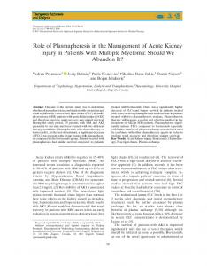
Fig. 1. Expression of Xenopus Sall1, Sall3, and Sall4 during hindlimb regeneration at the regeneration-complete stage and the regenerationincomplete stage. A: Top; whole-mount in situ hybridization (WMISH) of Sall4 in stage-53 and -57 limbs and in stage-53 and -57 regenerating hindlimbs at 0, 1, 3, and 5dPA. Bottom; RT-PCR comparison of Sall1, Sall3, and Sall4 expression patterns in stage-53 and -57 regenerating hindlimb blastemas and pseudoblastemas at 0, 1, 3, and 5dPA. Amputation was through the mid-zeugopodia and blastemas (from stage-53 limbs) or pseudoblastemas (from stage-57 limbs) were collected for analysis at 0, 1, 3, and 5dPA. At 0dPA about 1 to 2 mm of tissue from the distal end of the amputated limb was used as the internal tissue control. Odc (ornithine decarboxylase) was used as an internal control. WMISH data was taken from Neff et al. (2005) with the authors’ permission. B: QPCR comparison of Sall1, Sall3, and Sall4 expression patterns in stage-53 and -57 regenerating hindlimb blastemas and pseudoblastemas at 0, 1, 3, and 5dPA. Sall gene expression levels are normalized to odc and then shown relative to stage-53 0dPA levels of expression, which is arbitrarily set to 1.0. RT-PCR and QPCR were performed as described in King et al. (2009).
limb buds before Shh is expressed, it is expressed in amputated limb blastemas at the regenerationcompetent stage before Shh is expressed, and it is expressed in pseudoblastemas where there is no Shh expression and A/P patterning does not occur (Endo et al., 1997). Christen et al. (2010) further investigated regeneration and reprogramming during Xenopus hindlimb regeneration at regenera-
tion-complete (stage 52) and regeneration-incomplete stages (stage 57) by assaying (qPCR) the expression of pluripotency factors associated with iPSC reprogramming, including Oct4, Sox2, c-Myc, Lin28, and Sall4 in 0d, 1dPA, 3dPA, and 5dPA blastemas and pseudoblastemas. The developing and regenerating limb expression profile of these genes differed from the ESC-like blastomere gene expression profile.
Stage-52 limb regeneration blastemas did not express Oct4 or Lin28 and other genes such as Sox2 and Sall4 were up-regulated at low, statistically insignificant levels. Despite the low level of Sall4 expression measured by qPCR after hindlimb amputation at stage 52, the WMISH experiments presented by Christen et al. (2010) showed expression of Sall4 in stage-52 1dPA and 3dPA blastemas. These
Developmental Dynamics
DEDIFFERENTIATION, SALL4, AND LIMB REGENERATION 985
investigators amputated stage-52 limbs through the presumed zeugopodia and at this stage the expression of pluripotency markers is seen more broadly along the P/ D axis than at stage 53 or later. The amputation plane through the stage-52 hindlimb indicated by Christen et al. (2010) is through the middle of the Sall4 expression domain, which would give the 0dPA control high levels of expression. Neff et al. (2005) used stage53 hindlimbs, which are larger and can be more precisely amputated through the mid zeugopodia, which is proximal to the Sall4 expression domain (Neff et al., 2005). Surprisingly, Christen et al. (2010) found a significant increase in c-Myc, Sox2, and Sall4 expression in 1dPA and 3dPA pseudo-blastemas at stage 57. However, the overall level of expression was lower than that observed at stage 53. They concluded that some but not all iPSC reprogramming factors are expressed during epimorphic regeneration and that blastema cells are not like either ESC or reprogrammed pluripotent iPSCs. Christen et al. (2010) speculate that amphibian limb regeneration blastemas contain partially reprogrammed cells and that there may be a common mechanism between dedifferentiation and reprogramming.
COMPARISON OF SALL1, SALL3, AND SALL4 EXPRESSION DURING LIMB REGENERATION. Sall gene family members (Sall1, 2, 3, and 4) show similar and unique expression patterns in different systems such as organogenesis, limb development, and neurogenesis (reviewed de Celis and Barrio, 2009). For example, both Sall1 and Sall4 are expressed in ESC (Sakaki-Yumoto et al., 2006) and in early developing limb bud mesenchyme (Hollemann et al., 1996; Neff et al., 2005). SALL4 can heterodimerize with other family members such as SALL1 (Sakaki-Yumoto et al., 2006) and Sall1, Sall2, and Sall3 have essential redundant functions during murine neurulation (Bohm et al., 2008). Sall1 and Sall3 are involved in Shh linked A/P
patterning of developing mouse limbs (Kawakami et al., 2009) and human Sall1 mutations produce TownesBrocks Syndrome, most likely caused by the dominant-negative effect of truncated SALL1, which includes among its defects thumb abnormalities (Kohlhase et al., 1998; de Celis and Barrio, 2009). Functional gene knockout studies of mouse autopod development showed that Sall1 and Sall3 are partially redundant. While Sall1 can compensate for loss of Sall3 and vice versa, double knockouts have no digit 1, similar to Sall4 loss-offunction mutants (Kawakami et al., 2009). Both Sall1 and Sall3 are negatively regulated by Sall4 in ESCs (Yang et al., 2010). Cole et al. (2008) found that while SALL4 can bind the Tcf3, Oct4, and Nanog promoters, SALL1 only binds the Nanog promoter, SALL3 binds only the Oct4 promoter, and SALL2 does not bind to any of the mentioned t-factors (Cole et al., 2008). Sall1 is part of the core pluripotency network that suppresses ectodermal and mesodermal differentiation (Karantzali et al., 2010). These complex interactions between Sall gene family members prompted us to look at the expression profile of the Sall gene family during Xenopus hindlimb regeneration at the regeneration-complete and-incomplete stages utilizing RT-PCR and qPCR assays. RT-PCR (Fig.1A) and qPCR (Fig. 1B) were used to determine the expression of Sall1, Sall3, and Sall4 during Xenopus hindlimb regeneration at stage 53 and stage 57. Sall2 was not assayed because previous experiments demonstrated that Sall2 is not expressed during Xenopus hindlimb regeneration (data not shown). Amputation was through the midzeugopodia and blastemas (from stage-53 limbs) or pseudoblastemas (from stage-57 limbs) were collected for analysis at 0dPA, 1dPA, 3dPA, and 5dPA. At 0dPA, about 1 to 2 mm of tissue from the distal end of the amputated limb was used as the internal tissue control. Ornithine decarboxylase (odc) expression was used as the normalizer for gene expression. Expression of Sall1 and Sall3, like that of Sall4, was higher in stage-53 0dPA than stage-57 0dPA tissue just proximal to the amputation plane. In stage-53 1dPA blastemas, Sall4
showed a 5.2-fold increase while the levels of Sall1 and Sall3 were about the same as the 0dPA controls. Sall4 expression is up-regulated very early after amputation, being detected by WMISH in the dedifferentiation zone proximal to the amputation plane at the earliest time point assayed at 4 hr PA (Neff et al., 2005) and in a separate experiment at 6 hr PA by qPCR (unpublished data). The 3dPA blastema data was similar, with only Sall4 expressed at higher levels. At 5dPA, expression of all three Sall genes was higher than the 0dPA controls (Sall1, 9-fold; Sall3, 6.5-fold; Sall4, 5.4-fold). These data are consistent with the following interpretation: Sall4 alone is up-regulated at 1dPA because it, but not Sall1 nor Sall3, is involved in dedifferentiation/ reprogramming. In addition, Sall4 is the only Sall gene up-regulated in blastemas before Shh is up-regulated (unpublished data), which suggests that Sall4 expression correlates with dedifferentiation while Sall1 and Sall3 expression correlates with patterning. The early Sall4 expression after amputation is probably not a wound response because wounding of a Xenopus limb without amputation does not result in local Sall4 expression (Neff et al., 2005, unpublished data). We assume that reprogramming, growth of the blastema, and patterning are probably not all-ornone phenomena, considering the complexity of the events occurring during limb regeneration, but rather overlapping events such as reprogramming at 1 to 3dPA, maintenance of the undifferentiated cells in the growing blastema at 5dPA, and patterning at 7dPA. We speculate that by 5dPA when a definitive morphological blastema is established, Sall4 along with Sall1 and possibly Sall3 are involved in maintaining the blastema stem cells in the undifferentiated state. Both Sall1 and Sall3 may also be involved in AP patterning at this stage in limb regeneration. The expression of Sall1 and Sall3, like Sall4, is higher in stage-53 0dPA controls than stage-57 0dPA controls. In stage-57 1dPA pseudoblastemas, Sall4 was expressed 9.9-fold higher than the 0dPA controls while Sall1 and Sall3 were not up-regulated. This level of Sall4 expression is below the
Developmental Dynamics
986 NEFF ET AL.
level detectable by WMISH (Neff et al., 2005). In stage-57 5dPA pseudoblastemas, Sall4 expression was 27fold higher than the 0dPA control, while Sall1 and Sall3 expression remained unchanged. The increased expression of Sall4 in stage-57 pseudoblastemas after amputation is supported by data from Christen et al. (2010) who found significant but low levels of Sall4 expression 1dPA and 3dPA in stage-57 pseudoblastemas compared to 0dPA control levels. These data are consistent with the interpretation that in response to amputation at stage 57, dedifferentiation and reprogramming of local cells begin, but the response is diminished compared with that at stage 53 because a smaller population of cells is involved or a reduced response in cells capable of reprogramming. In addition, Sall1 and Sall3 are not expressed at a higher level than 0dPA after amputation of regeneration-incomplete limbs because there is no Shh expression and no AP patterning. We speculate that at 5dPA in stage-57 limbs, Sall4 expression in the pseudoblastemas is restricted to a population of undifferentiated mesenchymal cells that will eventually give rise to the cartilage cells of the hypomorphic spikes.
DEDIFFERENTIATION AND Sall4 REGULATION OF CHROMATIN STRUCTURE Embryonic and other stem cells maintain an epigenetic chromatin structure that determines their level of pluripotency, self-renewal, and differentiation. Genome-wide maps of global chromatin structure show very few differences between ESC and iPSC (Guenther et al., 2010). SALL4 protein is primarily localized to heterochromatin (Bohm et al., 2008) and has been linked to chromatin structure remodeling in ESCs (Yang et al., 2008; Bard et al., 2009; Lu et al., 2009; van den Berg et al., 2010). The mammalian ESC SWI/SNF ATPdependent chromatin remodeling complex, esBAF, is essential for the core ESC transcriptional network that binds SALL4 (Ho et al., 2009a,b; Lessard and Crabtree, 2010). Reprogramming to form blastema stem cells and maintenance of the blastema cells
within the limb regeneration blastema also most likely involves chromatin remodeling. Reprogramming is facilitated by chromatin remodeling components of the BAF complex (Singhal et al., 2010). Nuclear proteins of stem cells that can reprogram mouse embryonic fibroblasts include SALL4 and pluripotent cell-specific components of the SWI/SNF complex (Singhal et al., 2010). Overexpression of Smarca4 and Smarcc1 was sufficient to enhance reprogramming. Our microarray analyses showed that Smarca4 RNA was expressed at higher levels in stage-53 5dPA limb blastemas than stage-57 5dPA limb pseudoblastemas, while Smarcc1 RNA was expressed at higher levels in stage-53 3dPA blastemas compared to stage-53 1dPA blastemas (Grow et al., 2006 and unpublished data). The SMARCA4 protein is also expressed at much higher levels in stage-53 3dPA limb blastemas than in stage-57 3dPA limb blastemas (King et al., 2009). Other components of the pluripotency-associated SWI/SNF complex (Singhal et al., 2010) were also differentially expressed in our limb regeneration microarray screens, including Smarce1, Aridia, Smarcc1, SmarchA5, SmarcA6, Smarcd1, and Actl6a (Grow et al, 2006; unpublished data) and SMARCD1 protein (King et al., 2009). In contrast to SMARCA4, the SMARCC2 protein is a differentiation- (not pluripotency-) associated SWI/SNF complex component (Singhal et al., 2010) and was expressed abundantly in stage-57 3dPA limb pseudoblastemas. These data are consistent with the view that stage-53 3dPA blastemas possess a pluripotency-associated SWI/SNF complex that includes Sall4, while stage-57 limb pseudoblastemas contain a differentiated state SWI/SNF complex, which may or may not include Sall4. Epigenetic covalent histone tail modifications that activate, repress, or pose repressed genes for activation (bivalent histone modification is a hallmark of pluripotent cells) are involved in regulating stemness, development, differentiation, and reprogramming (Meissner, 2010). Dynamic changes in histone modifications occur during dedifferentiation of the dorsal and ventral irises during newt lens regeneration (Maki et al., 2009). A crucial early
event in the initiation of zebrafish fin regeneration involves histone modifications (Stewart et al., 2009). SALL4 is one of the pluripotency factors that regulate epigenetic modifiers in human ESC (Yang et al., 2008; Hemberger et al., 2009). Lineage-specific SALL4 promoter occupancy correlates with specific histone modifications in two distinct stem cell lineages (Lim et al., 2008). SALL4 has been linked to the epigenetic histone modification mechanism for regulation of Bmi-1 expression in hematopoietic and leukemic stem cells (Yang et al., 2007). SALL4 in association with Whsc1 is associated with histone modification that regulates the expression of their target genes in ESC (Nimura et al., 2009).
SIGNALING PATHWAYS REGULATING Sall4 EXPRESSION DURING LIMB REGENERATION Limb regeneration in Xenopus involves numerous signaling pathways (Beck et al., 2009), including BMP (Beck et al., 2006), Fgf (Yokoyama et al., 2000), and Wnt (Kawakami et al., 2006). Regulation of Sall4 expression during reprogramming and maintenance of pluripotency in ESC and somatic stem cells and probably during patterning also involves numerous context-dependent signaling pathways (Li et al., 2010). For example, in Drosophila melanogaster spalt/Sal expression is regulated during embryogenesis by Shh, dpp (BMP), and EGF signaling pathways, depending on the context (de Celis and Barrio, 2009). Xenopus Sall4 was originally identified in a differential display screen looking at genes expressed in animal caps exposed to activin (a TGF-beta family member) and retinoic acid (Onuma et al., 1999). Yang et al. (2008) used ChIP-chip assays to determine that SALL4 binding genes (genes with SALL4 binding sites) encode components in over 30 different signaling pathways, the most prominent being the Wnt, PTEN, NF-kB, and p53 signaling pathways. Sall4 expression is directly activated by the canonical Wnt signaling pathway (Bohm et al., 2008; Al-Qattan, 2010). Sall4 activates Fgf10 synergistically with Tbx5 and
Developmental Dynamics
DEDIFFERENTIATION, SALL4, AND LIMB REGENERATION 987
Tbx4 (Koshiba-Takeuchi et al., 2006). Unpublished data in our lab indicate that in cultured stage-55 Xenopus hindlimb autopods, Sall4 is up-regulated in a dose-dependent manner by Fgf10. Signal transducer and activator of transcription 3 (STAT3) regulates the expression of Sall4 (Bard et al., 2009). Among the growth factors that signal via STAT3 are LIF, which is necessary to maintain mouse ESC, and IL-6, which is required for compensatory liver regeneration (Tiberio et al., 2008). LIF/Stat3 signaling contributes to the maintenance of the undifferentiated state by preventing lineage-specific differentiation programs, including Sall4 expression (Bourillot et al., 2009). ESCs that are induced to differentiate by eliminating STAT3 signaling express Sall4 in the early phases of their differentiation (Bourillot et al., 2009). In Xenopus limb regeneration, Stat3 is up-regulated in both 3dPA stage-53 blastemas and 3dPA stage-57 pseudoblastemas relative to 1dPA limbs (Grow et al, 2006 and unpublished data), observations consistent with the finding that Sall4 is expressed in dedifferentiating multipotent cells. Proteomic analysis during the early phase of Xenopus limb regeneration revealed that the overall protein expression profile of 3dPA blastemas at stage 53 more closely resembles that of multipotent adult stem cells than that of totipotent ESCs (King et al., 2009). In summary, Sall4 responds and regulates numerous signaling pathways that are involved in both stemness and limb patterning.
PERSPECTIVES In 2005, we postulated that Sall4 may be playing a role in dedifferentiation (reprogramming) and in maintaining limb blastema cells in an undifferentiated state. This hypothesis was based on the observed differential pattern of SALL4 expression during Xenopus hindlimb regeneration at the regeneration-complete stage and -incomplete stage. Since then it has been shown that Sall4 is involved in maintenance of ESC pluripotency, is a major repressor of differentiation, and plays a major role in reprogramming differentiated cells into iPSC. Sall4 is a component of the stemness regulatory circuit involv-
ing Nanog, Oct4, Sox2 and others in pluripotent ESC and iPSC. However, unlike many such stemness factors, Sall4 is also expressed in multipotent embryonic, fetal, and adult somatic stem cells. In addition, Sall4 plays a role in epigenetic modulation of chromatin structure. These functional qualities make Sall4 a major player in regulating the dedifferentiation/ reprogramming events that produce and maintain the kinds of multipotent stem cells that comprise epimorphic regeneration blastemas. Functional analysis of genes activated and repressed by Sall4, of the signaling pathways that regulate Sall4 expression, and of Sall4 function in modulating chromatin structure during epimorphic regeneration has the potential to yield large dividends in the field of regenerative biology and medicine.
ACKNOWLEDGMENTS Research by the authors on dedifferentiation and Sall4 was supported by the National Science Foundation (grant IOS-0814399) and benefitted from the expert technical assistance of Elizabeth Osborne.
REFERENCES Al-Qattan MM. 2010. Wnt pathways and upper limb anomalies. J Hand Surg Eur Vol 36:9–22. Bard JD, Gelebart P, Amin HM, Young LC, Ma Y, Lai R. 2009. Signal transducer and activator of transcription 3 is a transcriptional factor regulating the gene expression of SALL4. Faseb J 23: 1405–1414. Barembaum M, Bronner-Fraser M. 2010. Pax2 and Pea3 synergize to activate a novel regulatory enhancer for spalt4 in the developing ear. Dev Biol 340: 222–231. Barger PM, Tassava RA. 1985. Kinetics of cell cycle entry in innervated and denervated forelimb stumps of larval Ambystoma. J Exp Zool 233:151–154. Beck CW, Christen B, Barker D, Slack JMW. 2006. Temporal requirement for bone morphogenetic proteins in regeneration of the tail and limb of Xenopus tadpoles. Mech Dev 123:674–688. Beck CW, Izpisua Belmonte JC, Christen B. 2009. Beyond early development: Xenopus as an emerging model for the study of regenerative mechanisms. Dev Dyn 238:1226–1248. Bohm J, Heinritz W, Craig A, Vujic M, Ekman-Joelsson BM, Kohlhase J, Froster U. 2008. Functional analysis of the novel TBX5 c.1333delC mutation result-
ing in an extended TBX5 protein. BMC Med Genet 9:88. Bourillot PY, Aksoy I, Schreiber V, Wianny F, Schulz H, Hummel O, Hubner N, Savatier P. 2009. Novel STAT3 target genes exert distinct roles in the inhibition of mesoderm and endoderm differentiation in cooperation with Nanog. Stem Cells 27:1760–1771. Brockes JP, Kumar A. 2002. Plasticity and reprogramming of differentiated cells in amphibian regeneration. Nature Rev Mol Cell Biol 3:566–574. Brockes JP, Kumar A. 2005. Appendage regeneration in adult vertebrates and implications for regenerative medicine. Science 310:1919–1923. Carinato ME, Walter BE, Henry JJ. 2000. Xenopus laevis gelatinase B (Xmmp-9): development, regeneration, and wound healing. Dev Dyn 217:377–387. Carlson BM. 2007. Principles of regenerative biology. Amsterdam: Elsevier Academic Press. Christen B, Robles V, Raya M, Paramonov I, Belmonte JC. 2010. Regeneration and reprogramming compared. BMC Biol 8:5. Cole MF, Johnstone SE, Newman JJ, Kagey MH, Young RA. 2008. Tcf3 is an integral component of the core regulatory circuitry of embryonic stem cells. Genes Dev 22:746–755. de Celis JF, Barrio R. 2009. Regulation and function of Spalt proteins during animal development. Int J Dev Biol 53: 1385–1398. Del Rio-Tsonis K, Tsonis PA, Zarkadis IK, Tsagas AG, Lambris JD. 1998. Expression of the third component of complement, C3, in regenerating limb blastema cells of urodeles. J Immunol 161:6819–6824. Dent JN. 1962. Limb regeneration in larvae and metamorphosing individuals of South African Clawed toad. J Morphol 110:61–78. Endo T, Yokoyama H, Tamura K, Ide H. 1997. Shh expression in developing and regenerating limb buds of Xenopus laevis. Dev Dyn 209:227–232. Forte A, Schettino MT, Finicelli M, Cipollaro M, Colacurci N, Cobellis L, Galderisi U. 2009. Expression pattern of stemness-related genes in human endometrial and endometriotic tissues. Mol Med 15:392–401. Fukazawa T, Naora Y, Kunieda T, Kubo T. 2009. Suppression of the immune response potentiates tadpole tail regeneration during the refractory period. Development 136:2323–2327. Godwin JW, Liem KF Jr, Brockes JP. 2010. Tissue factor expression in newt iris coincides with thrombin activation and lens regeneration. Mech Dev 127:321–328. Goss RJ. 1969. Principles of regeneration. New York: Academic Press. Grow MW, Neff AW, Mescher AL, King MW. 2006. Global analysis of gene expression in Xenopus limbs during stage-dependent complete and incomplete regeneration. Dev Dyn 235:2667–2685. Guenther MG, Frampton GM, Soldner F, Hockemeyer D, Mitalipova M, Jaenisch R, Young RA. 2010. Chromatin structure
Developmental Dynamics
988 NEFF ET AL.
and gene expression programs of human embryonic and induced pluripotent stem cells. Cell Stem Cell 7:249–257. Gurtner GC, Werner S, Barrandon Y, Longaker MT. 2008. Wound repair and regeneration. Nature 453:314–321. Han J, Yuan P, Yang H, Zhang J, Soh BS, Li P, Lim SL, Cao S, Tay J, Orlov YL, Lufkin T, Ng HH, Tam WL, Lim B. 2010. Tbx3 improves the germ-line competency of induced pluripotent stem cells. Nature 463:1096–1100. Heissig B, Nishida C, Tashiro Y, Sato Y, Ishihara M, Ohki M, Gritli I, Rosenkvist J, Hattori K. 2010. Role of neutrophilderived matrix metalloproteinase-9 in tissue regeneration. Histol Histopathol 25:765–770. Hemberger M, Dean W, Reik W. 2009. Epigenetic dynamics of stem cells and cell lineage commitment: digging Waddington’s canal. Nat Rev Mol Cell Biol 10:526–537. Ho L, Jothi R, Ronan JL, Cui K, Zhao K, Crabtree GR. 2009a. An embryonic stem cell chromatin remodeling complex, esBAF, is an essential component of the core pluripotency transcriptional network. Proc Natl Acad Sci USA 106: 5187–5191. Ho L, Ronan JL, Wu J, Staahl BT, Chen L, Kuo A, Lessard J, Nesvizhskii AI, Ranish J, Crabtree GR. 2009b. An embryonic stem cell chromatin remodeling complex, esBAF, is essential for embryonic stem cell self-renewal and pluripotency. Proc Natl Acad Sci USA 106: 5181–5186. Hockemeyer D, Soldner F, Cook EG, Gao Q, Mitalipova M, Jaenisch R. 2008. A drug-inducible system for direct reprogramming of human somatic cells to pluripotency. Cell Stem Cell 3:346–353. Hollemann T, Schuh R, Pieler T, Stick R. 1996. Xenopus Xsal-1, a vertebrate homolog of the region specific homeotic gene spalt of Drosophila. Mech Dev 55:19–32. Imokawa Y, Brockes JP. 2003. Selective activation of thrombin is a critical determinant for vertebrate lens regeneration. Curr Biol 13:877–881. Karantzali E, Lekakis V, Ioannou M, Hadjimichael C, Papamatheakis J, Kretsovali A. 2010. Sall1 regulates embryonic stem cell differentiation in association with Nanog. J Biol Chem 286:1037–1045. Kawakami Y, Rodriguez Esteban C, Raya M, Kawakami H, Marti M, Dubova I, Izpisua Belmonte JC. 2006. Wnt/beta-catenin signaling regulates vertebrate limb regeneration. Genes Dev 20:3232–3237. Kawakami Y, Uchiyama Y, Rodriguez Esteban C, Inenaga T, Koyano-Nakagawa N, Kawakami H, Marti M, Kmita M, Monaghan-Nichols P, Nishinakamura R, Izpisua Belmonte JC. 2009. Sall genes regulate region-specific morphogenesis in the mouse limb by modulating Hox activities. Development 136:585–594. Khan PA, Liversage RA. 1990. Ultrastructural comparison between regenerating and developing hindlimbs of Xenopus laevis tadpoles. Growth Dev Aging 54: 173–181.
Kimura Y, Madhavan M, Call MK, Santiago W, Tsonis PA, Lambris JD, Del Rio-Tsonis K. 2003. Expression of complement 3 and complement 5 in newt limb and lens regeneration. J Immunol 170:2331–2339. King MW, Nguyen T, Calley J, Harty MW, Muzinich MC, Mescher AL, Chalfant C, N’Cho M, McLeaster K, McEntire J, Stocum D, Smith RC, Neff AW. 2003. Identification of genes expressed during Xenopus laevis limb regeneration by using subtractive hybridization. Dev Dyn 226:398–409. King MW, Neff AW, Mescher AL. 2009. Proteomics analysis of regenerating amphibian limbs: changes during the onset of regeneration. Int J Dev Biol 53:955–969. Kohlhase J, Wischermann A, Reichenbach H, Froster U, Engel W. 1998. Mutations in the SALL1 putative transcription factor gene cause Townes-Brocks syndrome. Nat Genet 18:81–83. Kohlhase J, Heinrich M, Liebers M, Frohlich Archangelo L, Reardon W, Kispert A. 2002. Cloning and expression analysis of SALL4, the murine homologue of the gene mutated in Okihiro syndrome. Cytogenet Genome Res 98:274–277. Korneluk RG, Liversage RA. 1984. Tissue regeneration in the amputated forelimb of Xenopus laevis froglets. Can J Zool 62:2383–2391. Koshiba-Takeuchi K, Takeuchi JK, Arruda EP, Kathiriya IS, Mo R, Hui CC, Srivastava D, Bruneau BG. 2006. Cooperative and antagonistic interactions between Sall4 and Tbx5 pattern the mouse limb and heart. Nat Genet 38:175–183. Kragl M, Knapp D, Nacu E, Khattak S, Maden M, Epperlein HH, Tanaka EM. 2009. Cells keep a memory of their tissue origin during axolotl limb regeneration. Nature 460:60–65. Kumar A, Velloso CP, Imokawa Y, Brockes JP. 2000. Plasticity of retrovirus-labelled myotubes in the newt limb regeneration blastema. Dev Biol 218:125–136. Lessard JA, Crabtree GR. 2010. Chromatin regulatory mechanisms in pluripotency. Annu Rev Cell Dev Biol 26:503–532. Li Y, Zhao H, Lan F, Lee A, Chen L, Lin C, Yao Y, Li L. 2010. Generation of human-induced pluripotent stem cells from gut mesentery-derived cells by ectopic expression of OCT4/SOX2/NANOG. Cell Reprogram 12:237–247. Lim CY, Tam WL, Zhang J, Ang HS, Jia H, Lipovich L, Ng HH, Wei CL, Sung WK, Robson P, Yang H, Lim B. 2008. Sall4 regulates distinct transcription circuitries in different blastocyst-derived stem cell lineages. Cell Stem Cell 3:543–554. Lo DC, Allen F, Brockes JP. 1993. Reversal of muscle differentiation during urodele limb regeneration. Proc Natl Acad Sci USA 90:7230–7234. Loyd RM, Tassava RA. 1980. DNA synthesis and mitosis in adult newt limbs following amputation and insertion into the body cavity. J Exp Zool 214:61–69. Lu J, Jeong HW, Kong N, Yang Y, Carroll J, Luo HR, Silberstein LE, Yupoma, Chai L. 2009. Stem cell factor SALL4
represses the transcriptions of PTEN and SALL1 through an epigenetic repressor complex. PLoS One 4:e5577. Maki N, Suetsugu-Maki R, Tarui H, Agata K, Del Rio-Tsonis K, Tsonis PA. 2009. Expression of stem cell pluripotency factors during regeneration in newts. Dev Dyn 238:1613–1616. Mallanna SK, Rizzino A. 2010. Emerging roles of microRNAs in the control of embryonic stem cells and the generation of induced pluripotent stem cells. Dev Biol 344:16–25. Manfredi AA, Capobianco A, Bianchi ME, Rovere-Querini P. 2009. Regulation of dendritic- and T-cell fate by injury-associated endogenous signals. Crit Rev Immunol 29:69–86. Markiewski MM, Lambris JD. 2007. The role of complement in inflammatory diseases from behind the scenes into the spotlight. Am J Pathol 171:715–727. Martin P, D’Souza D, Martin J, Grose R, Cooper L, Maki R, McKercher SR. 2003. Wound healing in the PU.1 null mouse: Tissue repair is not dependent on inflammatory cells. Curr Biol 12:1122–1128. Mathew LK, Sengupta S, Kawakami A, Andreasen EA, Lohr CV, Loynes CA, Renshaw SA, Peterson RT, Tanguay RL. 2007. Unraveling tissue regeneration pathways using chemical genetics. J Biol Chem 282:35202–35210. Matzinger P. 2007. Friendly and dangerous signals: is the tissue in control? Nat Immunol 8:11–13. Medzhitov R. 2008. Origin and physiological roles of inflammation. Nature 454: 428–435. Meissner A. 2010. Epigenetic modifications in pluripotent and differentiated cells. Nat Biotechnol 28:1079–1088. Melton C, Judson RL, Blelloch R. 2010. Opposing microRNA families regulate self-renewal in mouse embryonic stem cells. Nature 463:621–626. Mescher AL. 1976. Effects on ault newt limb regeneration of partial and complete skin flaps over the amputation surface. J Exp Zool 195:117–127. Mescher AL. 1996. The cellular basis of limb regeneration in urodeles. Int J Dev Biol 40:785–795. Mescher AL, Tassava RA. 1975. Denervation effects on DNA replication and mitosis during the initiation of limb regeneration in adult newts. Dev Biol 44:187–197. Morgan TH. 1901. Regeneration. New York: Macmillan Co. Mori R, Shaw TJ, Martin P. 2008. Molecular mechanisms linking wound inflammation and fibrosis: knockdown of osteopontin leads to rapid repair and reduced scarring. J Exp Med 205:43–51. Morrison JI, Loof S, He P, Simon A. 2006. Salamander limb regeneration involves the activation of a multipotent skeletal muscle satellite cell population. J Cell Biol 172:433–440. Morrison JI, Borg P, Simon A. 2010. Plasticity and recovery of skeletal muscle satellite cells during limb regeneration. Faseb J 24:750–756.
Developmental Dynamics
DEDIFFERENTIATION, SALL4, AND LIMB REGENERATION 989
Mott JD, Werb Z. 2004. Regulation of matrix biology by matrix metalloproteinases. Curr Opin Cell Biol 16:558–564. Nakamura K, Maki N, Trinh A, Trask HW, Gui J, Tomlinson CR, Tsonis PA. 2010. miRNAs in newt lens regeneration: specific control of proliferation and evidence for miRNA networking. PLoS One 5:e12058. Neff AW, King MW, Harty MW, Nguyen T, Calley J, Smith RC, Mescher AL. 2005. Expression of Xenopus XlSALL4 during limb development and regeneration. Dev Dyn 233:356–367. Nimura K, Ura K, Shiratori H, Ikawa M, Okabe M, Schwartz RJ, Kaneda Y. 2009. A histone H3 lysine 36 trimethyltransferase links Nkx2–5 to Wolf-Hirschhorn syndrome. Nature 460:287–291. Odelberg SJ. 2005. Cellular plasticity in vertebrate regeneration. Anat Rec 287B:25–35. Ohgo S, Itoh A, Suzuki M, Satoh A, Yokoyama H, Tamura K. 2010. Analysis of hoxa11 and hoxa13 expression during patternless limb regeneration in Xenopus. Dev Biol 338:148–157. Oikawa T, Kamiya A, Kakinuma S, Zeniya M, Nishinakamura R, Tajiri H, Nakauchi H. 2009. Sall4 regulates cell fate decision in fetal hepatic stem/progenitor cells. Gastroenterology 136:1000–1011. Onuma Y, Nishinakamura R, Takahashi S, Yokota T, Asashima M. 1999. Molecular cloning of a novel Xenopus spalt gene (Xsal-3). Biochem Biophys Res Commun 264:151–156. Pacini S, Carnicelli V, Trombi L, Montali M, Fazzi R, Lazzarini E, Giannotti S, Petrini M. 2010. Constitutive expression of pluripotency-associated genes in mesodermal progenitor cells (MPCs). PLoS One 5:e9861. Putta S, Smith JJ, Walker JA, Rondet M, Weisrock DW, Monaghan J, Samuels AK, Kump K, King DC, Maness NJ, Habermann B, Tanaka E, Bryant SV, Gardiner DM, Parichy DM, Voss SR. 2004. From biomedicine to natural history research: EST resources for ambystomatid salamanders. BMC Genomics 5:54. Rao S, Shao Z, Roumiantsev S, McDonald LT, Yuan GC, Orkin SH. 2010. Differential roles of Sall4 isoforms in embryonic stem cell pluripotency. Mol Cell Biol 286:1037–1045. Sakaki-Yumoto M, Kobayashi C, Sato A, Fujimura S, Matsumoto Y, Takasato M, Kodama T, Aburatani H, Asashima M, Yoshida N, Nishinakamura R. 2006. The murine homolog of SALL4, a causative gene in Okihiro syndrome, is essential for embryonic stem cell proliferation, and cooperates with Sall1 in anorectal, heart, brain and kidney development. Development 133:3005–3013. Shafa M, Krawetz R, Rancourt DE. 2010. Returning to the stem state: epigenetics of recapitulating pre-differentiation chromatin structure. Bioessays 32:791–799. Shih YR, Kuo TK, Yang AH, Lee OK, Lee CH. 2009. Isolation and characterization of stem cells from the human parathyroid gland. Cell Prolif 42:461–470.
Singhal N, Graumann J, Wu G, ArauzoBravo MJ, Han DW, Greber B, Gentile L, Mann M, Scholer HR. 2010. Chromatin-remodeling components of the BAF complex facilitate reprogramming. Cell 141:943–955. Smith JJ, Putta S, Walker JA, Kump DK, Samuels AK, Monaghan JR, Weisrock DW, Staben C, Voss SR. 2005. Sal-Site: integrating new and existing ambystomatid salamander research and informational resources. BMC Genomics 6:181. Stamenkovic I. 2003. Extracellular matrix remodelling: the role of matrix metalloproteinases. J Pathol 200:448–464. Stewart S, Tsun ZY, Izpisua Belmonte JC. 2009. A histone demethylase is necessary for regeneration in zebrafish. Proc Natl Acad Sci USA 106:19889–19894. Stocum D. 1995. Wound repair, regeneration and artificial tissues. Austin: R.G. Landes Co. Stocum DL, Zupanc GK. 2008. Stretching the limits: stem cells in regeneration science. Dev Dyn 237:3648–3671. Straube WL, Tanaka EM. 2006. Reversibility of the differentiated state: regeneration in amphibians. Artif Organs 30:743–755. Sweetman D, Munsterberg A. 2006. The vertebrate spalt genes in development and disease. Dev Biol 293:285–293. Takahashi K. 2010. Direct reprogramming 101. Dev Growth Differ 52:319–333. Takahashi K, Tanabe K, Ohnuki M, Narita M, Ichisaka T, Tomoda K, Yamanaka S. 2007. Induction of pluripotent stem cells from adult human fibroblasts by defined factors. Cell 131:861–872. Takahashi K, Yamanaka S. 2006. Induction of pluripotent stem cells from mouse embryonic and adult fibroblast cultures by defined factors. Cell 126:663–676. Tamura K, Ohgo S, Yokoyama H. 2010. Limb blastema cell: a stem cell for morphological regeneration. Dev Growth Differ 52:89–99. Tanaka EM, Drechsel DN, Brockes JP. 1999. Thrombin regulates S-phase reentry by cultured newt myotubes. Curr Biol 9:792–799. Tassava RA, Mescher AL. 1975. Roles of injury, nerves, and wound epidermis during initiation of amphibian limb regeneration. Differentiation 4:23–24. Tiberio GA, Tiberio L, Benetti A, Cervi E, Montani N, Dreano M, Garotta G, Cerea K, Steimberg N, Pandolfo G, Ferrari-Bravo A, Mazzoleni G, Giulini SM, Schiaffonati L. 2008. IL-6 Promotes compensatory liver regeneration in cirrhotic rat after partial hepatectomy. Cytokine 42:372–378. Tsonis PA. 2008. Stem cells and blastema cells. Curr Stem Cell Res Ther 3:53–54. Tsubooka N, Ichisaka T, Okita K, Takahashi K, Nakagawa M, Yamanaka S. 2009. Roles of Sall4 in the generation of pluripotent stem cells from blastocysts and fibroblasts. Genes Cells 14:683–694. Uez N, Lickert H, Kohlhase J, de Angelis MH, Kuhn R, Wurst W, Floss T. 2008. Sall4 isoforms act during proximal-distal and anterior-posterior axis formation in the mouse embryo. Genesis 46:463–477.
Ulloa-Montoya F, Kidder BL, Pauwelyn KA, Chase LG, Luttun A, Crabbe A, Geraerts M, Sharov AA, Piao Y, Ko MS, Hu WS, Verfaillie CM. 2007. Comparative transcriptome analysis of embryonic and adult stem cells with extended and limited differentiation capacity. Genome Biol 8:R163. van den Berg DL, Snoek T, Mullin NP, Yates A, Bezstarosti K, Demmers J, Chambers I, Poot RA. 2010. An Oct4-centered protein interaction network in embryonic stem cells. Cell Stem Cell 6:369–381. Vinarsky V, Atkinson DL, Stevenson TJ, Keating MT, Odelberg SJ. 2005. Normal newt limb regeneration requires matrix metalloproteinase function. Dev Biol 279:86–98. Wong CC, Gaspar-Maia A, Ramalho-Santos M, Reijo Pera RA. 2008. High-efficiency stem cell fusion-mediated assay reveals Sall4 as an enhancer of reprogramming. PLoS One 3:e1955. Yang EV, Bryant SV. 1994. Developmental regulation of a matrix metalloproteinase during regeneration of axolotl appendages. Dev Biol 166:696–703. Yang EV, Gardiner DM, Carlson MRJ, Nugas CA, Bryant SV. 1999. Expression of Mmp-9 and related matrix metalloproteinase genes during axolotl limb regeneration. Dev Dyn 216:2–9. Yang J, Chai L, Liu F, Fink LM, Lin P, Silberstein LE, Amin HM, Ward DC, Ma Y. 2007. Bmi-1 is a target gene for SALL4 in hematopoietic and leukemic cells. Proc Natl Acad Sci USA 104:10494–10499. Yang J, Chai L, Fowles TC, Alipio Z, Xu D, Fink LM, Ward DC, Ma Y. 2008. Genome-wide analysis reveals Sall4 to be a major regulator of pluripotency in murine-embryonic stem cells. Proc Natl Acad Sci USA 105:19756–19761. Yang J, van Oosten AL, Theunissen TW, Guo G, Silva JC, Smith A. 2010. Stat3 activation is limiting for reprogramming to ground state pluripotency. Cell Stem Cell 7:319–328. Yokoyama H, Yonei-Tamura S, Endo T, Belmonte JCI, Tamura K, Ide H. 2000. Mesenchyme with fgf-10 expression is responsible for regenerative capacity in Xenopus limb buds. Dev Biol 219:18–29. Young HE. 2004. Existence of reserve quiescent stem cells in adults, from amphibians to humans. Curr Top Microbiol Immunol 280:71–109. Yu H, Zhu S, Zhou B, Xue H, Han JD. 2008. Inferring causal relationships among different histone modifications and gene expression. Genome Res 18:1314–1324. Yu J, Vodyanik MA, Smuga-Otto K, Antosiewicz-Bourget J, Frane JL, Tian S, Nie J, Jonsdottir GA, Ruotti V, Stewart R, Slukvin, II, Thomson JA. 2007. Induced pluripotent stem cell lines derived from human somatic cells. Science 318:1917–1920. Zhang J, Tam WL, Tong GQ, Wu Q, Chan HY, Soh BS, Lou Y, Yang J, Ma Y, Chai L, Ng HH, Lufkin T, Robson P, Lim B. 2006. Sall4 modulates embryonic stem cell pluripotency and early embryonic development by the transcriptional regulation of Pou5f1. Nat Cell Biol 8:1114–1123.