Amethod is presented for definition ofsurface-exposed epitopes using polyclonal ... Other surface-exposed epitopes have been located in the region of the fourth.
203
Biochem. J. (1991) 279, 203-212 (Printed in Great Britain)
Definition of surface-exposed epitopes on the (Ca2+-Mg2+)-ATPase of sarcoplasmic reticulum Richard E. A. TUNWELL,* J. Wayne CONLAN,t Ian MATTHEWS,* J. Malcolm EAST* and Anthony G. LEE* *Department of Biochemistry and tDepartment of Microbiology, University of Southampton, Southampton S09 3TU, U.K.
Epitopes for monoclonal antibodies binding to the native (Ca2+-Mg2+)-ATPase have been defined by studying binding to sets of hexameric peptides synthesized on plastic pegs. Epitopes have been confirmed by demonstrating the binding of antipeptide antibodies to the ATPase. A method is presented for definition of surface-exposed epitopes using polyclonal antibodies. Three surface-exposed epitopes have been defined in the nucleotide-binding domain of the ATPase, suggesting considerable surface exposure of this region. Other surface-exposed epitopes have been located in the region of the fourth stalk domain.
INTRODUCTION
Molecular biological techniques have provided the amino acid sequences of many membrane transport proteins, including that of the (Ca2+-Mg2+)-ATPase of sarcoplasmic reticulum (SR) and related ion-transport ATPases (Brandl et al., 1986; Rao et al., 1989; Serrano & Portillo, 1990). However, structures predicted from such sequence information are very uncertain in the absence of further information. Predictions of the number of transmembranous a-helices are of uncertain validity (Fasman & Gilbert, 1990; Jahnig, 1990). Thus for the (Na-K)-ATPase, Ovchinikov et al. (1987, 1988) have predicted the presence of seven transmembrane a-helices from a combination of hydropathyplots and immunological and proteolytic studies. In contrast, despite much sequence similarity with the (Na-K)-ATPase, the (Ca2+-Mg2+)-ATPase has been predicted to have either eight (Shull et al., 1985; Serrano, 1988; Jorgensen & Andersen, 1988) or 10 (MacLennan et al., 1985; Brandl et al., 1986) transmembrane helices. An even number of transmembrane helices is consistent with immunological data showing that the Cand N-terminal segments are on the cytoplasmic surface of the SR (Matthews et al., 1989), and chemical labelling has also shown a cytoplasmic location for the N-terminus (Reithmeier & MacLennan, 1981). The 10-helical model proposed by Brandl et al. (1986) locates 11 ofthe 13 tryptophan residues of the ATPase within the membrane, consistent with the observed maximum quenching of 85 % of the fluorescence intensity by hydrophobic quenchers. Furthermore, six of the tryptophan residues are close to the lipid-water interface, consistent with the quenching observed with fatty acids bound to the membrane (Froud et al., 1986); an alternative 10-helical model proposed by Green (1989), in which the fourth transmembrane a-helix is re-assigned, locates only 10 tryptophan residues within the membrane. Electron microscopy has shown that the bulk of the extramembranous region of the ATPase is on the cytoplasmic side of the membrane and consists of a pear-shaped lobe connected to the membrane by a narrow stalk (Stokes & Green, 1990). It has been suggested that the stalk region consists of five a-helical regions connected to three large domains: (1) a nucleotidebinding domain where ATP binds, (2) a phosphorylation domain containing the aspartine residue (Asp-35 1) on the ATPase that is phosphorylated by ATP, and (3) a transduction domain which serves to link phosphorylation of the ATPase to the transport of
Ca2+ (MacLennan et al., 1985). Comparison of the sequences of a large number of ATPases has located a number of conserved regions that have been postulated to be involved in phosphorylation of the ATPase and in binding ATP (Brandl et al., 1986; Rao et al., 1989; Taylor & Green, 1989; Serrano & Portillo, 1990). Further detail about the three-dimensional structure of the ATPase has been obtained using the fluorescence energy transfer technique. Such studies have suggested that the binding sites for Ca2+ and ATP on the ATPase are widely separated (Scott, 1985), with the ATP-binding site being on the uppermost surface of the ATPase (Gutierrez-Merino et al., 1987), and the Ca2+-binding sites being located either close to the membrane surface (Munkonge et al., 1989) or within the membrane (Clarke et al., 1989). Another technique for defining structure is to use monoclonal antibodies (mAbs) and anti-peptide antibodies to locate surface-exposed regions of the ATPase. In general, the definition of the epitope for an antibody is difficult. Although most X-ray crystallographic studies have shown epitopes to be discontinuous, with the amino acids contributing to the epitope being widely separated in space (Laver et al., 1990), studies with synthetic peptides suggest that continuous epitopes also exist (Atassi, 1975; Geysen et al., 1987; Van Regenmortel et al., 1988; Novotny et al., 1989) and, indeed, it has been suggested that for discontinuous epitopes, only five or six residues, which may be continuous, contribute the bulk of the binding energy (Novotny et al., 1989). It is believed that continuous epitopes generally correspond to mobile loops and ridges on the surface of the protein (Van Regenmortel et al., 1988; Van Regenmortel, 1989). In a previous paper we have described the preliminary mapping of a library of mAbs to the ATPase, studying binding to proteolytic fragments of the ATPase separated on SDS/ polyacrylamide gels (Colyer et al., 1989). From these and competitive binding studies, it was shown that mAbs binding to denatured ATPase (and thus presumed to have continuous epitopes) bound to three spatially distinct sites on the ATPase, all located beyond the T2 tryptic cleavage site at Arg- 198 (Colyer et al., 1989). The assignment of epitopes has been confirmed by studying binding to fusion proteins generated from cDNA fragment libraries of the ATPase (Tunwell et al., 1991). Here we define epitopes more closely by studying binding to series of hexameric peptides synthesized on plastic pegs.
Abbreviations used: SR, sarcoplasmic reticulum; PBS, phosphate-buffered saline; mAb, monoclonal antibody; KLH, keyhole limpet haemocyanin; HRP, horseradish peroxidase; DMF, NN'-dimethylformamide; Fmoc, fluorenylmethoxycarbonylamine; FITC, fluorescein isothiocyanate.
Vol. 279
204
MATERIALS AND METHODS (Ca2+-Mg2+)-ATPase was purified from rabbit skeletal muscle SR as described previously (Colyer et al., 1989) and gave a preparation which, on polyacrylamide gels stained with Coomassie Blue, was > 97 % pure (Gould et al., 1987). mAbs were prepared as described in Colyer et al. (1989) and were purified from ascites fluid by precipitation with 40 %-satd. (NH4)2SO4 followed by dialysis against 2 x 1 litre of phosphate-buffered saline (PBS; 137 mM-NaCl, 2.7 mM-KCI, 8.1 mM-Na2HPO4, 1.5 mM-KH2P04, pH 7.2), giving a preparation containing approx. 50% mAb by weight at a protein concentration of 1-5 mg/ml. Synthesis and use of hexapeptides Hexapeptides were synthesized on blocks of polyethylene pins designed to fit into 96-well microtitre plates, using kits supplied by Cambridge Research Biochemicals, as described by Geysen et al. (1987) by stepwise chain elongation using Fmoc (fluorenylmethoxycarbonylamine) chemistry (Van Regenmortel et al., 1988). NN'-Dimethylformamide (DMF; Romil Chemicals Ltd.) was stored over molecular sieve 4A (BDH) until assay for free amino groups using fluorodinitrobenzene showed it to be free of dimethylamine. All other reagents were AnalaR grade, and water was distilled. The pre-derivatized pins were supplied with Fmoc-alanine linked via hexamethylenediamine to the pins. The Fmoc N-terminal protecting group was removed from the pins and from subsequent coupled Fmoc-amino acids by washing with 20 % (v/v) piperidine in DMF for 30 min at room temperature with shaking. The pins were then washed for 5 min with DMF, four times with methanol for 2 min, air-dried for 10 min and washed for a further 5 min with DMF. Fmoc-amino acid active esters (Fields & Noble, 1990) were dissolved in DMF containing 1-hydroxybenzotriazole and the mixtures were dispensed into the appropriate wells of a polypropylene e.l.i.s.a. plate according to a computer-generated synthesis schedule. The pins were inserted into the wells and coupling was allowed to proceed at room temperature for at least 18 h. The blocks were then removed and washed as above. After synthesis of the hexapeptides and de-protection of the N-terminal amino acid, the N-terminal amino acid was acetylated by placing the block of pins into a mixture of DMF, acetic anhydride and di-isopropylethylamine (50: 5: 1, by vol.) and incubating for 90 min at room temperature. The blocks were then washed as above. Sidechain-protecting groups were removed by placing the block of pins in a mixture containing trifluoroacetic acid, phenol and ethanedithiol (38: 1: 1, v/w/v) for 4 h at room temperature. The pins were then washed twice with dichloromethane for 2 min, twice with 50% di-isopropylethylamine in dichloromethane for 5 min, and once with dichloromethane for 5 min before airdrying and finally washing in water for 2 min and methanol for 18 h before drying in vacuo. Enzyme immunoassays were carried out by first precoating the pins for I h by insertion into wells of e.l.i.s.a. plates containing buffer A (1 % ovalbumin, 1 % BSA, 0.1 0% Tween 20 and 0.05 % NaN3 in PBS). Pins were then incubated overnight at 4 °C in e.l.i.s.a. plates containing mAb at a 1:500 dilution with the buffer mixture described above. The pins were then washed three times for 10 min with PBS containing 0.05 % Tween 20, with shaking. Bound antibody was detected by incubation for 1 h at 25 °C in e.l.i.s.a. plates containing second antibody conjugated to horseradish peroxidase (HRP; Sera-Tec, diluted 1:1000 in buffer A but in the absence of NaN3). The pins were washed as before and then incubated in substrate solution [50 mg of azino-di-3ethylbenzthiazodinosulphate, 35 ,ul of 100 volume H202in 100 ml of citrate/phosphate buffer (80 mM), pH 4.0] in the wells of an
R. E. A. Tunwell and others
e.l.i.s.a. plate. The colour was allowed to develop in the dark for 30 min and then the plates were read at 410 nm on a Dynatech MR588 Microelisa Auto Reader. To allow re-use, antibodies were removed from the pins by cleaning in a sonication bath with 1 % SDS/0. 1 % 2-mercaptoethanol/0. 1 M-NaH2PO4, pH 7.2, at 60 °C for 30 min, followed by washing in hot distilled water (55-60 °C). Pins were then immersed in boiling methanol for 2 min and dried in air. Preparation of polyclonal antibodies Polyclonal antibodies to the purified ATPase were raised in sheep by intramuscular injections of 0.5 mg of ATPase in Freund's complete adjuvant followed by boosts in Freund's incomplete adjuvant. Antibodies were purified from antisera by precipitation with 40 % (NH4)2SO4 followed by dialysis against 2 x 1 litre of PBS. Antibodies were immunoprecipitated as follows. Samples (0.2 ml, containing 0.2 mg of antibody) were diluted to 10 ml with PBS. Purified ATPase (1 mg in 100,al) was then added to give a ratio of ATPase/antibody of 5: 1 (w/w), and the sample was incubated for 1 h at room temperature with shaking to allow precipitation of the antibody-antigen complex. Samples were then spun at 37000 g for 30 min at 20 'C, and the supernatant was removed and assayed as described above for the mAbs. Preparation of peptides and anti-peptide antibodies Peptides are named according to the residues to which they correspond in the ATPase. Peptides were synthesized by the
0 0.5
0.4 0.3 0.2 0.1 0 1 0.01
0.001
0.1
1
10
(,ug/well) Binding of mAbs to native and denatured ATPase using competitive ATPase
Fig.
1.
e.l.i.s.a. mAbs
Y/1F4 (a)
or
Y/2E9 (b)
were incubated
overnight
with the
given
amount of native (0) or heat-denatured (0) ATPase. The incubation mixture was then transferred to e.l.i.s.a. plates coated with native ATPase (1 #tg) to determine the amount of free mAb by
e.l.i.s.a.
1991
Epitopes on a calcium pump
205
0.8 (a)
0.6
-
0 .4
....
0 .2
ILm [fI.. LJL.a
0.
1
.
- ....I........
...........
.......................................................................................................................
................................................................................................................................................
...
. ...
.
.........................................................................
..........................I..................
....... 1-
..
...................
......I..
... ..
-
.I.JI
-xsuS laLasLEJa1a . a
on aIJB
JII IfII
it
I.IL. EaUEU
EFSRORKSMSVYCSPAKSSRAAVCNKUFVKCAPEGVIDRCNYVRVCTTRYPUTCPVKEKILSVlKEWCTCROTLACLALATrDTPPKREEUVLOOss5ru
I
I
500
520
58
560
540
580
0.14
(b)
0.12 .
...........
0.10
.........
.0 qw0
... .......
.0
.............. ....
.042
I., .....
.
........................ . ..............................................................................I .......... ......
.... ........... ..
. .... .......................................... .
.................
.....
.
...........................................................................................I
........
........
.
........................................
...
Li
0.7 .6
0
(c)
560
540
520
500
580
.. 0.5
............ .
.. ......................... . .......
.4
.....................
...
.............................................................
........................
.................................
.23 ............................................... ........................................ . . . . . . . 0.1
...... .. ..... ...
..
. . ....
....
0 EFSR0RKSSVYCSPAKSSRAAVGNK5FVKCAPECV80CCNYVRVCTTRvTGcVKEKILSVIKEWG GRDTLRCLLAATRo rK;ECWVL 500
0.
50
560
540
"r
580
5r
0.4
(d)
l
0.31 0.2 0.1
aYa _2c_ cIEYETOLTFVCVVCULDPPRKEVUCSIOLCU3A 0
600
-a
I RV U
620
V_
Al.U r-
IIvI.IL II.T
(A--III(
K aKA T R( A(O ICRRIC TCVNKCTAIA IFC[NEIVADRAYTGREFOO[LPLAOREACRRACCfARVEPSNKS LC
640
Residue
L
660
680
no.
Fig. 2. Binding of mAbs to hexapeptides corresponding to residues 486-751 Scans are shown of hexapeptides of the ATPase between amino acids 486 and 751 with the mAbs (a) Y/ I F4, (b) 1/3D2, (c) 1/2H7 and (d) Y/2E9. The letters on the horizontal axis correspond to the N-terminal amino acid of the hexapeptide, and the vertical axis shows the absorbance at 410 nm in the e.l.i.s.a. assay. In (a)H(c), only the binding profiles for hexapeptides with N-terminal amino acids between residues 486 and 585 are shown, and in (d) only those with N-terminal amino acids between residues 586 and 685 are shown. Binding to all other hexapeptides was at background levels.
Vol. 279
206
method of Merrifield (1986) and checked for purity by h.p.l.c. on a reversed-phase C8 column. Peptides were coupled to the carrier protein keyhole limpet haemocyanin (KLH) before immunization according to the method of Green et al. (1982). Primary immunizations were carried out with approx. 0.5 mg of peptide-KLH in Freund's complete adjuvant which was injected by the intramuscular route into New Zealand white rabbits on day 1. A booster injection of 0.25 mg of peptide-KLH was given in Freund's incomplete adjuvant by the same route after 28 days, and blood was taken for the production of antisera 7-14 days later. Antisera were stored at -70 'C. Antibody was purified from antisera by precipitation with 40 % (NH4)2S04, as described above. Further purification was carried out using an ATPase affinity column made by reacting SR vesicles with CNBractivated Sepharose. Antiserum was passed through the column at pH 8.0 and bound antibody was eluted with glycine/HCI (0.1 M, pH 2.5). The eluent was immediately neutralized with a few drops of Tris/HCl (2 M, pH 7.4) and dialysed against 2 x 1 litre of PBS. To study effects of the affinity-purified anti-peptide antibody on the activity of the ATPase, the enzyme was incubated for 30 min with antibody at a 3: 1 molar ratio of antibody to ATPase. ATPase activity was determined by a coupled enzyme assay (Colyer et al., 1989). All anti-peptide antibodies were mapped against the sets of pins as described above for the mAbs, and all showed recognition of the appropriate hexameric peptides, with no significant binding to other hexameric peptides.
Competitive e.l.i.s.a. A modified e.l.i.s.a. technique was used to demonstrate that the monoclonal and polyclonal antibodies bound to native ATPase (Colyer et al., 1989). Antibody (0.7,ug) was incubated overnight at 4 'C in PBS/Tween in uncoated e.l.i.s.a. plates with the ATPase (2-160 ,ug/ml) either in the native form or after heat denaturation (100 'C for 3 min). Samples were then transferred to e.l.i.s.a. plates coated with the ATPase (1 ug/well) and e.l.i.s.a. was performed using second antibody conjugated to HRP (Colyer et al., 1989). RESULTS Competitive e.l.i.s.a. was used to show that mAbs bound to the native ATPase (Fig. 1). Native and heat-denatured ATPases were incubated overnight with mAb, and the incubations were transferred to e.l.i.s.a. plates coated with native ATPase to determine the amount of free mAb remaining in the incubations. Fig. 1 shows that although mAbs Y/1F4 and Y/2E9 bound more strongly to denatured than to native ATPase, the difference was rather small. For example, for Y/ 1 F4, twice as much of the native ATPase than of the denatured ATPase was required to lower the e.l.i.s.a. reading by 50 %. Similar results were obtained with the other mAbs (results not shown), confirming that the epitopes for these mAbs are surface-exposed in the native ATPase. To define the exact epitopes for the mAbs, a set of overlapping hexameric peptides was synthesized on polyethylene pins arranged to fit the wells of standard microtitre plates (Geysen et al., 1987). On the basis of our preliminary mapping of the epitopes (Colyer et al., 1989), we chose to synthesize hexapeptides corresponding to amino acid regions 1-208, 277-381 and 486-751. Our previous studies suggested that the epitope for mAb Y/ 1 F4 lay between amino acids 500 and 615 (Colyer et al., 1989; Tunwell et al., 1991). The results of the binding analysis for mAb Y/1F4 with the set of hexapeptides from amino acids 486-751 are shown in Fig. 2(a). mAb Y/ 1 F4 showed strong recognition of hexapeptide NKMFVK, corresponding to residues 510-515,
R. E. A. Tunwell and others
indicating that this is the epitope for the antibody. The adjacent peptides VGNKMF, KMFVKG and MFVGAP gave lower signals, suggesting that these peptides contain only part of the epitope, with the sequence MF (Met-Phe) being the most critical part of the epitope. As shown in Fig. 2(d), mAb Y/2E9 gave a strong signal for hexapeptides LPLAEQ and PLAEQR, suggesting that the epitope was PLAEQ, corresponding to amino acids 662-666; the very weak binding to the neighbouring hexapeptides suggest that in this case the sequence PL (Pro-Leu) is critical for binding. mAb A/4H3 showed binding very similar to that of mAb Y/2E9, and mAb B/4H3 showed strong binding to peptides PLAEQR and LAEQRE, suggesting that their epitopes are the same as that of mAb Y/2E9 (Table 1). As shown in Fig. 2(c), mAb 1/2H7 bound strongly to hexapeptides DDSSRF, DSSRFM, SSRFME and SRFMEY, so that the major component of its epitope is probably SRF (Ser-Arg-Phe), corresponding to residues 583-585. Also, as shown in Fig. 2(b), the epitope for mAb 1/3D2 is SSRFME, corresponding to amino acids 583-588. From our previous epitope mapping experiments (Colyer et al., 1989; Tunwell et al., 1991), epitopes for mAbs B/3D6, Y/1G4, Y/1G6, Y/2D8, Y/2Dl1 and Y/3H2 were all thought to lie between amino acid residues 486 and 750, but for none of these mAbs was any clear pattern of binding to the hexameric peptides observed (results not shown), so that the epitopes for these mAbs cannot be defined using this approach. Epitopes for mAbs Y/3H5, Y/lH12, Y/2A2 and Y/4D6 have all been suggested to lie between residues 320 and 376 (Colyer et al., 1989; Tunwell et al., 1991). Binding of these mAbs was therefore tested with a set of hexapeptides corresponding to amino acids 277-381. The results for mAb Y/4D6 are shown in Fig. 3 and are considerably less clear cut than those described above. Binding to hexapeptides. AAIPEG, AIPEGL and IPEGLP was strong, suggesting an epitope whose main binding determinant is IPE (Ile-Pro-Glu), corresponding to amino acids
Table 1. Epitopes for mAbs defined by binding to hexapeptides and by binding to peptides in e.l.i.s.a.
Epitopes were defined by binding of mAbs or anti-peptide antibodies to overlapping sequences of hexameric peptides. Where indicated, these epitopes were confirmed by studying binding of mAbs to
peptides synthesized by the Merrifield method (Merrifield, 1986). Numbers in parentheses indicate the corresponding residues in the ATPase sequence. mAb
Y/1F4 Y/2E9 B/4H3 A/4H3 Y/3G6 1/2H7 1/3D2 Y/4D6
Epitope
Peptide binding
VGNKMFVKGA (508-517) PLAEQ (662-666) DDLPLAEQRE (659-688) PLAEQ (662-666) PLAEQ (662-666) PLAEQ (662-666) DDSSRFMEY (580-588) VLDDSSRFMEY (578-588) SSRFME (582-587) IPE (307-309)* VRSLPS (333-338) IPE (307-309)* Y/3H5 VRSLPS (333-338) TEPVPDPRAVNQDKK Polyclonal VPDPRAVNQDK (191-205) (194-204) AAVGNKM (506-512) VLDDSSRFMEY (578-588) DDSSRFM (580-587) WGMLD (595-600) IFGENEEVA (641-649) * Experiments with anti-peptide antibodies suggest that these residues are unlikely to be part of the epitope for this mAb (see the text). NKMFVK (510-515)
1991
207
Epitopes on a calcium pump 0.51 0.3
.....
..
..
....................... . ........ . . ......................
. ......... ... ......
°'° 04I 3 ...- ...-...
................
..
Itlll o.1~~~I
IIILilll
czr"oFvhGeSWl OA IVYrK I AVAtAVAA I PtoLr AV I T TCLALTRRMAKt(NA I
280
~
~
300
R- 11Ibdll'll
IIE iI 11,.1
VRSLStVE TtCCT*SV I CSSDK T CT t I * NOU5VCKWf II OKvocorCS Ll
~~~~I I
~
320
340
Residue
360
380
no.
Fig. 3. Binding of mAb Y/4D6 to hexapeptides corresponding to residues 277-381 Scans are shown of hexapeptides of the ATPase between amino acids 277 and 381 with mAb Y/4D6. 1.4
1.2 1.0
,s 0.8 ;
0.6 0.4 0.2
0 1.4 1.2
(d)
1.0 0.8 0.6 0.4 0.2
o--
0
0.001 ATPase (,ug/well)
0.01
0.1
1
10
100
ATPase (,ug/well)
Fig. 4. Binding of anti-peptide antibodies to native and denatured ATPase using competitive e.l.i.s.a. Antipeptide antibodies raised to peptides containing residues (a) 191-205, (b) 324-339, (c) 508-517 and (d) 643-654 of the ATPase were incubated overnight with the given amount of native (-) or heat-denatured (0) ATPase. The incubation mixture was then transferred to e.l.i.s.a. plates coated with native ATPase (1 ,tg) to determine the amount of free mAb by e.l.i.s.a.
307-309. However, equally strong binding was seen to hexapeptide VRSLPS (residues 333-338) which could suggest that the epitope for the mAb is an assembled one, containing both of these regions. Slightly weaker binding is also observed to hexapeptides PVHGGS (residues 282-287) and VHGGSW (residues 283-288); since hydropathy plots suggest that either residues 288-307 (MacLennan et al., 1985) or residues 295-315 (Green, 1989) make up a transmembrane a-helix, these could not be part of the same epitope as residues 307-309 and 333-338, and binding to these two hexapeptides would then have to be artifactual. Alternatively, one set of reactive peptides might be mimotopes of the true epitope. The pattern of binding observed with mAb Y/3H5 was very similar to that shown in Fig. 3 for
Vol. 279
mAb Y/4D6. mAbs Y/ 1 H 12 and Y/2A2 only gave weak binding to any of the hexapeptides, and so could not be clearly mapped. To check the epitope assignments made above, the peptides shown in Table I were synthesized using the method of Merrifield (1986), with all peptides containing an N-terminal Cys. E.l.i.s.a. was performed using peptide coated on to e.l.i.s.a. plates, and, as shown in Table 1, Y/ 1F4, Y/2E9 and 1/2H7 all bound to their respective peptides. No binding of mAb Y/3H5 was observed in e.l.i.s.a. to peptides AVAAIPEGLPAV (residues 303-314) or RRMAKKNAIVRSLPSV (324-339), which contain the two possible contributory epitopes for this mAb (see Table 1); a negative result in this experiment does not, however, prove that the epitope for the mAb is not contained in the peptides and
R. E. A. Tunwell and others
208 Table 2. Binding of anti-peptide antibodies to ATPase in e.l.i.s.a.
Numbers in parentheses indicate the corresponding residues in the ATPase sequence. Peptide
Binding to ATPase
TEPVPDPRAVNQDKK-Cys (191-205) AVAAIPEGLPAV-Cys (303-314) RRMAKKNAIVRSLPSV-Cys (324-339) VGNKMFVKGA-Cys (508-517) VLDDSSRFMEY-Cys (578-588) GENEEVADRAYT-Cys (643-654) DDLPLAEQRE-Cys (659-668)
+ + + + + +
could, for example, simply indicate that the peptides are coated on to the plastic plate in conformations unsuitable for binding. Peptides were coupled to the carrier protein KLH using the method of Green et al. (1982) and polyclonal antibodies were
raised in rabbits. Immunization with all the peptides produced antibodies which were capable of recognizing the immunizing peptide in e.l.i.s.a. (results not shown). Binding of antipeptide antibodies to the native ATPase was tested by competitive e.l.i.s.a. (Fig. 4, Table 2). Fig. 4 shows that antibodies directed against peptides containing residues 191-205, 324-339, 508-517 and 643-654 can bind to both native and denatured ATPase and that, as with the corresponding mAbs, although binding is stronger to the denatured ATPase, the difference is rather small. Results for the other anti-peptide antibodies are summarized in Table 2. Sera containing the anti-peptide antibodies were found to have no effect on the ATPase activity of the enzyme, although a number of mAbs are known to be inhibitory (Colyer et al., 1989). The anti-peptide antibodies were therefore purified on an SR affinity column in order to increase the proportion of antibodies in the mixture able to bind to the native ATPase. It was then found that one antiserum containing the anti-peptide antibodies against peptide 508-517 inhibited ATPase activity by 20 % when
0.8
------ ........
0.6
-0 .4
....
........
..............I....
.................
...........................
1 I
0.4
.......................................
RH
-.
EFSRDRKSMSVYCSPAKSSRAAVCNKMFVKCAPECVIORCNYVRVGTTRVPMTCPVKEKILSVIKEW@TOROTLRCLALATROTPPKREEMVLSOSSORf
I 500
~ 520
~
~~~~I I
~ 540
560
580
0.8 0.6 L 0 .4
...
......................................................................................
0.2[
....
..............................
EYETDLTFVCVVG
...
.....................................
......................
4LOPPRKEVMGSIQLCRDACIRVIMITGDNKGTAIAICR81ClFCENEEVADRAVTCREFDDLLAtEOREACRRACCFARVEPSHKS
620
600
"T
...........
660
640
680
0.4 0 .2 0
.....
...
.............
.................
-..
..............................................................
KtVEYLQSYDEI TAMTCDGVNDAPALKKAEICIAUGCSCTAAKTASEIVLADDNrST IVAAVEECR
700
740 Residue no. corresponding to residues 486-751 720
Fig. 5. Binding of polyclonal antibody p85 to hexapeptides Scans are shown of hexapeptides of the ATPase between amino acids 486 and 751 with sheep polyclonal antibody raised to the ATPase.
1991
Epitopes on a calcium pump
209
0.25
0o201
!
.1 0 0
...
........................
.............
............
..........
ELSKORK3U5VYCSFAKSSKAAVGNKMrVKCAPECVI DRCNYVRVGTTRVPSJTCPVKESCI LSVI KEWC TOROTLECLALATUDTPPKICEMVLGOSSIrV
I
I
500
i
560
540
52-0
580
0.35 0.30
0.25 [ 0
..
..
....
.
.......
..............
0.20 L -.--
"K 0.15
..
0.10
..................
0.05 0
.....
...
............................................
................................
I.......
..I............................................
if I I Ill. II . . . .
Ill
-1I' .1 l i ffil . . . . . . "" I.. i
LLA _ X s_s___ LAR _-LJRX"L A ILJL -& . aAAA RL--AA -JA XAJL EYETOLTFVCVVCULOPPRKEVYCSIOLCRDACIRVIUITGDNKCTAIAICRRICIFCENEEVAORAYTCREFDDLPLAEOREACRRACCFARVEPSHKS ---
--
-
600 600
640I
620 620
640
-
-
I68
660
680
U-JO I
0.25 -
.............
0.201---- -
..
0 .1 5
..........
[
...
--
........
...
KI0KIVEYLOSYOEITrAUTGDCYNDAPALKKAEICIAUCSGTAYAKTASELUVLADONFSTIVAAVEECR LOS ITTG,.NOPIAI A1M'STYAT, illl IOISTllA YO
700
740
720
Residue
no.
Fig. 6. Difference binding profiles for hexapeptides corresponding to residues 486-751 Difference spectrum of scans of hexapeptides of the ATPase between amino acids 486 and 751 recorded with sheep polyclonal antibody (p85) raised to the ATPase and with polyclonal antibody precipitated with ATPase.
incubated at a 3:1 molar ratio of antibody to ATPase for 30 min; under the same conditions mAb Y/ I F4 inhibited activity by 17 % (Tunwell et al., 1991). We also explored the potential of polyclonal antibodies raised against the ATPase for defining surface-exposed epitopes by comparing the binding of a polyclonal antibody to hexapeptides with the binding of a sample of polyclonal antibody that had been precipitated with native ATPase to remove antibodies that recognize surface-exposed epitopes. Fig. 5 shows that polyclonal antibody raised to the ATPase, when screened against hexapeptides containing residues in the region 486-751, shows strong binding to hexapeptides AAVGNK (residues 506-511) and AVGNKM (507-512), TGRDTL (554-559), DDSSRF (580-585) and DSSRFM (581-586), VVGMLD (595-600), IFGENE (641-646) and ENEEVA (644-649). The hexapeptides were then screened against polyclonal antibody which had been precipitated with native ATPase, and the difference between the unprecipitated and precipitated antibody was calculated (Fig. 6). In the difference plots, peaks indicate that incubation of the Vol. 279
with ATPase has resulted in the loss of antibodies that bind to that hexameric peptide, whereas peaks do not appear in the difference plots for antibodies that are not depleted by incubation with ATPase. In the difference plot, the peak corresponding to the hexapeptide TGRDTL (residues 554-559) disappears, suggesting that this is an epitope buried in the native ATPase. However, the other peaks remain, suggesting that these are surface-exposed epitopes (Table 1). A second polyclonal antibody to the ATPase raised in sheep showed strong binding to hexapeptides PAKSSA (499-504), AAVGNK (506-511) and DDSSRF (580-585), and weak binding to the other epitopes recognized by the polyclonal antibody used to generate the data in Figs. 5 and 6. Screening unprecipitated and precipitated polyclonal antibody against hexapeptides corresponding to residues 277-381 showed little binding above background levels, but difference binding plots for hexapeptides corresponding to residues 1-213 suggest surface exposure of the sequence VPDPRAVNQDK (194-204) in the region of the T2 tryptic cleavage site (Fig. 7). serum
R. E. A. Tunwell and others
210
O..3 O. .2
s°'1 l il 1 80
120
ll
d1~~~~~~~-41- O1VAC
1I1li II
1989; Green, 1989; Serrano & Portillo, 1990). Serrano ,s.llllllllllllllllllllll l .......&.&Green, Portillo (1990) have suggested that residues in the regions GESVSVKHTE
.DRlSIKS TlRV
160 140 Residue n(
...
must be close together in the native structure of the ATPase (Yamamoto et al., 1988). It has been proposed that Cys-471, although not part of the ATP-binding site, undergoes ATPdependent conformational changes (Kison et al., 1989). Sequence comparisons between ATPases have identified a I number of conserved regions that could be important for their lii function (Jorgensen & Andersen, 1988; Rao et al., 1989; Taylor ~l l l
180
00
200
Fig. 7. Difference binding profiles for hemxapeptides corresponding to residues 101-213 Difference spectrum of scans of hexapepttides of the ATPase between amino acids 101 and 213 recorded with sheep polyclonal antibody (p90) raised to the ATPase and with polycclonal antibody precipitated with ATPase. Binding to hexapeptides corresponding to residues 1-100 was at background levels.
L
DISCUSSION A variety of studies have suggesteci that continuous epitopes on proteins generally correspond to nnobile loops and ridges on the protein surface (Van Regenm ortel et al., 1988; Van Regenmortel, 1989). Such surface,loops commonly connect elements of secondary structure such as helices and sheets, and often are functionally important, forrming parts of active sites (Tramontano et al., 1989). The defiinition of surface-exposed epitopes for the (Ca2+-Mg2+)-ATPasse can, therefore, provide structural information about the AT]Pase, and the definition of epitopes for mAbs that affect activiity can help to define the functional role(s) of particular regic)ns of the surface of the ATPase. has been shown to be The cytoplasmic region of the ATI composed of a large pear-shaped lobe c,onnected to the membrane by a narrow stalk (Stokes & Green, 1990). MacLennan et al. (1985) have suggested that the ATPas4 e has 10 transmembranous a-helices and that the stalk region is miade up of five amphipathic to have a domain helices. The pear-shaped lobe is pic structure analogous to that of the kinaises, with movement of the nucleotide-binding and phosphorylatiion domains being necessary to bring the y-phosphate of bound ATP close to Asp-351, the residue on the ATPase which is phc)sphorylated (Petithory & changes conJencks, 1986; Brandl et al., 1986). Co current with or following phosphoryllation are transmitted to a Ca2l-binding gate, which controls mo)vement of Ca2+ across the membrane. In the model of MacL,ennan et al. (1985), the phosphorylation domain extends frorra the fourth stalk region to the first trypsin cleavage site (T1) at Airg-505, and the nucleotidebinding domain extends from Arg -505 to a hinge domain (residues 680-740) linking the nucleotide-binding domain to the fifth stalk region. Currently, functional roles of only simall regions of the ATPase have been defined. Lys-515, which is labelled by fluorescein isothiocyanate (FITC), is believed to be in the purine subsite of the ATP-binding site, since labelling with FITC is competitive with binding of ATP (Pick, 1981; Mitchinson et al., 1982). Although mutation of this residue imodifies ATPase activity, Lys-515 has been shown not to be es:sential for activity (Maruyama & MacLennan, 1988). Modificaation of Cys-344 and Cys364 in the neighbourhood of the phiosphorylation site blocks dephosphorylation of the acylphosphIo intermediate (Kawakita & Yamashita, 1987). Labelling the ATPase with adenosine triphosphopyridoxal has identified L)ys-684 as being part of the ATP-binding site, and has shown tIhat Asp-351 and Lys-684
'ase
ctured
L Informational
comprising residues 351-360, 515-519, 600-630 and 700-707 could be involved in binding ATP, with residues 515-519 constituting a 'swinging loop' undergoing a conformational change during the reaction cycle. Taylor & Green (1989) have suggested that residues 515-518 and 625-628 could constitute loops involved in binding ATP in an adenylate-kinase-like fold, with residues 510-515 being internalized as a fl-sheet making up part of the side of the binding cleft. In previous studies we have produced preliminary maps of epitopes for mAbs on the ATPase by studying binding to
proteolytic fragments separated on SDS/polyacrylamide gels (Colyer et al., 1989) and by studying binding to fusion proteins generated from ATPase cDNA fragment libraries (Tunwell et al., 1991). Here we define epitopes more closely by studying binding to hexameric peptides synthesized on plastic pegs by the method of Geysen et al. (1987). These epitopes (Table 1) have been confirmed, in most cases as described below, by studying the binding to peptides synthesized by the solid-phase Merrifield method (Merrifield, 1986). We have also shown that it is possible to use polyclonal antibodies raised against the ATPase to define surface-exposed epitopes. As shown in Fig. 5, the polyclonal antibody recognizes a number of hexameric peptides, but these could correspond to both surface-exposed and buried epitopes on the ATPase. We therefore precipitated the polyclonal antibody with native ATPase to deplete the polyclonal mixture of antibodies against the native ATPase, and calculated difference binding profiles (Fig. 6) to define likely surface-exposed epitopes the ATPase. Our mapping has defined three surface-exposed epitopes in the nucleotide-binding domain of the ATPase, residues 510-515, 580-588 and 622-666 (Table 1). The first of these is close to Arg505 at the first tryptic cleavage site on the ATPase, and suggests relatively unhindered surface exposure in this region, which also includes Lys-515, which is labelled by FITC and thus thought to be part of the ATP-binding site. Interestingly, the region beyond Lys-515 (residues 516-519) is highly conserved in all ATPases (Rao et al., 1989), although residues 510-515 are not, consistent with it being surface-exposed. In previous studies we have shown that an antibody raised to the sequence 504-511 was able to bind to the native ATPase (Matthews et al., 1989). Xu & Kyte (1989) have shown that binding of a polyclonal antibody raised to a peptide adjacent to the Lys residue on the (Na-K)-ATPase labelled by FITC inhibited activity, demonstrating surface exposure ofthis region on the (Na-K)-ATPase. mAb Y/ 1 F4 binding to the epitope comprising residues 510-515 is also inhibitory for the (Ca2+-Mg2+)-ATPase (Colyer et al., 1989; Tunwell et al., 1991), as is the anti-peptide to residues 508-517. Surface exposure of residues 510-515 would be inconsistent with their forming a fisheet structure in the binding cleft of ATP, as suggested by Taylor & Green (1989). Ohta et al. (1986) have reported that treatment of the (Na-K)-ATPase with trypsin releases four water-soluble peptides, which they therefore suggested were surface-exposed; one of these corresponds to residues 509-520 on the (Ca2l-Mg2+)-ATPase. The other regions of the (Na-K)ATPase suggested by Ohta et al. (1986) to be surface-exposed correspond to residues 361-377, 659-776 and 871-891 on the (Ca2+-Mg2+)-ATPase. Surface exposure of the second of these on
1991
Epitopes on a calcium pump
211
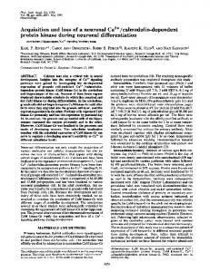
Nucleotide-binding /
domain
T,/
504-507SRAA
Ds
/
NKMFVK QEALP-
662-666~M--
R
580-58651
580-58
F
0674
Fig. 8. Suggested structure for the nucleotide-binding domain of the ATPase The suggested relative locations of the first tryptic cleavage site T1 at Arg-505 and the epitope-binding sites at residues 510-515, 580-588 and 662-666 are shown. Also shown are possible relative locations of Lys-515 labelled by FITC and two reactive cysteine residues (Cys670 and Cys-674).
regions would correspond to our exposed epitope containing residues 662-666. Labelling studies have identified Cys-670 and Cys-674 as being on the surface of the ATPase (Obara et al., 1988; Bishop et al., 1988). mAbs Y/2E9 and Y/3G6, whose epitopes are residues 662-666 (Table 1), strongly inhibit ATPase activity (Tunwell et al., 1991; Colyer et al., 1989), suggesting either a critical role for these residues in the function of the ATPase or that binding of a mAb to this region inhibits important conformational changes to the ATPase. The latter possibility is made more likely by the reports that covalent modification of Cys-670 and Cys-674 has no effect on ATPase activity (Squier et al., 1987; Bishop et al., 1988). Binding of mAbs Y/1F4 and Y/2E9 to the ATPase has been shown to be competitive (Colyer et al., 1989) suggesting that, in the native ATPase, residues 510-515 and 662-666 must be relatively close. Binding of mAb 1/2H7 with an epitope comprising residues 580-588 is not competitive with mAbs Y/ 1 F4 or Y/2E9, suggesting that this region of the ATPase is located away from residues 510-515 and 662-666. This is consistent with the observation that, whereas binding of mAbs Y/ 1F4 and Y/2E9 inhibits activity, binding of mAb 1/2H7 does not. The polypeptide chain of the ATPase between residues 510 and 666 would thus seem to be in the form of a large loop, bringing the two ends close together and away from residues 580-588 (Fig. 8). The loop would start close to the first site of trypsin cleavage (Arg-505), which presumably is also surface-exposed. The observation of three surface-exposed epitopes in the nucleotide-binding domain of the ATPase suggests that this region of the ATPase may be particularly exposed, consistent with a location on the upper surface of the ATPase, as suggested by fluorescence energy transfer experiments (Gutierrez-Merino et al., 1987). MacLennan et al. (1985) have suggested that residues 680-740 constitute a hinge region connecting the nucleotide-binding domain to the fifth stalk region. If the polypeptide chain from about residue 660 were to connect the upper surface of the nucleotide-binding domain to the stalk underlying the large pear-shaped lobe of the ATPase, this could explain the large separation (approx. 6 nm) between Cys-670 and Cys-674 and Lys-515 suggested from fluorescence energy transfer measurements (Fig. 8) (Squier et al., 1987; Bishop et al., 1988). Attempts to map the epitopes of mAbs Y/4D6 and Y/3H5 were less clear cut (Table 1). Binding to proteolytic fragments of Vol. 279
the ATPase and to fusion proteins had suggested that the epitopes for these mAbs were between residues 320 and 414 (Colyer et al., 1989; Tunwell et al., 1991). Binding to hexapeptides suggested that the epitope could include both residues 307-309 and residues 333-338 (Table 1) although, as described below, these regions are thought to be separated in the native ATPase structure, making this rather unlikely. The mAbs were unable to bind to the peptides 303-314 and 324-339 adsorbed to plastic wells in an e.l.i.s.a., but this could simply indicate that the peptides adopted conformations on the plastic that did not allow binding; the alternative possibility that the peptides were not adsorbed to the plastic can be eliminated, since it was found that antibodies raised to these peptides did recognize the peptides adsorbed in this way (results not shown). Surface exposure of residues 333-338 was confirmed by demonstrating that antibody raised to peptide 324-339 bound to native ATPase (Fig. 4b). Antibody raised to peptide 303-314, however, did not bind to either native or denatured ATPase in e.l.i.s.a. or to the ATPase on Western blots, although it did recognize the appropriate hexameric peptides on the set of pins containing residues 277-381, and also bound to peptide 303-314 in e.l.i.s.a. Thus, although no definitive statement can be made, the weight of evidence is against surface exposure of residues 307-309 and suggests that the true epitope for mAbs Y/4D6 and Y/3H5 is residues 333-338. In the original model of MacLennan et al. (1985) residues 307-309 were located at the interface between the fourth transmembranous a-helix and the fourth stalk region, with residues 333-338 being located at the interface between the top of the fourth stalk region and the start of the phosphorylation domain. The model was subsequently modified by Green (1989) such that residues 307-309 occurred towards the centre of the fourth transmembranous a-helix. Our data agree with the surfaceexposed nature of the fourth stalk region shown in our proposed model (Colyer et al., 1989). In previous studies (Matthews et al., 1989) we have shown that antibodies to peptide 381-400 could bind to the native ATPase, indicating surface exposure in this region of the phosphorylation domain, between the residue (Asp351) phosphorylated on the ATPase and the T1 cleavage site at Arg-505. mAbs Y/4D6 and Y/3H5 had no effect on ATPase activity (Colyer et al., 1989). The observed binding of anti-peptide antibody to peptide 191-205 to the native ATPase (Table 2) confirms the surface exposure of the region around the second tryptic cleavage site (T2) at Arg- 198 on the ATPase. Our failure to observe binding of polyclonal antibodies to any hexapeptides between the Nterminus and the region of the T2 site is consistent with our failure to obtain any mAbs binding to native ATPase with epitopes in this region (Colyer et al., 1989). A recent survey of antibodies to the ATPase by Molnar et al. (1990) also failed to find any mAbs binding in this region. This could either reflect a low immunogenicity for this region, or indicate that this region is buried in the native structure of the ATPase, as we have suggested (Colyer et al., 1989). We thank the Science and Engineering Research Council and the Wellcome Trust for financial support, and the Science and Engineering Research Council for a studentship (to R.E.A.T.) We also thank Dr. R. Sharma and Dr. M. E. Ward for their help and advice in peptide synthesis.
REFERENCES Atassi, M. Z. (1975) Immunochemistry 12, 423-438 Bishop, J. E., Squier, T. C., Bigelow, D. J. & Inesi, G. (1988) Biochemistry 27, 5233-5240 Brandl, C. J., Green, N. M., Korczak, B. & MacLennan, D. H. (1986) Cell 44, 597-607
212 Clarke, D. M., Loo, T. W., Inesi, G. & MacLennan, D. H. (1989) Nature (London) 339, 476-478 Colyer, J., Mata, A. M., Lee, A. G. & East, J. M. (1989) Biochem. J. 262, 439-447 Fasman, G. D. & Gilbert, W. A. (1990) Trends. Biochem. Sci. 15, 89-92 Fields, G. & Noble, R. (1990) Int. J. Peptide Res. 35, 161-214 Froud, R. J., East, J. M., Rooney, E. K. & Lee, A. G. (1986) Biochemistry 25, 7535-7544 Geysen, H. M., Rodda, S. J., Mason, T. J., Tribbick, G. & Schoofs, P. G. (1987) J. Immunol. Methods 102, 259-274 Gould, G. W., Colyer, J., East, J. M. & Lee, A. G. (1987) J. Biol. Chem. 262, 7676-7679 Green, N. M. (1989) Biochem. Soc. Trans. 17, 970-972 Green, N. M., Alexander, H., Olsen, A., Alexander, S., Shinnick, T. M., Sutcliffe, J. G. & Learner, R. A. (1982) Cell 28, 477-487 Gutierrez-Merino, C., Munkonge, F., Mata, A. M., East, J. M., Levinson, B. L., Napier, R. M. & Lee, A. G. (1987) Biochim. Biophys. Acta 897, 207-216 Jahnig, F. (1990) Trends. Biochem. Sci. 15, 93-95 Jorgensen, P. L. & Andersen, J. P. (1988) J. Membr. Biol. 103, 95-120 Kawakita, M. & Yamashita, T. (1987) J. Biochem. (Tokyo) 102, 103109 Kison, R., Meyer, H. E. & Schoner, W. (1989) Eur. J. Biochem. 181, 503-511 Laver, W. G., Air, G. M., Webster, R. G. & Smith-Gill, S. J. (1990) Cell 61, 553-556 MacLennan, D. H., Brandl, C. J., Korczak, B. & Green, N. M. (1985) Nature (London) 316, 696-700 Maruyama, K. & MacLennan, D. H. (1988) Proc. Natl. Acad. Sci. U.S.A. 85, 3314-3318 Matthews, I., Colyer, J., Mata, A. M., Green, N. M., Sharma, R. P., Lee, A. G. & East, J. M. (1989) Biochem. Biophys. Res. Commun. 161, 683-688 Merrifield, R. B. (1986) Science 232, 341-347 Mitchinson, C., Wilderspin, A. F., Trinnaman, B. J. & Green, N. M. (1982) FEBS Lett. 146, 87-92 Molnar, E., Seidler, N. W., Jona, I. & Martonosi, A. N. (1990) Biochim. Biophys. Acta 1023, 147-167
R. E. A. Tunwell and others Munkonge, F., East, J. M. & Lee, A. G. (1989) Biochim. Biophys. Acta 979, 113-120 Novotny, J., Bruccoleri, R. E. & Saul, F. A. (1989) Biochemistry 28, 4735-4749 Obara, M., Suzuki, H. & Kanazawa, T. (1988) J. Biol. Chem. 263, 3690-3697 Ohta, T., Nagano, K. & Yoshida, M. (1986) Proc. Natl. Acad. Sci. U.S.A. 83, 2071-2075 Ovchinikov, Yu. A., Arzamazova, N. M., Arystarkhova, E. A., Gevondyan, N. M., Aldanova, N. A. & Modyanov, N. N. (1987) FEBS Lett. 217, 269-274 Ovchinikov, Yu. A., Luneva, N. M., Arystarkhova, E. A., Gevondyan, N. M., Arzamazova, N. M., Kozhich, A. T., Nesmeyanov, V. A. & Modyanov, N. N. (1988) FEBS Lett. 227, 230-234 Petithory, J. R. & Jencks, W. P. (1986) Biochemistry 25, 4493-4497 Pick, U. (1981) Eur. J. Biochem. 121, 187-195 Rao, R., Nakamoto, R. K. & Slayman, C. W. (1989) in Ion Transport (Keeling, D. & Benham, C., eds.), pp. 35-54, Academic Press, London Reithmeier, R. A. & MacLennan, D. H. (1981) J. Biol. Chem. 256, 5957-5960 Scott, T. L. (1985) J. Biol. Chem. 260, 14421-14423 Serrano, R. (1988) Biochim. Biophys. Acta 947, 1-28 Serrano, R. & Portillo, F. (1990) Biochim. Biophys. Acta 1018, 195-199 Shull, G. E., Schwartz, A. & Lingrel, J. B. (1985) Nature (London) 316, 691-695 Squier, T. C., Bigelow, D. J., de Ancos, J. G. & Inesi, G. (1987) J. Biol. Chem. 262, 4748-4754 Stokes, D. L. & Green, N. M. (1990) J. Mol. Biol. 213, 529-538 Taylor, W. R. & Green, N. M. (1989) Eur. J. Biochem. 179, 241-248 Tramontano, A., Chothia, C. & Lesk, A. M. (1989) Proteins 6, 382-394 Tunwell, R. E. A., O'Connor, C. D., Mata, A. M., East, J. M. & Lee, A. G. (1991) Biochim. Biophys. Acta 1073, 585-592 Van Regenmortel, M. H. V. (1989) Philos. Trans. R. Soc. London B 323, 451-466 Van Regenmortel, M. H. V., Briand, J. P., Muller, S. & Plaue, S. (1988) Synthetic Polypeptides as Antigens, Elsevier, Amsterdam Xu, K. Y. & Kyte, J. (1989) Biochemistry 28, 3009-3017 Yamamoto, H., Tagaya, M., Fukui, T. & Kawakita, M. (1988) J. Biochem. (Tokyo) 103, 452-457
Received 28 February 1991/11 April 1991; accepted 29 April 1991
1991