43
Dental Journal
(Majalah Kedokteran Gigi) 2017 March; 50(1): 43–48
Research Report
Effects of citrus limon essential oil ( Citrus limon L. ) on cytomorphometric changes of Candida albicans Rina Prabajati,1 Iwan Hernawan,2 and Hening Tuti Hendarti2 Dinas Kesehatan Kabupaten Lumajang 2 Department of Oral Medicine, Faculty of Dential Medicine, Universitas Airlangga Surabaya - Indonesia 1
abstract
Background: The most common fungal infection found in oral cavity is oral candidiasis, largely caused by Candida species, particularly Candida albicans (C. albicans). Candida infection can get worse since it is difficult to be treated and resistant with antifungal drugs. Therefore, new drugs and compounds as well as alternative therapies involving natural sources that have antifungal activities have continually been developed. Limonene, β-pinene, and ɣ-terpinene contained in Citrus limon essential oil have been known to have quite good antifungal activities against C. albicans. Purpose: This research aimed to examine and analyze the effects of Citrus limon essential oil on cytomorphometric changes of C. albicans. Method: The research used post test only control group design. Based on the results of the pre-elementary research on antifungal activities of Citrus limon essential oil against C. albicans, Citrus limon essential oil used in this research was on concentrations of 1.56%, 1.37%, 1.17%, 0.98%, and 0.78%. Citrus limon essential oil by C. albicans inoculum and incubated for 24 hours and 48 hours. After the incubation, those C. albicans cells were fixed, dried, and then observed using a scanning electron microscopy. Result: The most effective concentrations of Citrus limon essential oil triggering cytomorphometric changes of Candida albicans were at 1.37% and 1.56% with the incubation period of 48 hours. Conclusion: C. albicans can undergo necrosis process through cytomorphometric changes after the administration of Citrus limon essential oil at concentrations of 1.56% and 1.37% with the incubation period of 48 hours. Keywords: Citrus limon; Candida albicans; necrosis; cytomorphometric changes Correspondence: Rina Prabajati, Department of Oral Medicine, Faculty of Dental Medicine, Universitas Airlangga. Jl. Mayjend. Prof. Dr. Moestopo no. 47 Surabaya 60132, Indonesia. E-mail:
[email protected].
introduction
Candida is an opportunistic organism in oral cavity, triggering no disease in healthy people, but leading to an infection in the body with low immune. Candida albicans (C. albicans) is a commensal organism colonizing on the skin and mucosal tissue of gastrointestinal and genitourinary tracts. When there is an imbalance between C. albicans and other oral microbial components, C. albicans will proliferate, colonize, and invade mucosal tissues to trigger opportunistic infection.1 C. albicans infection in people with HIV/ AIDS is a type of infection most commonly found at around 70-80% and also considered as a major cause of oral candidiasis, followed by Candida guilliermondii approximately at around 11.11%.2 In recent
years, many reported cases of oral candidiasis infections are caused by Candida glabrata.3 Population of Candida glabrata is almost half of the total population of non-C. albicans.4 However, C. albicans is still considered as the largest species of oral candidiasis in both HIV/ AIDS patients and other immunocompromised patients.2 Antifungal compounds widely used in the medical field for the treatment of fungal infections are derived from the polyene group, such as nystatin, amphotericin, and natamisin, as well as from the azole group, such as imidazole and triazole. Nevertheless, this conventional treatment of fungal infection is not considered to be beneficial in fungal infections treatment that have been resistance to antifungal compounds.5
Dental Journal (Majalah Kedokteran Gigi) p-ISSN: 1978-3728; e-ISSN: 2442-9740. Accredited No. 56/DIKTI/Kep./2012. Open access under CC-BY-SA license. Available at http://e-journal.unair.ac.id/index.php/MKG DOI: 10.20473/j.djmkg.v50.i1.p43-48
44
Prabajati, et al./Dent. J. (Majalah Kedokteran Gigi) 2017 March; 50(1): 43–48
Lemon rinds, contain essential oil, formed within the endoplasmic reticulum of the plant cells and then obtained from steam distillation or extraction process of the fruit, flowers, wood, roots, leaves, and seeds of the plant.7 The essential oil has anti bacterial, anti-oxidant, and anti-fungal functions. The essential oil contained on the outer part (pericarp) of lemon rinds is largely composed of limonene (90%), citral (5%), terpinol, linodylα-pinene, camphene, β-pinene, sabinene, myrcene, γ-terpinene, linalool, β-bisabolene, trans-α-bergamotene, and geranyl acetate.8 A previous research even reveals that limonene component found in the essential oil has good anti-fungal effects on Trichophyton rubrum.9 In another previous research, lemon rinds, moreover, contain some anti-fungal compounds classified into terpenoids, namely limonene, β-pinene, and γ-terpinene, which have strong anti-fungal activities against C. albicans. Terpenoids inhibit ergosterol synthesis that occurs in the cell membrane of C. albicans.10 Consequently, the synthesis of nucleic acids is disrupted, resulting in increased cell membrane permeability.11 Limonene, β-pinene, and γ-terpinene also can inhibit the metabolism of C. albicans, interfering organelles balance. The imbalance in organelles then makes intracellular components of the organelles disrupted as well as DNA damaged, resulting in the death of C. albicans.12 Another previous research even reveals that the extract of lemon rinds combined with 96% petroleum ether has inhibitory effects on the growth of C. albicans in vitro.10 Cell necrosis is morphologically characterized by increased cell volume (oncosis), organelle swelling, and plasma membrane rupture, followed by intracellular component secretion. Some ingredients even can trigger the death of fungal cells, such as H2O2, acetic acid, as well as some metals and materials/ anti-fungal drugs. Anti-fungal ingredients at low concentrations can lead to apoptosis, but at high concentrations can cause necrosis as a result of radical damage to the cellular structure and integrity.13 A research conducted by Kim et al on the death process of C. albicans given Amphotericin B and flucytosine shows that the morphology of C. albicans cells undergo the process of death through cell membrane damage.14 Similarly, a research conducted by Dai et al.12 finds that the administration of the root extract of Scutellaria baicaleinsis on C. albicans can trigger apoptosis in the cells of C. albicans, leading to death. Hao et al.16 argues that Caspofungin containing antifungal activities can trigger apoptosis and necrosis on the cells of C. albicans. Equol as a soy isoflavone also has antifungal activities against C. albicans by triggering the ultrastructural changes of the cells. For those reasons, this research aimed to analyze the effects of Citrus limon essential oil on cytomorphometric changes (necrosis process) of C. albicans using a scanning electron microscopy (SEM).
materials and method
This research was a laboratory research with a purely experimental approach. This research was conducted to observe changes in cell size and morphology (cytomorphometrics) of C. albicans treated with the essential oil of lemon rinds by using a SEM. Based on results of the preliminary research, the essential oil of lemon rinds has inhibitory effect at a concentration of 0.78%, and fungicidal effect at a concentration of 1.56%. Therefore, in this research the essential oil of lemon rinds used was at five different concentrations, namely 1.56%, 1.37%, 1.17%, 0.98%, and 0.78%. Those five groups of different concentrations as well as a control group without any treatment were incubated for 24 hours. Next, there were also five groups of different concentrations, namely 1.56%, 1.37%, 1.17%, 0.98%, and 0.78%, as well as a control group without any treatment, incubated for 48 hours. After that, fixation process and drying were performed, and then observation was conducted using a SEM at a magnification of 3500x. The images resulted from the observation using a SEM were calibrated to measure the cell size of C. albicans, six of which were taken from each treatment group. The data obtained then were processed using a statistical analysis, a One-way ANOVA to determine differences between groups, followed by LSD test.17
results
Based on results of the measurement of the cell size of C. albicans, the mean and standard deviations of the cell size of C. albicans were obtained from each group. The normality of the data then was analyzed using KolmogorovSmirnov test. Result of the Kolmogorov-Smirnov test on the groups with the incubation period of 24 hours showed a significance value of 0.05. This result indicated that the data in each group were distributed normally. Meanwhile, result of the homogeneity test using Levene’s test on the groups with the incubation period of 24 hours showed a significance value of 0.193. This result illustrated that the data were homogeneous. Next, result of the One-way ANOVA test on the groups with the incubation period of 24 hours showed a significance value of 0.583 (p>0.05). This result demonstrated that there was no significant difference between the control group and the five groups of different concentrations with the incubation period of 24 hours. Result of the Kolmogorov-Smirnov test on the groups with the incubation period of 48 hours showed a significance value 0.05. This result indicated that the data in each group were distributed normally. Result of the homogeneity test using Levene’s test on the groups with the incubation period of 48 hours showed a significance value of 0.073. This result illustrated that the data were homogeneous. Result of the
Dental Journal (Majalah Kedokteran Gigi) p-ISSN: 1978-3728; e-ISSN: 2442-9740. Accredited No. 56/DIKTI/Kep./2012. Open access under CC-BY-SA license. Available at http://e-journal.unair.ac.id/index.php/MKG DOI: 10.20473/j.djmkg.v50.i1.p43-48
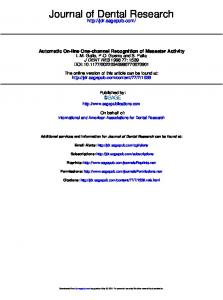
incubation periods, 24 hours and 48 hours indicate the morphological changes in the cell24wall. incubation periods, hours and 48 hours indicate the morphological c
Prabajati, et al./Dent. J. (Majalah KedokteranMoreover, Gigi) 2017results March; of 50(1): 45 of different the43–48 observation using a SEM on all the groups
concentrations, 1.56%, 1.37%, 1.17%, 0.98%, 0.78%, and the control group with both of the
Table 1. Results of the LSD test on the five groups of different concentrations with the incubation period of 48 hours
incubation periods, 24 hours and 48 hours indicate the morphological changes in the cell wall.
Group
(1.56%)
(1.56%)
-
(1.37%)
(1.17%)
(0.98%)
(0.78%)
Control
0.045*
0.015*
0.014*
0.136
0.066
0.022*
0.021*
-
0.079
0.386
0.377
-
0.619
0.606
0.8551µm=7,45mm 0.097
(1.37%)
-
(1.17%)
Moreover, results of the observation using a SEM on all the groups of different 1µm=6,51mm
(0.98%)
concentrations, 1.56%, 1.37%, 1.17%, 0.98%, 0.78%, and the control group with both of thewere exposed to the essential oil of lemon Figure 1. The cells of C. albicans (0.78%) 0.986 of 24 hours. (A) control: the surface incubation periods, 24 hours and 48 hours indicate the morphological changes in the cell wall. Control - of cells was smooth, round o
group with the concentration of 0.78%: the surface of some ce perfectly (). Some cells were separated from colonies (); (C) th 1µm=7,45mm of 0.98%: the surface of cells was rough and not round perfectly ( A B Cfrom colonies (); (D) the group with the concentration of 1.17% rough, not round (), and not colonizing (); (E) the group with t surface of cells was rough and not round (); (F) the group with t 1µm=6,51mm surface of cells was rough and not round perfectly (). Some cell (). Figure 1. The cells of C. albicans were exposed to the essential oil of lemon rind with the incubation period of 24 hours. (A) control: the surface of cells was smooth, round or oval, and colonizing; (B) the group with the concentration of 0.78%: the surface of some cells was rough and not round 1µm=5,78mm 1µm=13,56mm perfectly (). Some cells were separated1µm=5,78mm from colonies (); (C) the group with the concentration 1µm=7,45mm of 0.98%: the surface of cells was rough and not round perfectly (). Some cells were separated D Efrom colonies (); (D) the group with F the concentration of 1.17%: the surface of some cells was rough, not round (), and not colonizing (); (E) the group with the concentration of 1.37%: the surface of cells was rough and not round (); (F) the group with the concentration of 1.56%: the 1µm=6,51mm surface of cells was rough and not round perfectly (). Some cells were separated from colonies (). Figure 1. The cells of C. albicans were exposed to the essential oil of lemon rind with the incubation period of 24 hours. (A) control: the surface of cells was smooth, round or oval, and colonizing; (B) the group with the concentration of 0.78%: 1µm=6,23mm the surface of some cells was rough and not round 1µm=6,51mm 1µm=13,56mm 1µm=6,1mm 1µm=13,56mm perfectly (). Some cells were separated1µm=5,78mm from colonies (); (C) the group with the concentration 1µm=7,45mm Figure The cells were of C.period albicans were exposed to the essential oil of lemon 0.98%: surfacewere of cells was to rough and not round perfectly (). Some cells separated Figure 1. Theofcells of C.the albicans exposed the essential oil of lemon rind with1. the incubation of 24 hours. (A) control: of 24the hours. (A) control: surface of cells was smooth, round o colonies (D) the group the concentration of(B) 1.17%: the surface of concentration some cells wasof the the from surface of cells(); was smooth, roundwith or oval, and colonizing; the group with 0.78%: the surface groupfrom withof the concentration ofgroup 0.78%: the surface of some c rough,cells not was round (),and andnot notround colonizing ();(). (E) the group with theseparated concentration 1.37%: the (C) the of some rough perfectly Some cells were colonies (); with perfectly (). Some cells were separated from colonies (); (C) t surface of cells was rough and not round (); (F) the group with the concentration of 1.56%: the the concentration of 0.98%: the surface of cells was rough and not round perfectly (). Some cells were separated from 0.98%: from the surface of cells was rough and not round perfectly ( surface of cells was rough and not round perfectly (). Some cells were of separated colonies colonies (); (D) the group with the concentration of 1.17%: the surface of some cells was rough, not round (), and from colonies (); (D) the group with the concentration of 1.17% (). F 5 not colonizing (); (E) the group with the concentration of 1.37%: the surface 1µm=5,69mm of cells was rough and not round (); (F) rough, not round (), and not colonizing (); (E) the group with the group with the concentration of 1.56%: the surface of cells was rough and not round perfectly ().and Some surface of cells was rough notcells roundwere (); (F) the group with 1µm=6,23mm 1µm=6,65mm 1µm=6,23mm separated from colonies (). 1µm=6,1mm 1µm=13,56mm surface of cells was rough and not round perfectly (). Some cel 1µm=5,78mm ().
Note: * There was 1µm=7,45mm a significant difference
A
1µm=5,78mm
C
B
5
F
1µm=5,69mm 1µm=6,23mm 1µm=6,65mm
1µm=6,1mm 1µm=13,56mm
D
5
F 1µm=5,56mmm 1µm=5,95mm
F
E
1µm=6,1mm
1µm=5,69mm 1µm=6,23mm 1µm=6,65mm
5
F
1µm=5,56mmm 1µm=5,95mm
6
1µm=5,95mm
1µm=5,69mm Figure Thethe cells of C. albicans to the Figure 2. The cells of C. albicans were exposed to the essential oil of lemon rind 2. with incubation periodwere of 48exposed hours. (A) theessential oil of lemon of 48 hours. the control group: the surface control group: the surface of the cell was smooth and round in colonies; (B) the group with(A) the concentration of 0.78%: the of the cell was smo thegroup groupwith withthe theconcentration concentrationofof0.98%: 0.78%:the the surface of few cells surface of few cells was rough, not round (), and not colonizing (); (C) the not colonizing (); (C) the group with the surface of the cells was rough and not round (). Some cells were separated from colonies (); (D) the group with theconcentration of 0.98% rough and not round (). Some cells were separated from colonies concentration of 1.17%: the surface of the cells5 was rough, not round (), and not colonizing (); (E) the group with the F concentration of 1.17%: the surface of the cells was rough, not r concentration of 1.37%: the surface of the cells was rough, stood out, not round (), and not colonizing () (F) the group (); (E) the group with the concentration of 1.37%: the surface of with the concentration of 1.56%: The surface of the cells was rough, stood out, round (), andnot notcolonizing colonizing() ().(F) the group with the not not round (), and 6 1µm=5,95mm 1µm=5,95mm 1µm=5,56mmm surface of the cells was rough, stood out, not round (), and not co
Figure 2. The cells of C. albicans were exposed to the essential oil of lemon rind with the incubation period Dental Journal (Majalah Kedokteran Gigi) p-ISSN: 1978-3728; e-ISSN: 2442-9740. Accredited No. 56/DIKTI/Kep./2012. Open access under CC-BY-SA license. Available at http://e-journal.unair.ac.id/index.php/MKG of 48 hours. (A) the control group: the surface of the cell was smooth and round in colonies; (B) DISCUSSION DOI: 10.20473/j.djmkg.v50.i1.p43-48 the group with the concentration of 0.78%: the surface of few cells was rough, not round (), and 1µm=5,95mm
not colonizing (); (C) the group the concentration 0.98%:resistant the surface the cells was C.with albicans fungi areofoften to of antifungal therapy rough and not round (). Some cells were separated from colonies (); (D) the group with the Thus, more therapy nee concentration of 1.17%: amphoterisin. the surface of the cells another was rough, not effective round (),antifungal and not colonizing (); (E) the group with the concentration of 1.37%: the surface of the cells was rough, stood out, Cons alternative by considering the death process of C. albicans cells. 6 not round (), and not colonizing () (F) the group with the concentration of 1.56%: The
46
Prabajati, et al./Dent. J. (Majalah Kedokteran Gigi) 2017 March; 50(1): 43–48
One-way ANOVA test on the groups with the incubation period of 48 hours showed a significance value of 0.038 (p