EBT manuscript for IEEE special issue on exhaled breath
1
Development of an Individual Device for Exhaled Breath Temperature Measurement Todor A. Popov, Tanya Z Kralimarkova, Christo T. Tzachev, Stefan S. Dunev, Vasil D. Dimitrov, Jas Gill* Clinic of Allergy & Asthma Sofia; *Delmedica Investments Singapore
Abstract—Different thermometers have been constructed over the decades to measure the temperature of the body to help detect and monitor morbid states. They yield slightly different estimates of the core body temperature depending on the proximity of the measurement site to the internal milieu of the organism, the principle of temperature assessment and the specific characteristics of the gauging devices. Evaluation of the exhaled breath temperature (EBT) has been recently suggested as a new method to detect inflammatory processes in the conducting airways due to changes in the blood flow perfusion of their walls and adjacent structures. While the first reported EBT experiments required sealed laboratory environment and sophisticated equipment, we designed a simple hand-held instrument for EBT measurement and proven its precision, reproducibility and validity in subjects with asthma. We now describe the construction principles of our instrument, the procedure to test the fitness for purpose of the separate units and the novel features of the newest prototypes outfitted with microprocessor and memory. We also outline the potential clinical applications of an individual device for EBT measurement. Index Terms—Core body temperature, thermometry, exhaled breath temperature, airway inflammation, clinical applications. I. INTRODUCTION human organism needs to maintain the temperature of Titsrangevital organs (core body temperature) within a narrow to allow essential enzymatic reactions to occur. HE
Changes outside this range are indicative of pathologic states Manuscript received February 15, 2008. Much of this work was supported by a research grant from Delmedica Investments in Singapore. T. A. Popov is with the Clinic of Allergy and Asthma, Medical University, Sofia, 1431, Bulgaria. Phone: +359-2-9230297; Fax: +359-2-9230621; email:
[email protected]. T. Z. Kralimarkova s with is with the Clinic of Allergy and Asthma, Clinic of Allergy and Asthma, Medical University, Sofia, 1431, Bulgaria. (e-mail:
[email protected]). C. T. Tzachev is with the Faculty of Pharmacy, Medical University, Sofia, 1431, Bulgaria (e-mail:
[email protected]). S. S. Dunev is retired construction engineer of electromedical equipment (e-mail:
[email protected]). V. D. Dimitrov is with the Clinic of Allergy and Asthma, Clinic of Allergy and Asthma, Medical University, Sofia, 1431, Bulgaria. (e-mail:
[email protected]). J. Gill is engineer with Delmedica Investments Singapore (e-mail:
[email protected]).
and reactions and have made temperature measurement an integral part of routine patient examination [1]. Historically, temperature measurement has been used to assess fever and its course as a diagnostic and prognostic tool in infectious diseases. Modern day anesthesia uses controlled hypothermia to allow prolonged surgical interventions of vital organs requiring accurate core body temperature monitoring [2]. For practical purposes thermometry at traditional body sites is performed giving estimates of the actual core temperature with reasonable degree of approximation [3]. Thus, rectal temperature is considered most representative of the core body temperature, but its measurement is disliked by patients and carries the risk of bacterial contamination. Oral temperature is generally 0.5ºC less than the rectal temperature and is more prone to influences from the ambient environment. Core temperature can be estimated with reasonable accuracy at the axilla (armpit) and by a catheter in the bladder. Because the eardrum is close to the carotid artery and the hypothalamus, tympanic membrane temperature, usually referred to as otic temperature, is another reliable estimate of core temperature and is often used as reference for other sites. All other conditions of measurement being equal, the differences between the temperature values at specific body sites are due to the influence of the “core-to-surface” interface. While this may be considered a confounding factor from point of view of evaluation of the “true” core temperature, differences due to the core-to-surface gradient may present an opportunity to get useful information about pathology associated with the interface itself. This type of reasoning became the starting point for attempts to measure exhaled breath temperature (EBT). The deep structures of the lung typically have temperature representative of the body core. It is determined by the blood flowing along the rich vascular network of the alveoli. During the breathing cycle gases but also thermal energy are exchanged between the inner milieu of the organism and the ambient environment. The temperature of the inhaled air is tempered during its flow in and out of the branching airways, which have a separate system of blood supply. As blood is the main carrier of thermal energy maintaining the core body temperature, processes that would modify its flow within the airway walls might reflect on the temperature of the outgoing air, i.e. EBT. High precision gauging devices may pick this signal and give ground for clinical inferences.
EBT manuscript for IEEE special issue on exhaled breath
Thus, some 8 years ago, Paredy and colleagues in London [4] and Piacentini and colleagues in Verona [5] started measuring the exhaled breath temperature (EBT) of their asthmatic patients. Their reasoning was that tissue vascularization and temperature increase are inherent to the process of inflammation, which is nowadays believed to be the hallmark of asthma. Both teams used fast reacting thermocouples in front of the mouths of the tested subjects and analyzed the rise in temperature during single breath maneuvers on the screen of computers. This required sealed laboratory environment with constant temperature, minimal air movement and subject training to allow the record of comparable exhaled temperature curves. While the London team considered the rate of increase of the exhaled breath temperature as indicative of asthma [4, 6], the researchers in Verona made a series of experiments proving that the top of the plateau of the exhaled breath temperature curve is the variable, distinguishing asthmatics from healthy controls [7, 8].
2
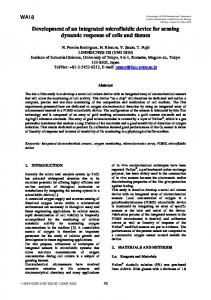
We believed that the capacity of the thermoinsulated chamber to preserve heat from spilling into the ambient environment was a key to getting an accurate estimate of the temperature of the air exhaled by the subject. We used for our first prototypes 0.5 L vacuum flasks (known as Dewar’s vessels) available on the market as containers to preserve the temperature of beverages or thermoses (Figure 2) [9].
Some 10 years ago we also began work on EBT measurement using a simple portable device, which makes the evaluation process less dependent on ambient factors and allows individual home measurements in patients with diseases requiring daily / frequent control. II. BASIC DESIGN A. Designing of the first prototypes The idea we developed when constructing our EBT device was to accumulate the thermal energy of the exhaled air of a tested subject into an insulated vessel containing a metal core with high heat capacity (Figure 1):
Figure 2. First prototype of EBT device. We needed to select from the existing diversity of models the ones with best capacity to preserve heat, so we devised a protocol to test their thermal resistance (R). It consisted of placing a resistor (heater) in the lower part of Dewar's vessel and connecting it to external stabilized 12V power (P) source, emitting 0,5 Watt (W) heat inside the thermos (input power,PIN = constant = 0,5 W). In the upper part of the flask, a temperature sensor was placed and connected to a temperature reading device (thermometer). The passage of connecting wires through the thermos cap was sealed. Switching on of the heater initiated temperature rise in the thermos. Convection of heat from inside to outside (output power,POUT) proportional to temperature difference (TIN TOUT) occurred: POUT = (TIN - TOUT) / R, where R is the thermal insulation resistance of the thermos TIN rose in time to reach a maximum value TIN MAX, when the output power became equal to consumed power: POUT MAX = PIN = (TIN MAX - TOUT) / R. With PIN constant = 0,5 W, the difference TIN MAX - TOUT [K (kelvins)] was an indicator of the insulation of the thermos: R = 2 * (TIN MAX - TOUT) [K / W] Example: If TIN MAX - TOUT = 10 K, then R = 2 * 10 = 20 K/W.
Figure 1. Schematic of the concept of a portable EBT measuring device.
As it turned out, thermoses from the same make and model yielded a wide range of values for R, with less than 10%
EBT manuscript for IEEE special issue on exhaled breath
3
showing good insulating properties, making them fit for use as EBT measurement devices.
B. Generations of EBT measurement devices The first experiments to prove the feasibility of EBT measurement in subjects with inflammatory airway disease were made with only 2 devices giving identical readings (first generation devices) to minimize any bias due to inter-device variability (Figure 2). As the aim of the experiments to follow was to check the feasibility of making daily measurements at the homes of asthmatics in order to monitor the control of their inflammatory airway disease [10], we had to manufacture a bigger batch of individual instruments with slight change in the overall design (e.g. the volume of the thermoinsulated chamber was reduced to 0.3 L) to give to the patients (second generation devices). We needed, therefore, an additional procedure for testing of the relative accuracy of the next 40 devices we built to ensure that the values measured by each one fall within a very narrow range. Each EBT device was tested under the same ambient conditions in a sealed room with fixed temperature and no movement of people. Air with constant temperature and constant flow was passed through each device for 10 min. The temperature values at the 10th minute for each device were pooled together to calculate the average temperature for the whole series. Devices deviating by more than 0.15 K were excluded from the series. Using the first and second generations of EBT measurement devices was acceptable on the part of the tested subjects, but still lacked the level of convenience to make it usable for larger scale trials and eventually for everyday clinical practice. Thus investigator and tested subjects had to check the temperature reader of the EBT device every minute to identify the end of the measurement (reaching a plateau, i.e. two identical readings 1 minute apart). In addition to high precision and reliability, we had to consider putting in electronics to signal the end of the evaluation, to memorize the measured value, to identify the tested subject and to document the date and time of the measurement. We reckoned also that monitoring the progress of the attempt and analyzing the curve of the measurement on the screen of a computer would add extra value for physicians and investigators. With the help of professionals from Philips Singapore we have now at our disposal for testing a batch of devices offering simplicity of use combined electronic prompt and ergonomic design (Figure 3). However, one of the most important tasks ahead of us now would be to shorten the time of measurement from typically 5 to 7 minutes to less then 3 minutes. Presently we are exploring the possibility to predict with reasonable degree of precision the final result of the temperature measurement ahead of time [11].
Figure 3. Front and hind photographs of a third generation EBT measuring device with indication of components. III. APPLICABILITY OF THE EBT DEVICE IN MEDICINE One of the crucial questions we needed to answer at the beginning of our work, was whether EBT is just another surrogate measure of core body temperature, or whether it also captures the signal emitted by the airways. Pooling together 132 EBT measurements of healthy subjects and asthmatics, we did not find meaningful correlation between EBT and otic temperature, while there was a highly significant correlation between otic and axillary temperatures (Figure 4) [9].
Figure 4. There was a highly significant correlation between otic and axillary temperatures, and no correlation between otic and EBT.
Another important issue was whether moderate changes in the ambient conditions would bias measurement with the EBT device. Multiple regression analysis of the same 132 measurements made with EBT as dependent variable and room temperature (values on separate days in the range 18–25°C), atmospheric pressure (range 954–982 hPa) and humidity (ranges 22–72%) as independent variables, did not pick any of the ambient conditions as significant determinants [9]. Most studies on EBT so far have been done in asthmatic patients and have suggested the utility of this approach to assess non-invasively changes in the degree of airway inflammation [4-10]. Our portable device makes this method much more applicable in everyday clinical practice as it makes possible individual measurements possible in the home of
EBT manuscript for IEEE special issue on exhaled breath those suffering. The option with the newest third generation devices to have a long list of measurements accessible to the treating physicians could make decision making about treatment modalities more objective. EBT measurement appears to hold promise also in other lung diseases. There is a report that EBT is shifting downwards in patients with chronic obstructive pulmonary disease, in whom airways and pertaining vasculature are reduced [12]. We have preliminary yet unpublished data of increased EBT (but not axillary temperature) in cases with viral infections, tuberculosis and bronchopneumonia. IV. CONCLUSION The temperature of the exhaled breath turns out to be an unexplored area on the map of human physiology and disease. There is a vast territory to systematically cover in order to figure out the utility of this simple and cheap approach. Once data start poring in, applications may become evident also in other medical fields. REFERENCES [1]
D.L. Kasper, E.Braunwald, A.S. Fauci, S.L. Hauser, D.L.Longo, J.L. Jameson. Harrison's Principles of Internal Medicine. New York: McGraw-Hill, 2005. [2] S.R. Insler, D.I. Sessler. Perioperative thermoregulation and temperature monitoring. Anesthesiology Clin 24 (2006) 823–837. [3] M. Sund-Levander, C. Forsberg, L.K. Wahren. Normal oral, rectal, tympanic and axillary body temperature in adult men and women: a systematic literature review. Scand J Caring Sci 2002;16(2):122–8. [4] P.Paredi, S.A. Kharitonov, P.J. Barnes. Faster rise of EBT in asthma: a novel marker of airway inflammation? Am J Respir Crit Care Med 2002; 165: 181–184. [5] G.L. Piacentini, A. Bodini, L. Zerman, et al. Relationship between exhaled air temperature and exhaled nitric oxide in childhood asthma. Eur Respir J 2002; 20: 108–111. [6] P. Paredi, S.A. Kharitonov, P.J. Barnes. Correlation of exhaled breath temperature with bronchial blood flow in asthma. Respiratory Research 2005; 6, 15: 1-10. [7] G.L. Piacentini, D. Peroni, E. Crestani, F. Zardini, A. Bodini, S. Costellaw, A.L. Boner. Exhaled air temperature in asthma: methods and relationship with markers of disease. Clin Exper Allergy 2007; 37: 415–419. [8] M. Pifferi, V. Ragazzo, A. Previti, G. Pioggia, M. Ferro, P. Macchia, G.L. Piacentini, A.L. Boner. Exhaled air temperature in asthmatic children: a mathematical evaluation. Pediatr Allergy Immunol 2008, online: Mar 10 2008; DOI: 10.1111/j.1399-3038.2008.00742.x. [9] T.A. Popov, S. Dunev, T.Z. Kralimarkova, S. Kraeva, L.M. DuBuske. Evaluation of a simple, potentially individual device for exhaled breath temperature measurement. Respiratory Medicine, 2007; 101: 2044-2050. [10] T. Kralimarkova, C. Lazarova, V. Dimitrov, T.A. Popov. Ability of an individual device for measurement of the temperature of exhaled breath to detect changes in patients recovering from mild exacerbations of asthma. ERS Berlin 2008: 186s. [11] T.A. Popov, T.Z. Kralimarkova, C.T .Tzachev, V.D. Dimitrov, K.K .Mun, J. Gill. Exhaled breath temperature measurement made easy. Pediatr Allergy Immunol 2008, online: Nov 10 2008; DOI: 10.1111/j.1399-3038.2008.00837.x. [12] P. Paredi, G. Caramori, D. Cramer, S. Ward, A. Ciaccia, A. Papi, S.A. Kharitonov, P.J. Barnes. Slower rise of exhaled breath temperature in chronic obstructive pulmonary disease. Eur Respir J. 2003 Mar;21(3):439-43.
4