Korean Journal of Pediatrics Vol. 49, No. 1, 2006
□ Case Report □ 1)
Development of Crohn disease in patients with myelodysplastic syndrome : report of two children Jeong Ok Shim, M.D., Jeong Kee Seo, M.D., Hye Ran Yang, M.D. Jae Sung Ko, M.D., Hee Young Shin, M.D., Hyo Seop Ahn, M.D. * † Woo Sun Kim, M.D. and Gyeong Hoon Kang, M.D. *
†
Departments of Pediatrics, Radiology and Pathology , Seoul National University College of Medicine, Seoul, Korea
Crohn disease (CD) is rare, but the incidence of CD has been increasing over the past ten years. We found two cases of CD, associated with myelodysplastic syndrome (MDS), for the first time in children. In the first patient, MDS was diagnosed at three years of age and CD developed later at eight years of age. The patient presented with recurrent abdominal pain, diarrhea, bloody stools and failure to thrive. Colonoscopy revealed cobble stone like mucosa and mass like lesions with superficial ulceration and inflammatory exudates, observed from the cecum to ascending colon. Ileo-cecal biopsy samples showed ulcers with skipped areas and lymphoid infiltrations. The patient was started on treatment with mesalazine and deflazacort, and symptoms remitted. In the second patient, MDS was diagnosed at nine years of age and CD developed at 13 years of age. This patient has recurrent hematochezia, abdominal pain, vomiting and fever. Colonoscopy revealed a large, deep indurative ulceration on the cecal side of the ileo-cecal valve. Ileocecectomy was done, and histology revealed ulceration with transmural inflammation and lymphoid aggregates. Symptoms improved after ileocecectomy. (Korean J Pediatr 2006;49:107-111) Key Words : Crohn disease, Myelodysplastic syndrome, Children
Introduction
Case Report Patient 1
Crohn disease (CD) is rare. After the report of the 1)
twelve patients with CD by Seo et al. , the incidence of
A 7-year-old boy was brought to our hospital for recur-
2, 3)
CD appears to be increasing
.
rent abdominal pain. During the past several years, he had
In recent years, a few adult patients with CD asso-
bout of intensive periumbilical pain, nausea, vomiting and
ciated with myelodysplstic syndrome (MDS) have been
loose stools. Neither fever nor bloody stool was noted at
reported
4-12)
. These reports suggest that the association
the time of presentation.
between the two disorders is not fortuitous. We report
He was born at 40 weeks of gestational age, and admit-
here, the first observation of two children with CD asso-
ted because of the necrotizing enterocolitis at 22 days of
ciated with MDS.
life. Between the age of two and three years, he was admitted to a different hospital three times because of pneumonia, and at that time an atrial septal defect (secondum type) and pancytopenia were detected. Bone marrow aspirate revealed MDS. At four years of age, he was transfer-
접수 : 2005년 8월 11일, 승인 : 2005년 10월 6일 책임저자 : 서정기, 서울대학교 의과대학 소아과학교실 Correspondence : Jeong Kee Seo, M.D. Tel : 02)2072-3627 Fax : 02)743-3455 Email :
[email protected]
red to our hospital and has been followed up with intermittent transfusions. The family history was noted for an elder brother who was diagnosed as MDS at four years of age and a grand-
- 107 -
Jeong Ok Shim, et al. : Development of Crohn disease in patients with myelodysplastic syndrome
B
A
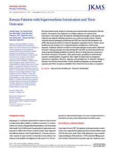
Fig. 1. Barium enema of the patient 1 showed shortening of the ascending colon (A), and deformity and pseudosacculation (B : arrow head) of the cecum. Thickening of the ileocecal valve (B : arrow), and wall thickening and mucosal nodularity of the ascending colon, cecum and ileum were also noted.
father who died of liver disease with unknown etiology,
enema showed shortening of the ascending colon, and de-
otherwise there was no family history of malignancies, in-
formity and pseudosacculation of the cecum. Thickening of
flammatory bowel disease and so on.
the ileocecal valve, and wall thickening and mucosal nodu-
The patient had a body weight of 15 kg (below 3 percentile), height 109 cm (below 3 percentile) and head cir-
larity of the ascending colon, cecum and ileum were also noted (Fig. 1). Mesalazine (200 mg/dose, twice per day) was started 1
cumference 47.3 cm (below 3 percentile). His face looked like an inverted triangle, and the cutaneous vessels were
month later because of the aggravated abdominal pain.
prominent. Periumbilical tenderness was noted. His oral
One year later, the patient had a recurrence of lower ab-
cavity and perianal area were clear and lymph nodes were
dominal pain, loose stools and vomiting. Colonoscopy re-
not enlarged.
vealed cobble stone like mucosa and mass like lesions from
Laboratory examinations showed pancytopenia and ma-
the cecum to the proximal portion of the ascending colon.
crocytic anemia (white blood cell 2,860/µL, hemoglobin 8.2
Superficial ulceration and inflammatory exudates were not-
g/dL, hematocrit 28.0%, mean corpuscular volume 63.6 fL
ed in the mucosa (Fig. 2). Colonoscopy biopsy of non-ul-
3
and platelet 147×10 /µL). Erythrocyte sedimentation rate
cerated areas situated near the ulcers showed a relatively
was 70 mm/hr, and C reactive protein was 2.95 mg/dL.
normal looking appearance but plasma cells and lympho-
Anti-ds DNA was 5.3 IU/mL, C3 191 mg/dL and C4 19
cytic infiltrations of the lamina propria, and those findings
mg/dL. Anti-neutrophil cytoplasmic antibody was negative,
were competent with Crohn disease (Fig. 3). The patient
immunoglobulin G, A and M were 2,559 mg/dL, 151 mg/dL
was treated with mesalazine (750 mg/day) and deflazacort
and 110 mg/dL repectively. HBs Ag, anti-HBs, anti-HCV,
(18 mg/day). He responded well to the treatment, and de-
anti-HIV, Ebstein-Barr virus and parvovirus B19 were all
flazacort was discontinued. He is now in remission.
negative. The bone marrow exam done 6 months ago revealed
myelodysplastic
syndrome/myeoproliferative
Patient 2
syn-
A 13-year-old boy presented to our hospital with com-
drome, and the karyotype was normal. Stool cultures were negative. An abdominal sonography
plaints of hematochezia and vomiting. Three weeks prior to
revealed wall thickening of the cecum and terminal ileum
the visit, left lower abdominal pain and hematochezia de-
and multiple mesenteric lymph node enlargement. Barium
veloped. He was not febrile.
- 108 -
Korean J Pediatr : 제 49 권 제 1 호 2006년
The past medical history revealed that at the age of two
right upper abdominal and epigastric pain. He denied
years pancytopenia was first detected when he was evalu-
smoking and a family history that might relate to these
ated for pneumonia. At the age of nine years, a bone mar-
problems.
row aspirate showed findings consistent with MDS. He
His body weight was 47.6 kg (between 50 and 75 per-
was treated with alfacalcidol and deflazacort, and has wait-
centile) and height 155.7 cm (between 50 and 75 percen-
ed for stem cell transplantation. Neutropenic fevers were
tile). His bowel sounds were increased and his abdomen
problems for this patient several times. Four months prior
was mildly tender. His oral cavity and perianal area were
to the visit, multiple hepatic adenomas were found due to
clear. Lymph nodes were not enlarged. Laboratory tests showed pancytopenia and macrocytic anemia (white blood cell 1,090/µL, hemoglobin 4.2 g/dL, hematocrit 11.9%, mean corpuscular volume 85.6 fL and 3
platelet 65×10 /µL). Erythrocyte sedimentation rate was 48 mm/hr, and C reactive protein was 1.76 mg/dL. A bone marrow aspirate done four months ago revealed hypoplastic MDS. The karyotype revealed trisomy 8 (46XY, inv(9) (p11q13)[8]/47XY, idem, +8[10]). Stool cultures were negative. He was treated conservatively and symptoms were relieved. Two months later, hematochezia and abdominal pain recurred. Severe anemia and thrombocytopenia (hemoglobin 3
3.9 g/dL, hematocrit 10% and platelet 10×10 /µL) were noted. Red blood cell and platelet transfusions were started. A bleeding scan was done, and there was suspected bleedings in the transverse colon. At CT colonography, a 3.5×2 cm sized large ulcer was detected in the cecum near ileocecal valve. The patient was treated with mesalazine (2,250 Fig. 2. Colonoscopic finding of patient 1 revealed cobble stone like mucosae with superficial ulcerations and inflammatory exudates.
mg/day), prednisolone (45 mg/day) and ciprofloxacin (1,000 mg/day for 2 weeks). Abdominal pain and bloody diarrhea improved, and after a month of treatment follow-up CT colonography revealed improvement of the cecal ulcer.
Fig. 3. Photomicrograph of ileocecal biopsy samples showed ulcerated areas (lower half of the figure) and non-ulcerated areas. Non-ulcerated areas demonstrated some alteration of villous configuration and increased lympho-plasma cell infiltration into lamina propria (H&E stain, ×100).
Fig. 4. Ileocecectomy specimen of the patient 2 showed a 2.5 ×2×0.6 cm sized large deep ulcer at cecum (medial aspect of ileocecal valve).
- 109 -
Jeong Ok Shim, et al. : Development of Crohn disease in patients with myelodysplastic syndrome
A
B
Fig. 5. Photomicrograph of the resected specimen of the patient 2 displayed a wide area of ulceration with increased transmural lympho-plasmocytic infiltration and a non-ulcerated portion with architectural distortion of crypts (H&E stain, ×40).
After two months of treatment with mesalazine, the
13, 14)
and exogenous or endogenous triggers
. There are also
patient developed massive hematochezia with hypotension.
hypotheses presented to explain a possible link between
Admission to the intensive care unit, and conservative
MDS and immunological disorders . Though the associa-
managements with transfusion and somatostatin infusion
tion between CD and MDS still remains unknown, this re-
followed. Colonoscopy showed a large deep indurative ul-
port could give some clues for elucidating the pathophysi-
ceration that was detected in the previous CT colonogra-
ology of these rare diseases.
15)
phy. There was no ulceration or inflammation in terminal ileum or other area of the colon. Capsule endoscopy was
한 글 요 약
performed to examine the small bowel, and there were no abnormalities.
골수이형성 증후군으로 진단받은 소아에서 발생한 크론병
Ileocecectomy was performed because there was no lesion except in the cecal side of ileocecal valve. The re-
서울대학교 의과대학 소아과학교실, * † 진단방사선과학교실 , 병리학교실
sected specimen showed a 2.5×2×0.6 cm sized ulceration in the cecal side of ileocecal valve (Fig. 4). Histology re-
심정옥·서정기·양혜란·고재성 * † 신희영·안효섭·김우선 ·강경훈
vealed ulceration with transmural inflammation and lymphoid aggregates and adjacent mucosa with mild cryptitis and crypt architecture distortion (Fig. 5). These findings were
크론병은 매우 드문 질환이나 지난 10년간 발생률이 꾸준히
competent with Crohn disease. Symptoms disappeared after
증가하고 있다. 저자들은 골수 이형성 증후군을 가진 환아에서
surgical resection.
크론병이 발병한 2례를 소아에서는 최초로 보고하는 바이다. 첫 번째 환아는 3세에 골수 이형성 증후군으로 진단받았고, 수 년 간 지속된 반복적인 복통 및 설사, 혈변, 성장 부전이 있어 8세
Discussion
에 크론병으로 진단받았다. 대장 내시경 검사에서는 맹장에서 오 The association between CD and MDS has previously 4-12)
름 결장에 걸쳐 조약돌상 점막과 표재성 궤양 및 염증성 삼출이
. In most of the reported cases,
있었으며, 조직 소견은 궤양 사이에 정상 점막을 포함하고 있으
CD was diagnosed first or CD and MDS were coexistent.
면서 림프구 침윤을 보였다. Mesalazine과 deflazacort로 치료
In just one case of an 87-year-old woman, MDS was
후 증상은 호전을 보였다. 두 번째 환아는 9세에 골수 이형성
been reported in adults
9)
diagnosed first four years prior to CD . To the best of our
증후군으로 진단받았으며, 13세에 반복되는 혈변과 복통, 구토,
knowledge, our patients represent the first report of CD
발열로 크론병으로 진단받았다. 대장 내시경 검사에서 크고 깊은
with MDS in children.
경화성 궤양이 회맹판에서 맹장 쪽 주위에서 발견되었다. 이 외
The etiology of CD is unclear. It is hypothesized that
의 부위에는 병변이 없어 병변을 절제하였고, 조직은 경벽 염증
chronic immune-mediated intestinal injury results from
과 림프구 집합을 동반한 궤양 소견을 보였다. 절제술 후 증상
complex interactions between predisposing genetic factors
은 호전을 보였다.
- 110 -
Korean J Pediatr : 제 49 권 제 1 호 2006년
References 1) Seo JK, Yeon KM, Chi JG. Inflammatory bowel disease in children?clinical, endoscopic, radiologic and histopathologic investigation. J Korean Med Sci 1992;7:221-35. 2) Hildebrand H, Finkel Y, Grahnquist L, Lindholm J, Ekbom A, Askling J. Changing pattern of paediatric inflammatory bowel disease in northern Stockholm 1990-2001. Gut 2003; 52:1432-4. 3) Cosgrove M, Al-Atia RF, Jenkins HR. The epidemiology of paediatric inflammatory bowel disease. Arch Dis Child 1996;74:460-1. 4) Harewood GC, Loftus EV Jr, Tefferi A, Tremaine WJ, Sandborn WJ. Concurrent inflammatory bowel disease and myelodysplastic syndromes. Inflamm Bowel Dis 1999;5:98103. 5) Hebbar M, Kozlowski D, Wattel E, Mastrini S, Dievart M, Duclos B, et al. Association between myelodysplastic syndrome and inflammatory bowel disease. Report of seven new cases and review of the literature. Leukemia 1997;11: 2188-91. 6) Eng C, Farraye FA, Shulman LN, Peppercorn MA, Krauss CM, Connors JM, et al. The association between the myelodysplastic syndromes and Crohn's disease. Ann Intern
Med 1992;117:661-2. 7) Sahay R, Prangnell DR, Scott BB. Inflammatory bowel disease and refractory anaemia (myelodysplasia). Gut 1993;34: 1630-1. 8) Boberg KM, Brinch L, Vatn M. Crohn disease and the myelodysplastic syndrome. Ann Intern Med 1995;122:395403. 9) Tani T, Sakai Y, Shirai Y, Ohtake M, Hatakeyama K. Simultaneous development of Crohn's disease and myelodysplastic syndrome progressing to acute myelocytic leukemia in a patient with normal karyotype. J Gastroenterol 1996;31:599-602. 10) Lee-Elliott C, Alexander J, Gould A, Talbot R, Snook JA. Langerhan's cell histiocytosis complicating small bowel Crohn's disease. Gut 1996;38:296-8. 11) Halme L, Von Knorring J, Elonen E. Development of acute myelocytic leukemia in patients with Crohn's disease. Dig Dis Sci 1990;35:1553-6. 12) Dombret H, Marolleau JP. De novo acute myeloid leukemia in patients with Crohn's disease. Nouv Rev Fr Hematol 1995;37:193-6. 13) Podolsky DK. Inflammatory bowel disease. N Engl J Med 1991;325:928-37, 1008-16. 14) Shanahan F. Crohn's disease. Lancet 2002;359:62-9. 15) Hamblin TJ. Immunological abnormalities in myelodysplastic syndromes. Semin Hematol 1996;33:150-62.
- 111 -