Development of intrinsic optical fiber pH sensors for industrial applications T. Hien Nguyen, Thillainathan Venugopalan, Tong Sun and Kenneth T.V. Grattan School of Engineering and Mathematical Sciences, City University London Northampton Square, London, EC1V 0HB, UK E-mail:
[email protected] inhomogeneity of the material[11, 12]. The second comprises dyes electrostatically deposited in a polymeric thin film using the layer-by-layer (LbL) electrostatic self-assembly technique which may offer a better solution to the above problems to a certain degree [13, 14]. However, due to the nature of electrostatic interactions, the effect of ionic strength can be an important issue and extreme pH values may cause an irreversible damage to the sensitive films. Hence, they are not suitable for sensing in very high and low pH ranges, which are important in a number of applications. The third group can be fabricated and is based on the covalent binding of the indicators to the supporting material or directly to the fiber. The preparation of these optrodes is relatively complicated and time-consuming but they are more reliable and durable as the indicators are virtually bonded to the substrate therefore they are unlikely to leach out under normal conditions[15].
Abstract— The development of intrinsic optical fiber pH sensors based on fluorescence from novel coumarin dyes which are covalently immobilized onto the distal end of the fiber is described. The sensors provide response in the pH range of 0.5 − 6 with good stability over 24 h. The response time of the sensors is approximately 25 s. The sensors show no sensitivity to ionic strength (IS) and have excellent photostability.
I.
INTRODUCTION
A. Optical Fiber Chemical Sensors A number of different schemes proposing fiber optic sensors for a range of different chemical sensing applications have been proposed over the last two to three decades, with some showing more success than others. The attraction of the fiber optic approach lies in particular in the advantages offered in terms of small size, immunity to electromagnetic interference, remote sensing capability, resistance to chemicals and biocompatibility[1, 2]. Among all the various types of chemical sensors discussed, optical fiber pH sensors have received the most attention because of the importance of in situ and in vivo pH measurements in various scientific research and practical applications, in particular where available conventional glass electrodes are not suitable[3-6]. Most pH optrodes (the optical fiber analog of electrode) function through monitoring the changes in the absorbance or fluorescence properties of certain pH sensitive indicators which are immobilized on/in proton-permeable solid substrates[7].
B.
Dye Immobilization and Optical Fiber Chemical Sensors To date, many different approaches to covalent immobilization have been reported. Ensafi et al.[16] attached alizarine yellow and naphthyl red on cellulose membrane using thio-urea as the cross-linking agent whereas Kostov et al.[17] immobilized congo red, neutral red and phenol red to an activated diacetylcellulose. Fluoresceinamine was also bound to cellulose using cyanuric acid by Saari et al.[18] Duong et al.[19] attached the same molecule in a sol-gel matrix made from two precursors methyltriethoxysilane and 3glycidoxypropyltrimethoxysilane (GPTMS) employing the covalent binding between the epoxy group of GPTMS and the amine group of fluoresceinamine. Baldini et al.[20] covalently bound phenol red to a glass surface by means of the Mannich reaction. Uttamlal et al.[8, 12] reported the covalent immobilization of fluorescein derivatives by photo- and electropolymerisation. In all of the above examples, although covalent binding is employed, the stability of the pH indicators themselves, the stability of the substrates and the linking bonds should also be considered. The commonly used ester linkage and acidamide linkage are not very stable in acidic or alkaline aqueous conditions[4, 7].
The reasons for limited success with many previous designs are quite varied but the effective immobilization of the appropriate pH indicators is probably the key step in the development of an optimum optical pH sensor, as it governs the lifetime and signal stability of the sensor. Previous work has shown that poor immobilization results in dye leaching and consequently a drifting of the signal which leads to the gradual breakdown of its sensing ability[7, 8]. The majority of pH optrodes can be classified into three main groups. The first comprises dyes entrapped or absorbed within a solid support which is mostly a sol-gel matrix[2, 9, 10]. pH optrodes of this type are easy to make but suffer from numerous disadvantages such as cracking, dye leaching and This research is funded by the Engineering & Physical Sciences Research Council (EPSRC), UK under grants EP/D030196/1 and EP/F016395/1
978-1-4244-5335-1/09/$26.00 ©2009 Crown
89
IEEE SENSORS 2009 Conference
OH OH OH OH OH OH
H2SO4/H2O2
O O Si O
Fiber surface COOH
COOH
=
O O
or
7-SCC
N H
O O
7-VBACC
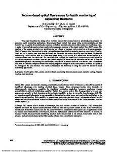
Figure 1. Preparation of a pH sensor tip: schematic of the processes involved.
B. Synthesis of fluorescent dyes In order to create appropriate pH-sensitive dyes for the applications considered, it was decided to synthesize the sensitive dyes needed rather than to use ‘off-the-shelf’ indicators. Two fluorescent dyes were synthesized and used in this work: 7-Styrylcoumarin-4-carboxylic acid (7-SCC) and 7-Vinylbezylaminocoumarin-4-carboxylic acid (7-VBACC). The synthetic routes to the compounds are described in detail elsewhere[30] and are beyond the scope of this paper but their chemical structures are shown in Fig. 1.
In this work, new pH sensing systems that overcome the common disadvantages of optical fiber pH sensors mentioned above have been developed. Novel polymerisable coumarin dyes bearing a carboxylic acid group were designed and synthesized. Coumarins in many cases are used as laser dyes for single-molecule fluorescence so they were expected to be photo-stable[21, 22]. The dissociation of the carboxylic acid group allows for the determination of pH in the acidic region of the pH scale which is suitable for gastric measurements[23, 24], acidic soil measurements[25] as well as the measurement of pH in certain chemical reactors. The dyes were covalently bound to the fiber surface by polymerization, similarly to the method reported by Uttamlal et al.[8, 26] but allyltriethoxysilane (ATES) was used to functionalize the fiber surface with polymerisable groups rather than 3(trimethoxysilyl) propyl methacrylate to avoid the unstable ester linkage. Fluorescence detection method was employed rather than the simpler commonly used method based on colorimetric measurements as fluorescent sensors are usually more precise and sensitive than their colorimetric counterparts[5, 27].
C.
pH probe fabrication Building on the work done to create an appropriate indicator dye, the next step in the development of the sensor was the creation of an appropriate pH sensing probe incorporating the dye developed. This required a multi-step process and the fabrication of the pH sensing probes used in the work is shown schematically in Fig. 1.
The distal end of a 1000 μm diameter UV multimode fiber purchased from Thorlabs was polished in succession with 5 μm, 3 μm and 1 μm polishing pads (Thorlabs) and washed with acetone. The distal end was then immersed in 10% KOH in isopropanol for 30 min with subsequent rinsing in copious amounts of distilled water and dried with compressed nitrogen. After that, it was treated in a 30:70 (v/v) mixture of H2O2 (30%) and H2SO4 (conc.) (Piranha solution) for 60 min, rinsed in distilled water for 15 min and dried in an oven at 100°C for 30 min. This procedure leaves the surface with exposed hydroxyl groups which facilitate bonding of ATES.
The primary applications-focus of the work has been for the determination of pH changes in acidic conditions for a range of industrial applications and the interest ranges from chemical processing to biomedical applications where precise and continuous pH monitoring is critical for the process control and medical diagnosis. II.
EXPERIMENTAL
The fiber surface was then modified by silanizing for 2 h in a 10% solution of ATES in ethanol. The fiber was washed with methanol and distilled water respectively in an ultrasonic bath. Subsequently, it was dried in an oven at 50°C for 2 h. This procedure functionalizes the fiber surface with polymerizable allyl groups.
A. General All chemicals were of analytical grade, purchased from Sigma-Aldrich and were used without further purification. All solvents used were of HPLC grade from Fisher Scientific. All aqueous solutions were prepared using distilled water. Absorption and fluorescence measurements of aqueous solutions containing fluorophores were carried out on a PerkinElmer Lambda 35 spectrophotometer and a Horiba Jobin Yvon Fluoromax-4 spetrofluorometer system with FluorEssenceTM as driving software, respectively. Refractive indices were measured on an Abbe refractometer. Quantum yields of fluorescence were determined using quinine sulfate as the standard (Φ = 0.55)[28, 29].
Monomer stock solution of 7-SCC was prepared by dissolving 7-SCC (5.8 mg, 0.02 mmol), 1,4bis(acryloyl)piperazine cross linker (19.4 mg, 0.1 mmol), acrylamide co-monomer (2.8 mg, 0.04 mmol), ammonium persulphate initiator (5 mg) in a mixture of 250 μL H2O, 250 μL MeCN and 250 μL 50 mM pH 7.5 phosphate buffer. Monomer stock solution of 7-VBACC was prepared slightly different as preliminary experiments suggested that 7-VBACC
90
Mini-spectrometer
1x2 optical fiber coupler LED light source
pH probe
Figure 3. Experimental set-up used in the evaluation of the performance of the probe designed.
Figure 2. Typical pH sensor tip prepared in this work showing the active distal end of the sensor
III.
did not polymerize in solutions containing water. The solution of 7-VBACC contained 7-VBACC (6.4 mg, 0.02 mmol), 1,4bis(acryloyl)piperrazine cross linker (19.4 mg, 0.1 mmol), acrylamide co-monomer (2.8 mg, 0.04 mmol), AIBN initiator (4 mg) and 200 μL dimethylformamide (DMF). The stock solutions were purged thoroughly with nitrogen for 10 min. A small volume of each solution was drawn into a capillary tube and the distal end of the fiber was inserted. They were sealed quickly with PTFE tape and polymerized in an oven at 80 oC for 18 h. This procedure forms a thin polymer film of the dyes which is covalently bound to the surface of the fiber. A typical pH probe prepared by this procedure is shown in Fig. 2 where it can be seen that the distal end of the probe shows a distinctive coloration due to the presence of the dye. The sensor tip was placed in pH 7 buffer for 24 h to remove all unreacted materials and the excess amount of polymer formed which was not directly bound to the fiber. Two different types of probes were created using different dyes synthesized, i.e. 7Styrylcoumarin-4-carboxylic acid (7-SCC) and 7Vinylbezylaminocoumarin-4-carboxylic acid (7-VBACC), to create probes Type 7-SSC and Type 7-VBACC respectively.
RESULTS AND DISCUSSION
A. Properties of the fluorescent indicators 7-SCC and 7VBACC in solution The absorption spectra of indicators 7-SCC and 7-VBACC show only one main absorption band of each dye in the UV region (Fig. 4). 7-VBACC absorbs and emits at longer wavelengths compared to 7-SCC. Both dyes exhibit very large Stokes shifts (the difference in wavelength between the absorption and the fluorescence spectral peaks) of 170 nm for 7- SCC and 150 nm for 7-VBACC, which is very important for the sensor system design to minimize the interference of the excitation light source with the fluorescence emission. The quantum yield of 7-VBACC is reasonable good in both H2O and ethanol whereas that of 7-SCC is relatively low, especially in H2O. However, as 7-SCC proved to polymerize better than 7-VBACC, both dyes continue to be worth investigating for these applications. To determine the pKa values for the free dyes, a series of pH titration experiments was carried out using 50 mM citrate buffer solutions with different pH. In the titration, 50 μL of 0.8 mM dye stock solution was added to 3 mL of buffer in a cuvette, followed by measurement of emission spectra. The calculation of the pKa value for the free dye was performed using a nonlinear fitting method according to the following equation[31]:
D. Experimental set-up With the probes having been fabricated as described above, it was necessary to undertake a calibration of their performance prior to their use in the applications domain. The set-up used for the measurements undertaken to calibrate the probes is as presented in Fig. 3, where light from a LED, emitting at a center wavelength of 365 nm for the Type 7-SCC probe or 400 nm for the Type 7-VBACC probe, is coupled through a multimode UV/Visible fiber with hard polymer cladding, 1000µm silica core and numerical aperture (NA) of 0.37, using collimation and focusing lenses, into a 2x1 Y fiber coupler, made using two multimode UV/Visible fibers with hard polymer cladding, 600µm silica core and 0.37 NA, which is connected to the sensor probe with the active sensing region being located at the distal end of the fiber. Following pH interaction with the active region, a portion of the total light emitted from the sensing layer is collected and guided through the other end of the fiber coupler to an Ocean Optics USB2000 spectrometer, the output from which is then displayed on a computer screen.
F=
Fmax + Fmin × 10 ( pH − pK a ) 10 ( pH − pK a ) + 1
(1)
where F is a measured fluorescence intensity of the system, Fmax is the fluorescence intensity of the fully protonated system, Fmin is the fluorescence intensity of the deprotonated system, and the pKa is the variable fitting parameter, which is the pH where 50% of the dye population in solution is protonated. The data obtained for each dye are summarized in Table 1.
91
TABLE I.
0.012
0.4
SPECTRAL DATA AND PKA VALUES OF THE FLUORESCENT DYES
0.01
0.008 0.25 0.006 0.2 0.004 0.15 0.002
0.1 0.05
0
280
320
360
400
440
480
520
560
600
640
680
Wavelength (nm)
Fluorescence intensity (cps)
1.2E+05
1.1E+05
1.0E+05
9.0E+04
8.0E+04
7.0E+04
6.0E+04
5
UV max (nm)
Emission max (nm)
Quantum yield
7-SCC (EtOH)
342
470
0.121
7-SCC (1.5% EtOH in H2O)
346
518
0.023
7-VBACC (EtOH)
384
496
0.565
7-VBACC (1.5% EtOH in H2O)
387
534
0.146
pKa
2.24
2.34
response ranges of the free dyes. The VBACC probe exhibited an increase in fluorescence intensity with increasing pH whereas the SCC probe behaved in the opposite direction. Interestingly, the SCC probe and the free dye also responded differently to pH. This is probably due to the interchange of the excited state energy levels of the molecule when it is incorporated into the polymer network[32]. Data obtained for the VBACC probe were better than those for the SCC probe as the changes observed for the VBACC is greater than for the SCC, thus introducing less error. The pKa values calculated using Eq. (1) for the VBACC and the SCC are 3.6 and 3.0, respectively. However, the value for SCC was obtained with a big error as the result of the poor calibration curve. The values of pKa are slightly higher for the immobilized forms of the dyes than for their free forms in solution. This is attributed to the decrease in the polarity of the microenvironment[5].
Figure 4. Absorption and emission spectra of 7-SCC (solid lines) and 7VBACC (dotted lines) (14 μM) in 1.5% EtOH in H2O. Emission spectra recorded with excitation at 350 nm for both 7-SCC and 7-VBACC using the same slit widths.
0
Dye
10 15 20 25 30 35 40 45 50 55 60 65 70 75 80 Time (s)
Figure 5. Dynamic response of the VBACC pH sensor showing the 25 s response time (to 95%).
4000
4.E+03
Intensity at 540 nm (counts)
0.3
Normalised Emission
Absorbance (a.u.)
0.35
light source
3500
3000
Intensity (counts)
B. Response time of the sensors created using these dyes Before performing calibration measurements of the sensors, their response time was investigated. Fig. 5 shows the dynamic response obtained from the spectrofluorometer of the VBACC sensing probe to a step change from pH 0.5 to pH 6. The response time of SCC probe was assumed to be similar as the two probes were prepared using the same method. In this work, the response time is considered to be the time required for 95% of the total signal change and the measurement of the response time of the VBACC optrode was found to be 25 s. In comparison to other pH sensors such as the sensor reported by Wallace et al.[12] with the response time of around 500 s or the one reported by Netto et al.[23] with the response time of few minutes, this response time is much shorter. This is probably both due to the intrinsic sensor design and the thichkness of the polymer films as well as their hydrophilicity. This has obvious advantages where a rapid change of pH is to be monitored and a real-time measurement to be achieved.
4.E+03
3.E+03
pK a = 3.6
3.E+03
2.E+03
2.E+03
0
2500
1
2
3
4
5
6
7
8
9
10
pH
increasing pH 2000
7-VBACC 1500
1000
500
0
200
300
400
500
600
700
800
Wavelength (nm)
Intensity (counts)
Intensity at 530 nm (counts)
2100
2500
light source
2000
2000
pK a = 3.0
1900 1800 1700 1600 1500
Increasing pH
0
1
2
3
4
5
6
7
pH
1500
7-SCC 1000
500
C. Response of the sensors to different pH The calibration measurements of the sensors were performed in 50 mM citrate buffer at different pH (note: citrate does not act as a good buffer at pH higher than 6 and lower than 3, however citrate was used for all pH to avoid any differences in fluorescence caused by the difference in buffer composition). The titration curves for the VBACC and SCC probes are shown in Fig. 6. Both probes showed response to pH in the range from 0.5 to 6 which is wider than the dynamic
0
200
300
400
500
600
700
800
Wavelength (nm)
Figure 6. The evolution of fluorescence spectra of VBACC probe (top) and SCC probe (bottom) with pH in the range from 0.5 to 7. Insets show the titration plots at 540 nm and 530 nm respectively.
92
D. Effect of Ionic strength (IS) 4.0E+03
IS (mM) 0 10 50 100 200
PH MEASUREMENTS OF PH 4 BUFFER SOLUTIONS WITH DIFFERENT IS
VBACC sensor pH 4.13 4.20 4.20 3.95 4.25
difference 0.13 0.20 0.20 0.05 0.25
SCC sensor pH 4.5 3.7 3.5 4.8 4.4
Intensity at 540 nm (counts)
TABLE II.
difference 0.5 0.3 0.5 0.8 0.4
1st titration titration after 24 h 3.5E+03
3.0E+03
pK a = 3.6
2.5E+03
2.0E+03
1.5E+03
0
Sensitivity to IS can be a serious problem in the cases of optical fiber sensors as it affects pKa values, thus resulting in errors in pH determination. The effect of IS was investigated with the prepared pH 4 buffer solution adjusted with NaCl to different ionic strengths ranging from 10 mM to 200mM. The fluorescence intensity obtained for each solution was converted to pH using the calibration curve and presented in Table 2. As can be seen from the table, there appears to be no sensitivity to IS for both sensors. The errors caused are probably due to the system error rather than the change in IS.
1
2
3
4
5
6
7
8
pH
Figure 7. Titration curves for VBACC probe obtained between 24 h interval.
1.6E+05
SCC
1.4E+05
Intensity (cps)
1.2E+05
E. Reproducibility and photostability Critical to the successful application of these sensors is their stability both in terms of storage, their susceptibility to error due to intense irradiation of the sample and their reproducibility in use. A preliminary evaluation of these parameters was made in order to understand better the performance of the sensors. The stability of the VBACC sensor was tested by calibrating it with buffers at different pH values ranging from 0.5 to 7 and recalibrating it after 24 h. The titration curves are shown in Fig. 7. As can be observed from the figure, there is very little difference between this two calibrations and the pKa values calculated are almost the same, illustrating the high stability of the sensor scheme produced.
1.0E+05
VBACC
8.0E+04 6.0E+04 4.0E+04 2.0E+04 0.0E+00 0
500
1000
1500
2000
2500
3000
Time (s)
Figure 8. Fluorescence intensities of the pH probes at the emission wavelengths as functions of time during 60 min of continuous illumination.
IV.
CONCLUSIONS
The work has indicated what has been shown to be an effective approach to the development of pH optrodes with superior performance and fast response. The aim has been to work from first principles and to base the approach on the appropriate synthetic chemistry – to create as a result fibre optic probes with fast response, minimal photodegradation on exposure to light from intense uv/blue sources and good reproducibility in performance. All these are essential to meet the requirements of a range of today’s industrial applications for such sensors. Sensors of this type are potentially inexpensive to produce in quantity and the large Stokes shift shown allows for more accurate measurements due to the minimum interference between light source and fluorescence signals generated. For industrial applications, the sensors would require ‘packaging’ to withstand use by inexperienced operators but prior work with optical fiber relative humidity sensors by some of the authors and others has shown an effective model to protect the sensitive fiber tip[33]. In addition, the intrinsic sensor design discussed in this paper has enabled the direct light coupling between the fibre and the sensor material therefore there is a minimum loss caused by the exciation or the fluorescence signal collection.
Photostability is one of the critical properties of fluorescent indicators. In order to test the photostability ot the dyes, the two probes were coupled into the fluorimeter through a dichroic mirror using a fiber bundle. The excitation light (365 nm for SCC and 400 nm for VBACC) was launched to the distal end of the probe consisting the sensing material by the high power Xe lamp of the fluorimeter continuously for 1 h. The fluorescence intensity of the probe was dynamically collected. As can be seen from Fig. 8, no photobleaching was observed for both probes over the time investigated. Compared to the decrease in fluorescence intensity by 65% observed for carboxyfluorescein or by 1013% observed for iminocoumarin derivatives after 60 min of continuous illumination using a mercury lamp[5], the materials prepared using the coumarin fluorophores synthesized in this work possess superior photostability, a feature that is critically important with excitation by high intensity solid state sources.
93
Referencing schemes to allow for corrections due to fluctuations in the source light or environmental perturbation to the sensor system will be considered to be incorporated into such a sensor either through dynamic calibration or a built-in software correction algorithm.
[13]
[14]
There is thus considerable scope for development based on the achievements outlined. In addition, such sensors can readily be multiplexed along a single optical fibre or along a parallel optical network, using various techniques to identify each individual sensor probe. As various applications are considered, the sensor scheme can be tailored for different uses, thus emphasizing the versatility of the approach taken here.
[15] [16]
[17]
ACKNOWLEDGMENTS
[18]
The authors would like to acknowledge the support of the Engineering & Physical Sciences Research Council (EPSRC) in the UK through various schemes and the Royal Academy of Engineering/Leverhulme Trust for the Senior Research Fellowship for one of the authors.
[19]
[20] [21]
REFERENCES [1]
K. T. V. Grattan and B.T.Meggitt, "Chemical and environmental sensing," in Optical Fiber Sensor Technology, vol. 4: Kluwer Academic Publishers, 1999. [2] S. T. Lee, J. Gin, V. P. N. Nampoori, C. P. G. Vallabhan, N. V. Unnikrishnan, and P. Radhakrishnan, "A sensitive fibre optic pH sensor using multiple sol-gel coatings," J. Opt. A: Pure Appl. Opt., vol. 3, pp. 355-359, 2001. [3] S. A. Grant, K. Bettencourt, P. Krulevitch, J. Hamilton, and R. Glass, "In vitro and in vivo measurements of fiber optic and electrochemical sensors to monitor brain tissue pH," Sens. Actuators, B, vol. 72, pp. 174-179, 2001. [4] J. I. Peterson, S. R. Goldstein, R. V. Fitzgerald, and D. K. Buckhold, "Fiber optic pH probe for physiological use," Anal. Chem., vol. 52, pp. 864-869, 1980. [5] A. S. Vasylevska, A. A. Karasyov, S. M. Borisov, and C. Krause, "Novel coumarin-based fluorescent pH indicators, probes and membranes covering a broad pH range," Anal. Bioanal. Chem., vol. 387, pp. 2131-2141, 2007. [6] O. S. Wolfbeis, "Fiber-optic chemical sensors and biosensors," Anal. Chem., vol. 74, pp. 2663-2677, 2002. [7] Z. H. Liu, J. F. Liu, and T. L. Chen, "Phenol red immobilized PVA membrane for an optical pH sensor with two determination ranges and long-term stability," Sens. Actuators, B, vol. 107, pp. 311-316, 2005. [8] M. Uttamlal, W. D. Sloan, and D. Millar, "Covalent immobilization of fluorescent indicators in photo- and electropolymers for the preparation of fibreoptic chemical sensors," Polym. Int., vol. 51, pp. 1198-1206, 2002. [9] T. Fujii, A. Ishii, Y. Kurihara, and M. Anpo, "Multiple fluorescencespectra of fluorescein molecules encapsulated in the silica xerogel prepared by the sol-gel reaction," Res. Chem. Intermediat., vol. 19, pp. 333-342, 1993. [10] F. J. Arregui, M. Otano, C. Fernandez-Valdivielso, and I. R. Matias, "An experimental study about the utilization of Liquicoat solutions for the fabrication of pH optical fiber sensors," Sens. Actuators, B, vol. 87, pp. 289-295, 2002. [11] G. J. Mohr and O. S. Wolfbeis, "Optical sensors for a wide pH range based on azo dyes immobilized on a novel support," Anal. Chim. Acta, vol. 292, pp. 41-48, 1994. [12] P. A. Wallace, N. Elliott, M. Uttamlal, A. S. Holmes-Smith, and M. Campbell, "Development of a quasi-distributed optical fibre pH sensor
[22] [23]
[24]
[25]
[26] [27]
[28] [29] [30]
[31]
[32] [33]
94
using a covalently bound indicator," Meas. Sci. Technol., vol. 12, pp. 882-886, 2001. J. Goicoechea, C. R. Zamarreno, I. R. Matias, and F. J. Arregui, "Optical fiber pH sensors based on layer-by-layer electrostatic selfassembled Neutral Red," Sens. Actuators, B, vol. 132, pp. 305-311, 2008. Y. Egawa, R. Hayashida, and J. I. Anzai, "Multilayered assemblies composed of brilliant yellow and poly(allylamine) for an optical pH sensor," Anal. Sci., vol. 22, pp. 1117-1119, 2006. J. Lin, "Recent development and applications of optical and fiber-optic pH sensors," Trac-Trend. Anal. Chem., vol. 19, pp. 541-552, 2000. A. A. Ensafi and A. Kazemzadeh, "Optical pH sensor based on chemical modification of polymer film," Microchem. J., vol. 63, pp. 381-388, 1999. Y. Kostov, S. Tzonkov, L. Yotova, and M. Krysteva, "Membranes for optical pH sensors," Anal. Chim. Acta, vol. 280, pp. 15-19, 1993. L. A. Saari and W. R. Seitz, "pH sensor based on immobilized fluoresceinamine," Anal. Chem., vol. 54, pp. 821-823, 1982. H. D. Duong, O. J. Sohn, H. T. Lam, and J. I. Rhee, "An optical pH sensor with extended detection range based on fluoresceinamine covalently bound to sol-gel support," Microchem. J., vol. 84, pp. 50-55, 2006. F. Baldini, A. Giannetti, and A. A. Mencaglia, "Optical sensor for interstitial pH measurements," J. Biomed. Opt, vol. 12, 2007. C. Eggeling, J. Widengren, R. Rigler, and C. A. M. Seidel, "Photobleaching of fluorescent dyes under conditions used for singlemolecule detection: Evidence of two-step photolysis," Anal. Chem., vol. 70, pp. 2651-2659, 1998. K. H. Drexhage, "Fluorescence efficiency of laser-dyes," J. Res. Nat. Bur. Stand., Sect. A, vol. 80, pp. 421-428, 1976. E. J. Netto, J. I. Peterson, M. McShane, and V. Hampshire, "A fiberoptic broad-range pH sensor system for gastric measurements," Sens. Actuators, B, vol. 29, pp. 157-163, 1995. P. Wiczling, M. J. Markuszewski, M. Kaliszan, K. Galer, and R. Kaliszan, "Combined pH/organic solvent gradient HPLC in analysis of forensic material," J. Pharm. Biomed. Anal., vol. 37, pp. 871-875, 2005. M. Simek, L. Jisova, and D. W. Hopkins, "What is the so-called optimum pH for denitrification in soil?," Soil Biol. Biochem., vol. 34, pp. 1227-1234, 2002. W. D. Sloan and M. Uttamlal, "A fibre-optic calcium ion sensor using a calcein derivative," Luminescence, vol. 16, pp. 179-186, 2001. D. Staneva and R. Betcheva, "Synthesis and functional properties of new optical pH sensor based on benzo[de]anthracen-7-one immobilized on the viscose," Dyes Pigments, vol. 74, pp. 148-153, 2007. D. F. Eaton, "Reference materials for fluorescence measurement," Pure Appl. Chem., vol. 60, pp. 1107-1114, 1988. J. B. Birks, "Fluorescence quantum yield measurements," J. Res. Nat. Bur. Stand., Sect. A, vol. 80, pp. 389-399, 1976. T. H. Nguyen, T. Venugopalan, T. Sun, and K. T. V. Grattan, "Coumarin-based optical fiber pH sensors for in situ monitoring," Sens. Actuators, B, vol. unpublished, 2009. M. Szabelski, K. Guzow, A. Rzeska, J. Malicka, M. Przyborowska, and W. Wiczk, "Acidity of carboxyl group of tyrosine and its analogues and derivatives studied by steady-state fluorescence spectroscopy," J. Photochem. Photobiol., A, vol. 152, pp. 73-78, 2002. T. H. Nguyen and R. J. Ansell, "Fluorescent imprinted polymer sensors for chiral amines," Org. Biomol. Chem., vol. 7, pp. 1211-1220, 2009. T. L. Yeo, T. Sun, and K. T. V. Grattan, "Fibre-optic sensor technologies for humidity and moisture measurement," Sens. Actuators, A, vol. 144, pp. 280-295, 2008.