of Melbourne, just south of Christmas Hills on the Wat son's Creek to Yarra Glen ...... their outer end into a large club-shaped structure that extends distally and ...
PROC. R. SOC. VICT. vol. 95, no. 1, 1-21, March 1983
DEVONIAN AND ?LATE SILURIAN PALAEONTOLOGY OF THE WINNEKE RESERVOIR SITE, CHRISTMAS HILLS, VICTORIA By P. A. J e l l
and
D. J . H o l l o w a y
National Museum of Victoria, 285-321 Russell Street, Melbourne, Victoria 3000 A b s t r a c t : During construction of the Winneke Reservoir a number of Late Silurian or Early Devo nian fossils were discovered in the Dargile-Humevale Formation transition beds and as all the sites are now underwater, description of these fossils is presented as most are first records for Australia. They in clude a species of scyphomedusoid, Conularia comriei sp. nov. the trilobites Odontochile formosa Gill 1948, Reedops sp. nov. and a questionable record of the Subfamily Tropidocoryphinae, the crinoids Den drocrinus saundersi sp. nov., Codiacrinus rarus sp. nov., Kooptoonocrinus nutti gen. et sp. nov., and a new genus of Dimerocrinitidae, the solutan carpoid Rutroclypeus junori (Withers 1933) and the ophiuroids Urosoma glabridiscus Talent 1965 and Mausoleaster sugarloafensis gen. et sp. nov. Apart from the new genera it is the first Australian record for Reedops, Codiacrinus and Dendrocrinus.
son’s Creek to Yarra Glen Road (Fig. 1, inset). Fossils were recovered between 1979 and 1981 from the two now-flooded localities NMVPL260 & 261 (Fig. 1). NMVPL261 was a low cutting for an access track on the right bank of the stream gully not far above the stream level; Yan Yean 1:63 360 geological sheet, grid reference 500298. Fossil list: Odontochile form osa Gill 1948 Trilobita Reedops sp. nov. Tropidocoryphinae? gen. et sp. indet. straight nautiloid indet. Mollusca solitary Rugosa (moulds only) Coelenterata Conularia comriei sp. nov. Echinoderm ata Rutroclypeus ju n o ri Withers 1933 Mausoleaster sugarloafensis gen. et sp. nov. Brachiopoda Notoparm ella plentiensis G arratt 1980 NMVPL260 was in a small cutting for a track that ran up the side o f the ridge from the main creek gully; Yan Yean 1:63 360 geological sheet, grid reference 501295. Fossil list: Echinoderm ata Codiacrinus rarus sp. nov. Brachiopoda Notoparm ella plentiensis G arratt 1980 Notanoplia panifica G arratt 1980 Site geologists Ray Saunders and Wayne Regan made a collection during initial excavations at a site now buried beneath the right abutm ent o f the main dam wall, which is situated at grid reference 493293 on the Yan Yean 1:63 360 geological sheet. Fossil list: Echinoderm ata Dendrocrinus saundersi sp. nov. Mausoleaster sugarloafensis gen. et sp. nov. Urosoma glabridiscus Talent 1965 The only other fossils described are on a single slab o f siltstone that was removed from a quarry at the dam site to a distant location. A t the time of dumping Mr.
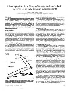
W hen the W inneke Reservoir filled in 1981 the area drowned was underlain by U pper Silurian a n d /o r Early Devonian sediments of the Dargile and Humevale For mations. Considerable earthw orks including quarrying for rock fill during construction o f the dam afforded ex cellent exposures of fresh rock. Fossils proved to be relatively rare in the exposed sediments but are never theless quite significant in several respects. One site NMVPL261 yielded enorm ous numbers of the large dalm anitinid trilobite Odontochile form osa but only five specimens o f Reedops, one of the carpoid Rutroclypeus ju n o ri and one o f the ophiuroid M ausoleaster sugarloafensis. NMVPL260 on the other hand produced large numbers o f brachiopods but only a single echinoderm. Taken overall the seven echinoderm species are known from only 10 specimens whereas one trilobite species is known from hundreds. The lithologies of the collecting localities are not vastly different, suggesting that conditions were similar. Such radically different faunas from similar sites, thought to be relatively close together temporally, suggest that they are not natural assemblages. Moreover, articulated trilobites and whole echinoderms suggest burial of live animals. Taken together these two indications suggest some sort of m ud flow deposition. The fauna itself provides several new generic records for A ustralia particularly among the echinoderms, new inform ation on R utroclypeus that shows its stele to have a structure much more in keeping with other solutan carpoids than was previously acknowledged, a firmer understanding of the species O dontochile form osa, and the first A ustralian record of Reedops. All material is housed in the Palaeontological Collec tions of the N ational Museum o f Victoria (prefixed NMVP) and localities appear on the Palaeontological Locality register o f the same institution (prefixed NM VPL). LOCALITIES The W inneke Reservoir is situated 33 km northeast o f M elbourne, just south o f Christmas Hills on the W at 1
Fig. 1 - Locality map. The dam walls are stippled and the flooded area is horizontally lined (in the inset also). The Humevale Forma tion (diagonally lined) is separated from the Dargile Formation (rest of map) by a dashed line representing a transitional boundary.
PALA EO NTO LO GY OF W INNEKE RESERVOIR Ron N utt split the rock exposing two crinoids identified as Kooptoonocrinus nutti gen. et sp. nov. and Dimerocrinitidae gen. et sp. nov.; as a consequence the exact site and horizon o f the specimens are not certain. Fossil fragments were noted at a num ber o f other localities within the dam site but no specimens were complete enough or well enough preserved for iden tification. STRA TIG RA PH Y A ND AGE O n the Yan Yean 1:63 360 geological map the strata at the site of the main dam wall are assigned to the Dargile Form ation o f Ludlovian age. N one of the fossils from the site contradict this age, and in fact one of the species, U rosom aglabridiscusTalent 1965, was original ly described from the type area o f the Dargile Form ation in the H eathcote district, where it occurs at a similar stratig rap h ic level as early Ludlow graptolites (VandenBerg & G arratt 1976). Localities NMVPL260 and 261 lie close together stratigraphically much higher in the sequence than the site o f the dam , but there has been some confusion as to their exact age and horizon. On the Yan Yean 1:63 360 geological map they are situated ju st above the base o f the Humevale Form ation which is shown as corresponding with the SilurianDevonian boundary, although in the Clonbinane area some 45 km to the northw est the Humevale Form ation is now known to extend down into the Ludlow (G arratt 1978). G arratt (1980) assigned the strata at NMVPL260 to the upper part o f the Dargile Form ation and sug gested a late Ludlow -Pridolian age, based on the presumed ages o f the strata at other localities in the Melbourne Trough where Notoparm ella plentiensis and N otanoplia panifica are known to occur. However, the fossils we describe from NMVPL261 suggest that the age may be somewhat younger than this. Odontochile form osa, Conularia comriei and Rutroclypeus junori all occur quite high in the Humevale F orm ation at Kinglake West, where the sediments are o f Early Devonian age. O f course these species may be long-ranging, but more compelling evidence for an Early Devonian age is pro vided by Reedops sp. nov. The oldest known species of this genus is R. deckeri Delo 1935 from the Gedinnian of O klahom a, and the next oldest species are all early Siegenian (Campbell 1977). SYSTEMATIC PALAEONTOLOGY Phylum
COELENTERATA
(P .A .J.)
Class S c y p h o z o a O rder C o n u l a r iid a Miller & Gurley 1896 Family C o n u l a r h d a e W alcott 1886 Geuns Conularia Sowerby 1821 Conularia comriei sp. nov. Fig. 2 E tym ology:
The species is named for Mr. Michael
3
Comrie, who, as site manager for the reservoir project, greatly facilitated our collecting. H o l o t y p e : The external mould NMVP74247 o f an in complete specimen from NMVPL261. P a r a t y p e s : NMVP79546 from a small quarry on the W hittlesea road 3.2 km from Kinglake West, and NMVP73828 from NMVPL261. D e s c r i p t i o n : The individual was very large (80 mm long and incomplete); regular transverse ridges (21 per 10 mm) bear prom inent tubercles which are not continuous across the interridge spaces but do line up in longitudinal regular columns; the transverse ridges are continuous across marked corner depressions; tubercules on the transverse ridges become quite crowded together in the corner depressions and become elongate off the ridges into the interridge spaces which they cross completely at many levels. On the faces, the transverse ridges very rarely bifurcate (3 examples known, see Fig. 2 A , B, D ). R e m a r k s : The holotype is larger than almost all describ ed conulariids. The state o f preservation with the two surfaces crushed against each other and a great deal of fragm entation suggests that the wall was relatively soft and, unlike that of m ost other conulariids, had little mechanical strength. Towards the apical end (i.e., down in Fig. 2C) are two long tapering scimitar-shaped objects that project from inside the ruptured individual. Such objects are common throughout the Silurian and Devo nian o f the M elbourne Trough but are generally not associated with conulariids. If they are not part of the conulariid it is difficult to imagine how they became lodged where they are in what are apparently rapidly buried fossils. It may be that they are part o f an organism that scavenged on and caused the extensive fragmentation of walls evident at that end o f the con ulariid. This association is ambiguous but may hold a clue to the identity of the relatively common scimitar shaped fossils. The bifurcations (or discontinuities) o f certain transverse ridges forming a loop in one case (Fig. 2D) are rare in conulariids outside the corners and midlines. Their significance is not apparent as functional inter pretation o f conulariids has not yet been accomplished. This species is readily distinguished from C. ornatissima Chapm an 1903 (see Talent 1965) by its fewer ridges per unit length and by its size; from C. chapmani Fletcher 1938 by its discontinuous longitudinal columns, size and occasional ridge bifurcations; from C. sowerbyi de Verneuil 1845 by its size, by its tubercles not being so close packed, and by its tubercles being in longitudinal alignment. It should be noted in passing that neither of the Victorian specimens attributed to C. sowerbyi by Chapm an (1903) belongs to C. chapmani as suggested by Fletcher (1938); they each represent separate species. T alent’s (1965) Conularia n. sp. appears to be conspecific with C. comriei but its very fragmentary nature precludes certain identification. Phylum A r t h r o p o d a Class T r il o b it a
(D .J.H .)
4
P. A. JELL AND D. J. HOLLOW AY
Fig. 2 —Conularia comriei sp. nov. NMVP74247. A, B, D, enlargements of latex cast of various parts of specimen fully illustrated in C; arrows indicate bifurcations in the transverse ridges, A, B, x 6; D, x 4; C,
x 1. Order P r o e t i d a Fortey & Owens 1975 Superfamily P r o e t a c e a Salter 1864 Family P r o e t i d a e Salter 1864 Subfamily T r o p id o c o r y p h i n a e Pribyl 1946 Tropidocoryphinae? gen. et sp. indet. Fig. 31 An incomplete thorax and pygidium from NMVPL261.
M a t e r ia l :
R e m a r k s : The most distinctive features of the pygidium are the narrow axis (the axial rings themselves are not preserved); the pleural furrows that are sharply impress ed, except for the posterior two, and have steep anterior slopes and gentle posterior slopes; the non-existent in terpleural furrows; the anterior pleural bands that are elevated above the posterior bands, especially distally; the very narrow border on the pleurae; and the orna ment o f fine striations. The specimen is tentatively assigned to the Tropidocoryphinae because the structure o f the pygidial pleurae is similar to the imbricate type found in that subfamily.
PALAEONTOLOGY OF W INNEKE RESERVOIR Order P h a c o p id a Salter 1864 Suborder P h a c o p in a Struve in M oore 1959 Superfamily P h a c o p a c e a Hawle & C orda 1847 Family P h a c o p id a e Hawle & C orda 1847 Subfamily P h a c o p in a e Hawle & C orda 1847 Genus Reedops R. & E. Richter 1925 y p e S p e c i e s (by original designation): Phacops bronni Barrande 1846 from the Dvorce-Prokop Limestone (Pragian) at Damil near Teti'n, Czechoslovakia. R e m a r k s : This genus has been discussed recently by Campbell (1977) and Chlupac (1977).
T
Reedops sp. nov. Fig. 3A-H M a t e r i a l : T w o i n c o m p le te c e p h a l a , a n in c o m p le te t h o r a x , a n d t w o t h o r a c e s w ith p y g id ia a t ta c h e d , f r o m
NMVPL261. D e s c r i p t i o n : Glabella very incomplete on available cephala. Occipital ring with well-defined lateral lobes 0.5 times as long (exsag.) as medial portion of ring. Lateral nodes of lp lobe distinctly larger than occipital lobes; lp furrow directed posteromedially as far as inner edge of node where it shallows abruptly and is deflected for wards. Sides of glabella diverge at approxim ately 60° in front of lp furrow; 2p and 3p furrows very weak, 2p ap parently situated level with posterior edge o f eye, 3p w ith convex-forw ard inner p a rt lying opposite midlength o f eye and outer part converging gently with axial furrow at front o f palpebral lobe. Cheek is gently convex in transverse and lateral profiles and slopes steeply anterolaterally. Eye situated well forw ard and low on cheek, with ventral margin lying in lateral border furrow; distance o f eye from junction o f lateral and posterior border furrows almost equal to its own length. Palpebral lobe relatively narrow, with outer rim defined by shallow marginal furrow; palpebral furrow only weakly curved and continuous posteriorly with a deeper postocular furrow. Visual surface composed of at least 21 dorsoventral lens files o f up to 13 lenses each; lenses very closely spaced so that each is almost in contact with its neighbours, even at top o f files. Posterior border well-rounded (exsag.) proximally, expanding abaxially beyond the fulcrum and becoming more flattened; posterior border furrow deep and sharp. Lateral border steeply inclined, narrowing slightly from genal angle to posterior edge of eye; lateral border furrow decreases in depth where it is deflected outwards around base o f eye. Entire dorsal surface of cephalon (except in furrows) covered with coarse granules ranging in diameter from 0.2 mm to less than 0.1 mm. Finest granules on glabella are just above preglabellar furrow, and densest concen tration is on upper surface o f frontal lobe. On cheek, granules decrease in size and density adjacent to axial furrow and on lateral border beneath eye; outer rim of palpebral lobe is densely covered with fine granules.
5
Lateral part o f cephalic doublure carries a deep, weakly notched vincular furrow bounded on the inside by a prominent, sharp ridge that weakens opposite posterior edge of eye. Posteriorly, vincular furrow is deflected inwards across doublure towards distal end of posterior border furrow, and anteriorly it shallows abruptly beneath midlength o f eye (the sharp line extending forward from this position in Fig. 3E is a crack). M edial part of doublure incompletely known, but fragments preserved show that anterior part of vin cular furrow is long (sag., exsag.), weak and poorly defined, and carries norm al ornam ent (Fig. 3H, arrow ed). O rnam ent on doublure consists o f granules that are smaller than those on dorsal surface, and that develop laterally into fine terrace lines on ridge along inside of vincular furrow. Inner, flattened part o f doublure below this ridge is smooth. Thoracic segments have strongly arched (tr.) axial rings with well-developed lateral nodes, and pleurae that rise gradually from axial furrow to fulcrum where they are turned strongly downwards. Axial rings contract slightly medially; in lateral profile they are rather flattopped posteriorly but curve strongly downwards anteriorly into a deep, sharp articulating furrow that is slightly recessed below front o f ring. Pleural furrows sharply impressed, with short, abrupt anterior slopes and more gradual posterior slopes; abaxially they curve forwards slightly onto articulating facets. On anterior segments, pleura curves gently forwards beyond fulcrum to broadly rounded tip; on posterior segments abaxial part of pleura is straighter and tip is almost orthogonally truncated. Doublure beneath pleural tips has welldeveloped panderian protuberances that are more obli que on anterior segments than posterior segments, and on posterior and distal margins of doublure are ventral projections that during enrollment overlap distal edge of succeeding segment (Fig. 3G). Granules on axial rings (excluding lateral nodes) and on downturned portion of pleurae beyond fulcrum are similar to those on glabella; on axial nodes and proximal portion of pleurae they are finer and less dense. Pleural doublure seems to be smooth. Pygidium with 9 axial rings successively decreasing in convexity (sag., exsag.) posteriorly; small pseudoarticulating half rings present on rings 2 and 3, and tiny vestiges of them possibly remaining on rings 4 and 5. Ring furrows 1 to 4 deep and sharply impressed, but subsequent ones much shallower; all except posterior few ring furrows have transverse medial portions and lateral portions that are deflected forwards slightly and are weakly concave backwards. There are 7 or possibly 8 pleural furrows, the first four deep and sharp, and the last few very weak; there are 4 interpleural furrows that are shallow but distinct and do not extend as far abaxial ly as pleural furrows. Doublure consists o f inner and outer bands that are concave in cross-section and separated by a marked change o f slope that swings sharply inwards anteriorly across doublure. Inner band more steeply inclined than outer band, more concave (tr.) and wider, except posteriorly where it contracts
6
P. A. JELL AND D. J. HOLLOW AY
Fig. 3 -A -H , Reedops sp. nov. All photographs except B are of latex casts. A, B, E, cephalon NMVP 82779, x2; A, E, dorsolateral and ventral views; B, internal mould in dorsal view prior to preparation to expose external mould of doublure. C, pygidium of specimen with articulated thorax NMVP82780, ven tral view, x 1.8. D, H, cephalon NMVP82781; D, dorsolateral view, x 2 .5; H, ventral view, x2.2(arrow indicates very weak anterior portion of vincular furrow; note that coarsely granulated object just behind doublure in centre of photograph is axial ring of a displaced thoracic segment). F, thorax and pygidium NMVP82782, dorsolateral view, x2.2. G, pleural tips of incomplete thorax NMVP82783, ventrolateral view, x2.5 (anterior at top of photograph). I, Tropidocoryphinae? gen. et sp. indet. NMVP82784. Dor sal view of incomplete thorax and pygidium with exoskeleton preserved, x5.3.
gradually behind axis. Granules on dorsal surface of pygidium similar to those on glabella; granules on doublure much finer. This species is clearly a member o f Reedops, as shown by the rather uniform size of the granules over the entire exoskeleton, including the glabella; the relatively small, anteriorly placed eye that lies with its lower edge in the lateral border furrow; the very weak 2p and 3p furrows; and the vincular furrow that is deep R em a rks:
laterally but almost non-existent medially. In the coarseness of the exoskeletal granulation and the deep axial and pleural furrows on the pygidium, it is most similar to R. deckeri Delo 1935 from the Gedinnian of O klahom a (Campbell 1977). It differs from that species, however, in having eyes that are shorter (exsag.) but have a greater number o f lenses per dorsoventral file, more weakly curved palpebral furrows, weaker furrows 2p and 3p, and a more poorly defined medial notch in the inner edge of the pygidial doublure.
PALAEONTOLOGY O F W INNEKE RESERVOIR Superfamily D a l m a n it a c e a Vogdes 1890 Family D a l m a n it id a e Vodges 1890 Subfamily D a l m a n it in a e Vodges 1890 Genus Odontochile Hawle & C orda 1847 (ICZN Opinion 537 (1959)): Asaphus hausmanni Brongniart 1822 from the Dvorce-Prokop Limestone (Pragian), Prague district, Czechoslovakia.
T y p e S p e c ie s
Odontochile formosa Gill 1948 Fig. 4 1948 Odontochile form osa Gill, p. 20, pi. 2, figs 1, 2. M a t e r i a l : Holotype M elbourne University Geology D epartm ent 882, internal mould o f very large, almost complete dorsal exoskeleton from quarry ap proximately 2.8 km southwest o f Kinglake West, Vic toria (vicinity of locality W1 on map o f Williams 1964, fig. 2); Humevale Form ation (Early Devonian). O t h e r M a t e r i a l : A bundant remains of cephala, thoraces, pygidia and hypostomes, mostly disarticulated and broken, from NMVPL261. D e s c r i p t i o n : Glabella expanding strongly forwards, widths at occipital ring, 3p lobe and frontal lobe approx imately in ratio 1:1.5:2. Occipital ring well-rounded in lateral profile; in transverse profile it is strongly convex medially but becomes gently concave abaxially; no me dian occipital tubercle. Occipital furrow moderately deep medially, deflected gently backwards abaxially and becoming sharper as it turns down into occipital apodeme, finally curving laterally or even slightly for wards distally. Apodem al pits lp and 2p directed slightly obliquely backwards abaxially, connected to axial fur row by distinct furrows lp and 2p, and tending to be joined medially across glabella by shallow transverse furrows. 3p furrow deep, inner portion gradually in creasing in length (exsag.) abaxially, outer portion ex panded, more oblique than inner portion and more evenly curved in cross section (exsag.). Glabellar lobe lp convex (tr.) medially but rising fairly steeply towards its anterolateral extremities; lobes 2p and 3p with inflated abaxial portions separated from gently convex (tr.) medial portions by broad and poorly defined longitudinal furrows. Frontal lobe approxim ately 1.6 times as wide as long, comprising more than half glabellar length and bearing a shallow longitudinal depression medially in its posterior half. Preglabellar furrow shallow abaxially and not impressed medially. Palpebral lobe in m ost specimens situated further from sagittal axis posteriorly than anteriorly, distance from posterior border furrow slightly greater than length (exsag.) o f posterior border; palpebral furrow shallowest in its posterior half. Eye with lenses arranged in approxim ately 50 dorsoventral lens files o f up to 14 lenses each. Beneath eye is strongly impressed furrow, along outside of which on librigena is a prom inent rounded (tr.) eye platform abutting lateral border fur row. A nterior branch o f facial suture lies close to axial T y pe
7
and preglabellar furrows, diverging strongly from y to /3; from e, posterior branch of suture runs ju st outside furrow beneath eye, before curving in a broad arc across cheek so that oi lies opposite 2p lobe. A nterior border of cephalon decreases slightly in length (sag., exsag.) adaxially, median process absent. Lateral border with epiborder furrow more distinct on librigena than on fixigena; lateral border furrow shallow and wellrounded in cross section on librigena but sharper on fixigena. Posterior border expands distally to more than twice its proximal length (exsag.). Genal spine rather long and slender. G ranular ornam entation very sparse over most of dorsal surface o f cephalon, but more dense on posterior and median parts o f occipital ring, on central part of frontal lobe, on anterior border, and on palpebral lobe. (On the latter, granules increase in density towards the lateral margin but decrease in size.) O rnam entation not preserved on lateral and posterior borders and on dorsal surface o f genal spine. Hypostom al suture gently convex forwards. In front o f hypostom al suture doublure gradually expands adaxially and is crossed by a deep arcuate depression running subparallel to cephalic margin; lateral to hypostomal suture doublure narrows slightly abaxially in front of eye and has a very low, upturned inner flange lying directly beneath eye platform . Posterior branch o f facial suture runs backwards across doublure in a slight curve, meeting inner margin beneath distal end o f posterior border furrow. Hypostome approxim ately as wide across anterior wings as long (sag.). A nterior border ex pands strongly towards anterior wings and anterior border furrow is very weak. Lateral margin curves sharply outwards from anterior wing to prom inent shoulder lying well in front o f hypostom al midlength. Behind shoulder, lateral border decreases in height and width, and lateral border furrow converges only weakly until level with medial part of middle furrow, where border becomes more flattened and expands gradually, and border furrow shallows and curves inwards. Posterior border has a shallow transverse, arcuate fur row running close to posterior margin, the latter deflected backwards medially and abaxially to three small spines; lateral border furrow contains a pair of in distinct depressions directly in front o f lateral pair o f spines. O uter portion of middle furrow meets lateral border furrow opposite shoulder and runs obliquely backwards to a pair of depressions lying just behind the hypostom al midlength; medial portion o f middle furrow transverse, shallower than outer portion. Maculae not observed. Cephalic doublure (with possible exception of inner flange) and hypostom e densely granulate. Thorax composed o f 11 segments. Axis increases in width (tr.) only weakly on first three segments and nar rows ju st as weakly on last six segments; axial rings rather flattened (sag., exsag.), contracting slightly medially and tending to be orthogonally truncated by axial furrow distally. Pleural tips not well preserved on more anterior segments; posterior segments curve backwards distally to a sharp point. Doublure on
8
P. A. JE L L AND D. J. HOLLOW AY
pleurae o f posterior segments broad (tr.), inner edge subangular in outline (Fig. 41, K); no trace of any panderian protuberance. Pygidium approxim ately as wide as long (including mucro) in relatively undistorted specimens; lateral margin gently curved but overall outline is subtriangular rather than semielliptical. Axis 0.2 times as wide as pygidium anteriorly and narrowing uniform ly back wards, composed o f 17-19 or possibly 20 axial rings plus a terminal piece. Successively diminishing pseudoarticulating half rings are developed on at least rings 2-8, and in some specimens oblique muscle impressions are present on posterior half o f rings lateral to pseudoarticulating half rings (Fig. 4B); ring furrows very short (exsag.) and sharp abaxially but longer sagittally and weak, even near front o f axis. There are 13-14 pleural furrows that are evenly curved on more anterior segments but almost straight on posterior ones. Pleural furrows expand slightly distally and become shallower but extend virtually to pygidial margin; interpleural fur rows term inate well inside margin. M ucro slender and extended horizontally backwards; its length measured from axial terminus is approxim ately half length o f axis. Doublure gently convex (tr.), lacking a steeply inclined inner flange. Granules on axis of thorax and pygidium are concen trated along posterior margins o f rings and are virtually absent elsewhere. Abaxial part of pygidial pleurae and mucro are covered with a uniform granulation, but closer to pygidial axis and on thorax granules are present only along posterior edges o f pleural bands. Doublure on pygidium and thoracic pleurae more densely granlated than dorsal exoskeleton. R e m a r k s : Gill’s description o f O. form osa was based only on the holotype, but there are a num ber of characters that cannot be determined from that specimen because it is an internal mould and is also broken in places. For example, there is no inform ation on surface sculpture, the shape o f the anterior cephalic border, or the length and shape o f the mucro, and the eyes are very poorly preserved. N o other specimens of O. form osa with these features preserved are known from the type locality. Gill (1948) also illustrated as O. fo rm osa an internal m ould of a cephalon belonging to a much smaller individual than the holotype and coming from a different locality in the same form ation and area. Numerous additional specimens from this locality are available in the N ational Museum o f Victoria collec tions, and they include external moulds of cephala and pygidia as well as articulated exoskeletons. They differ from the holotype in a few respects, mainly in having a
more weakly inflated glabella, less distinct furrows lp and 2p, and eyes that extend much closer to the posterior border furrow. However, I attribute the differences to the very much smaller size of these specimens. The specimens from the W inneke Reservoir site, which are o f intermediate size, fall within the range o f variation in these features, and in all other features closely resemble the specimens from both of the Kinglake West localities. Three other dalm anitinid species have been recorded from the Late Silurian to Early Devonian of southeastern Australia, but each is in need of revision as the published illustrations and descriptions are inade quate. Dalmanites wandongensis Gill 1948 from the Dargile Form ation (Ludlovian) near W andong, Vic toria, is based on a cephalon and an internal mould of a pygidium differing from O. fo rm o sa in that glabellar lobes 2p and 3p are not as inflated abaxially and the ax ial furrow is weaker; the 3p furrow is shallower; there is no eye platform ; the lateral cephalic border is narrower and more convex (tr.); there is almost no change in slope from the frontal glabellar lobe to the anterior border; the front o f the cephalon is deflected downwards medially in anterior view; and there are only 14 axial rings and 11 pleural furrows in the pygidium. Odontochile meridianus (Etheridge & Mitchell 1895) from the late Ludlovian-Gedinnian of the Yass Basin, New South Wales, is distinguished from O. fo rm o sa by its much smaller eyes th at do not extend as close to the lateral border furrow; a narrower lateral cephalic border; a pygidium with 15-16 axial rings and 11-12 pleural fur rows that are deflected more strongly backwards distal ly; anterior pleural bands on the pygidium th at are weaker, particularly abaxially; and a mucro that is relatively narrower and more convex (tr.) at its base and is connected to the axial terminus by a strong postaxial ridge. The anterior cephalic border is not preserved in any o f the types, but specimens of O. meridianus in the National Museum o f Victoria collections show that it differs from the anterior border of O. fo rm o sa in con tracting slightly in front o f the lateral part o f the frontal lobe and expanding medially to form a very short (sag.), broad projection. The types of O dontochile loomesi (Mitchell 1919), also from the late Ludlovian-Gedinnian o f the Yass Basin, appear to be indistinguishable from O. fo rm o sa but there is no inform ation on surface sculpture as the specimens are all internal moulds, and the only cephalon known lacks the anterior border. W hen more material o f loomesi becomes available it may be necessary to place fo rm o sa in synonymy with th at species.
Fig. A —Odontochile form osa Gill 1948. All photographs except H are of latex casts. A, cranidium NMVP82785, dorsal view, x l.5 . B, pygidium NMVP82786, dorsal view, x l . C, hypostome NMVP 82787, ventral view, x l.2 5 . D, cephalon NMVP82788, dorsal view, x 0 .9 . E, cephalon NMVP82789, ventral view, x 1.3. F, incomplete thorax NMVP82790, dorsal view, x 1. G, pygidium NMVP82791, dor sal view, x 1. H, internal mould of hypostome NMVP82792, ventral view, x 1.4. I, K, M, pygidium and articulated thorax NMVP82793; I, M, ventral and dorsal views, x0.9; K, enlargement of pleural tips on posterior thoracic segments in ventral view, x 1.4. J, L, incomplete cephalon NMVP82794 in ventral and dorsal views, x l.2 5 .
PALAEONTOLOGY OF W INNEKE RESERVOIR
9
10
P. A. JE L L AND D. J. HOLLOW AY Phylum E c h in o d e r m a t a
(P .A .J.)
Class H o m o io s t e l e a Order S o l u t a Jaekel 1901 Family R u t r o c l y p e id a e Gill & Caster 1960 Genus Rutroclypeus W ithers 1933 T y p e S p e c i e s (by original designation): Rutroclypeus ju n o ri W ithers 1933 from the Humevale Form ation (Early Devonian) at Kinglake West, Victoria. D ia g n o s is : A s given by Gill and Caster (1960) but the emphasis on flattened theca and proximal stele should be removed until proven. R e m a r k s : The specimen described below may be used to argue that the proximal stele was not flattened as described by Gill and Caster (1960) and suggests other alternative interpretations for various parts o f the stele. A closer correspondence with other solutan steles is in terpreted (see Remarks on the species, below).
Rutroclypeus junori Withers 1933 Fig. 5, 6 1933 Rutroclypeus ju n o ri Withers, p. 18, pi. 5, figs 1, 2. 1960 Rutroclypeus ju n o ri Withers; Gill & Caster, p. 30, pi. 1, figs 1-3, pi. 2, figs 2, 3. 1967 Rutroclypeus ju n o ri W ithers; Caster, p. S616. 1982 Rutroclypeus ju n o ri W ithers; Jell & Holloway, p. 42, fig. A. H o l o t y p e : NMVP13681 from NMVPL229, Collins Q uarry 2.4 km northwest o f Kinglake West Post Office on the west bank o f King P arrot Creek, Victoria. Humevale F orm ation—Early Devonian. M a t e r i a l : One specimen, NMVP73811 from NMVPL 261 D ia g n o s is (Gill & Caster 1960): “Rutroclypeus with spinose proximal stele having ten segments, the more distal o f which may be deflected distally”. D e s c r ip t io n o f f i g u r e d s p e c im e n : The theca is round, 30 mm in diameter, consists o f many polygonal plates reaching up to 4 mm in greatest dimension, and is preserved quite flat. Each plate has a short central spine. A round the thecal margin, plates curve back onto the other face very sharply and the median spine then ap pears to be marginal (as illustrated by Caster 1967, fig. 391-6). Marginal plates slightly dislodged on the right hand side (Fig. 5A) show the spines to be central with the marginal ridge linking the spines o f contiguous plates. The stele is relatively long (5.2 cm preserved) but is still incomplete. The proxistele consists o f 10 circlets each o f 4 plates arranged in four longitudinal series. O ther longitudinal fractures of individual plates are ir regular and when present may be near the axis or near
the outer margin and may be oblique, so thay are clearly not following lines o f weakness but were caused by com paction. The spines on the plates are much closer to the axis distally than they are near the theca, especially on the reverse side. The most proximal pair of obverse plates and the second and ninth plates in the lefthand obverse series lack spines. On the reverse the proximal pair o f plates also lack spines. Between the centre line of the stele and the spine the posterior margin of each plate extends distally in a broad lobe beyond the m ore abaxial margin. On the obverse side there is no deflection o f the distal plates. Each plate in the proxistele has an anterior ledge on which the next anterior plate overlaps. Otherwise there is very little overlap o f plates. The central suture on the reverse side becomes irregular to wavy distally with what appear to be small semicircular excavations (Fig. 5F, centre) in the plates o f the last pair at their midlength. The mesistele consists o f 1 plate on the obverse; the ap parent sutures are fractures caused by com paction. On the right o f the median stele (Fig. 5E, bottom right) is a spine that appears to be articulated. This suggests that all others on the proxistele may be articulated as well. O n the reverse side the mesistele also appears to be a single plate as the fractures across it are highly irregular and due to com paction. The distal stele (Figs 5D, 6) consists of an obverse and a reverse series o f elongate plates that become pro gressively longer away from the theca. Transverse sec tion o f the distal stele is roughly diam ond shaped with the margins o f the reverse series apparently sitting just inside or against the margins o f the obverse series. In lateral view plates of the obverse have an obtuse central elevation and their lowest point is at each end o f the suture with the next plate in the series. Reverse plates that bridge the sutures between obverse plates apparent ly have a prominence to fit into the dip in the obverse lateral margin. All plates opposite the obtuse pro minences have broadened lateral flanges upon which the obtuse prominences might fit b u t only the obverse plates are so situated in this specimen; reverse plates have all been displaced and lie inside the obverse series. Plates of the reverse series have a carinate median ridge, and are arranged so that there is one within the length o f an obverse plate and that none o f the junction sutures cor respond to those of the obverse side. Consequently there are twice as many in the reverse series as in the obverse series. In the incomplete part o f the distal stele preserved are 19 or 20 reverse plates b ut only nine and a half obverse plates. A n ornam ent o f fine reticulate ridges, apparent on the proximal stele and thecal plates, is in broad terms radial away from the spine. On the distal stele the or nam entation is longitudinal, less reticulate and more granulate.
Fig. 5—Rutroclypeus junori Withers 1933, NMVP73811. All of latex casts. A, B, obverse and reverse, x 1.7 and x 1.2 respectively. C, D, lateral oblique view of distele showing plate arrangement, x 8 . E, obverse view of prox istele, x5.5. F, reverse view of proxistele—distele junction, x 11. G, reverse view of proxistele, x 6 .
12
P. A. JELL AND D. J. HOLLOW AY
Fig. 6 —Sketch of lateral view of plate arrangement in proximal part of distele of Rutroclypeus junori. Mesistele is enlarging plate at right. Obverse plates are upper series, reverse plates below. Drawn from Fig. 5D.
The arm is poorly preserved with only a single series o f low wide brachial plates of the upper surface preserv ed. The inner side of the other series may be present beneath the upper series. R e m a r k s : This specimen offers details on the thecal margin that uphold Caster’s (1967) reinterpretation. Its greatest value is in the inform ation it offers on the struc ture o f the stele. The medial stele is seen to be form ed by a single rather convex plate on each o f the obverse and reverse sides. This in turn leads to the conclusion that the proximal stele had a subcircular or at least a transversely oval section and is flattened only by com paction. The com mon longitudinal fracture of plates is consistent with flattening an oval shape. Such a prox imal stele is unlikely to have joined into a theca as flat as th at suggested by Gill and Caster (1960, fig. 6) but such a section for the theca is not impossible. I cannot say what the thecal height was on available inform ation. A t the type locality o f R utroclypeus ju n o ri all other echinoderms with a sealed interior, including several with a considerable internal cavity such as blastoids, have been flattened w ithout sediment filling the thecal cavity; plates have accom modated by fracture as in Rutroclypeus instead of by dislocation. The structure of the distal stele is biserial and its cross-section is identical with that o f Dendrocystites sedgwicki Barrande (see Caster 1967, fig. 372). Moreover the lateral view o f the distal stele is very similar in these two genera in the way the plates fit together (Fig. 5D). This specimen and the recent note on R . withersi (Jell & Holloway 1982) suggest that a more detailed study o f Rutroclypeus is warranted. Class C r in o id e a Subclass C a m e r a t a Order D ip l o b a t h r i d a M oore & Laudon 1943 Suborder E u d ip l o b a t h r i d a Ubaghs 1953 Superfamily D i m e r o c r i n i t o id e a Zittel 1879 Family D im e r o c r in it t d a e Zittel 1879 Dimerocrinitidae gen. et sp. nov. Figs 7C-E, 8 a t e r i a l : One individual, NMVP74246, found in a block o f rock removed from the floor of the dam site during preflooding quarry works. It was noticed as the material was being dumped elsewhere so its original location and horizon are unknow n. The holotype of Kooptoonocrinus nutti sp. nov. occurs on the same
M
block of rock but whereas both part and counterpart of that specimen are available the counterpart o f the dimerocrinitid fell from the face o f the block, presumably at the time o f splitting. D e s c r i p t i o n : Cup small for family, low, conical; infrabasals 5, visible laterally but quite low; basals 5, hex agonal, approxim ately twice height o f infrabasals; CD basal slightly enlarged; radials 5, in contact with each other laterally except in posterior interray, with obtusely pointed lower margins; radial facet 0.8 o f plate width, with broadly rounded outer margin, sloping out and down; prim anal large, almost as large as CD basal upon which it sits, indirect contact with two posterior radials; further anal plates not known; interprim ibrachs present but very deeply recessed between arms, one larger plate adjoining both first and second primibrach and sup porting 2 smaller plates above. Arm s dividing only once on prim ibrach 2 into 10 ram i, uniserial, pinnulate, con sisting of approxim ately 45 brachial plates per ramus; primibrachs fixed in theca; pinnules long, relatively stout, arising on alternate sides of consecutive brachials. Stem very slender, at least 25 cm long, circular in sec tion, composed o f cirriferous nodals separated by very low internodals varying from 3 to more than 20 per in ternode, and one or two non-cirriferous or weakly cir riferous nodals midway between the cirriferous ones or dividing the distance between strong nodals into three; distal part of stem with elongate irregular cirri apparent ly used in holding onto the substrate. R e m a r k s : This specimen is assigned to the C am erata on the presence o f interbrachial plates which are however not readily apparent. It is clear that these interbrachials are sutured to the sides o f the brachial plates and that they are part of the tegmen, not simply fortuitously placed. Moreover, between the B and C rays may be seen the internal surface o f a ramus from the opposite side o f the theca (Fig. 7C, E) so that it would be very difficult for a long anal sac to be present w ithout in terfering with the mould of this ram us from the opposite side. O n the other hand a short anal tube might not reach as high as the point where the opposite ram us ap pears or might be enveloped within the rami o f the C and D rays. Such highy organised pinnulation is not known in inadunates of this age but is known in camerates. As a dicyclic camerate with radial plates adjoining each other except in the posterior interray it may be referred to the Dimerocrinitoidea. W ithout a bulged posterior, with infrabasals visible laterally and with relatively low basals and infrabasals it may be excluded from all the families except the Dimerocrinitidae. This specimen does appear relatively close to Ptychocrinus W achsmuth & Springer 1885 with its low infrabasals, depressed interprim ibrachials and uniserial arms but it differs from th at genus in having far less obtrusive inter primibrachials, in lacking medial ray ridges and anal ridge, in the relative sizes o f cup plates and in the struc ture of the stem. It is p art of the uniserial Ptychocrinus stock that apparently continued from the Ordovician and Lower Silurian (Witzke & Strimple 1981, Bret 1978)
PALAEONTOLOGY OF W INNEKE RESERVOIR
13
P tych o crin u s through the Silurian and into the Devo nian with M acarocrinus Jaekel 1895. Consequently
another group should be added to the three already pro posed in this family (Witzke & Strimple 1981); the fourth group should include uniserial members o f the Dimerocrinitidae namely P tychocrinus, M acarocrinus and the new genus described above. Although it is clear ly not assignable to any existing genus this specimen is left in open nomenclature until details of its posterior in terray and a better preserved cup are available. Subclass I n a d u n a t a Order C l a d i d a Moore & L audon 1943 Suborder C y a t h o c r i n i n a Bather 1899 Superfamily C y a t h o c r i n i t o i d e a Bassler 1938 Family E u s p i r o c r i n i d a e Bather 1980 Geuns Kooptoonocrinus nov. From the Y arra aboriginal word k o o p to o n meaning one. It refers to the fact that both species of the genus are known from only one specimen. T y p e S p e c ie s : K o o p to o n o crin u s n u tti sp. n o v . E ty m o lo g y :
O th e r
S p e c ie s I n c l u d e d :
K o o p to o n o c rin u s borealis
sp. nov. Its holotype is the specimen placed in A m pheristo crin u s typ u s Hall 1879 by Springer (1926, pi. 31, fig. 1), from the Beech River Form ation (Silurian) in D ecatur County, Tennessee. It is housed in the N ational Museum of N atural History, Smithsonian Institution, W ashington D .C ., U.S.A. D ia g n o s is : Cup conical with strong ray ridges; infrabasals 5; anal X in radial circlet, large, supporting 1 to 3 sac plates; radianal pentagonal, below and left of C radial; arms slender, primibrachs 3 or 5 axillary, bran ching isotomously 4 times to produce a characteristic branching pattern; stem transversely rounded, compos ed of short alternating nodals and internodals. R e m a r k s : Springer (1926) referred the type of K o o p to o n o crin u s borealis to A m p h e risto crin u s ty p u s , the
type species of that genus, but whereas he stated that A m p h e risto c rin u s was characterised by 3 infrabasals his
own specimen is now known to have 5. Weller (1900) also considered the possession of only 3 infrabasals as a critical generic character. In view o f the fact that im por tant features o f the arms and stem are not available from the type species of A m p h eristo crin u s I use the number o f infrabasals to separate the new genus from A m p h eristo crin u s. Springer (1926) said he was uncertain of the num ber o f infrabasals on his Tennessee specimen but Dr. P. M. Kier o f the National Museum of Natural Fig. 7 —A, B, Dendrocrinus saundersi sp. nov. NMVP74239. All of latex casts. A, enlargement of theca in left posterior view, x 7 . B, left posterior view of latex cast of entire animal showing inner side of anterior arms, x 2 . C-E, Dimer ocrinitidae gen. et sp. nov. NMVP74246. C, enlargement of theca and lower arms from left posterior, x 6 . D, posterior view of latex cast of theca, x 6. E, upper stem and crown, x 3.
P . A. JE L L AND D. J. HOLLOW AY
14
History has recently determined that it has five in frabasals (written com munication 17 June, 1982). It seems likely that M oore (1962) and M oore, Lane and Strimple (1978) used th at specimen for their representa tion of 5 infrabasal plates in Am pheristocrinus also. A lthough the family concept (M oore, Lane & Strim ple 1978) appears to need some revision that is not at tempted here. The new genus is assigned because it does not contradict the family concept and has some similarities with Am pheristocrinus. Its position in the phylogeny of the family is not clearly understood. K ooptoonocrinus borealis sp. nov. is congeneric with the Australian specimen described below since they have the same thecal plate arrangem ent, narrow radial facets, branching pattern and stem structure. They may be distinguished by the Australian species having primi brach 5 (not 3) axillary and having the broad ridge runn ing around its cup just beneath the radial facets. Kooptoonocrinus nutti sp. nov. Figs 8, 9A-D The species is named for Mr. Ron N utt of Healesville who found and donated the only specimen. M a t e r i a l : Only the holotype, NMVP74245 is known. It was found in the same block o f rock as the new dimerocrinitid crinoid described above. The block comes from the dam site but no m ore precise locality is known. D ia g n o s is : Infrabasals 5; one anal tube plate on top of anal X; strong ridge transversely on radial plates just beneath radial facets encircling theca; primibrachs 5 ax illary; arms branching isotomously at six different levels but each branch divides only four times; stem o f nodals and internodals near theca, ossicles becoming uniform in height distally. D e s c r i p t i o n : Cup conical with slightly attenuated lower part, approximately 5 mm high. Infrabasals 5, pen tagonal when viewed laterally, 1 mm high x 1.4 mm wide, with broadly obtuse central upper peak, or nam ented with broad low but distinct ridges in “Y” form with 2 arms norm al to the upper margins at their mid points. Basals 5, hexagonal, 2 mm high x 1.5 mm wide, ornam ent continuing from infrabasals and forming a narrow cross so that the four ends of ridges are norm al to plate margins at midpoints of sides. Radials 5, pen tagonal, with ornam ent o f two ridges continuing from basals to outer edge o f radial facet and a further ridge running transversely across the plate where the vertical ridges meet. The transverse ridges form a circlet around the cup at a level just beneath the midheight o f the plates. Articular facets 0.6 o f width o f plate, horseshoe shaped, sloping outwards. A nal X plate large, situated in radial circlet, pentagonal with transverse upper margin, ornam ented the same as radial plates except that a further ridge rises vertically from the central confluence of the ridges and continues onto the first tube plate. Radianal not fully preserved but apparently pen tagonal, below and left o f C radial. A nal X supporting one large anal tube and possibly one or m ore very small E ty m o lo g y :
Fig. 8 —Plan of slab with Dimerocrinitidae gen. et sp. nov. and Kooptoonocrinus nutti sp. nov. showing length of stem and appendages.
plates beside the large one, remainder of anal tube not preserved. Arm s very slender, long, branching isotomously; prim ibrach 5 axillary; first prim ibrach shorter than others. Secundibrach 5 axillary; all 10 secondary divi sions at same height above cup. Tertibrach 7 axillary on two outer branches o f each arm; tertibrach 12 axillary on two inner branches; outerm ost branches o f each arm with quaternary brachial 12 axillary and two central branches of each arm with quaternary brachial 12 ax illary. Branching regular and consequently at six different levels above cup but no arm divides more than 4 times; branching pattern apparently identical in all arms. Deep food groove on inner side of brachials with small triangular covering plates. Tips of arms 30 to 35 mm above cup. Stem circular in section, very long with 60 mm preserved and still incomplete; composed of alternating long and short nodals and internodals for proximal 10 mm but thereafter formed o f ossicles o f uniform height. R e m a r k s : A lthough thecal plates have been slightly dislodged so affording good illustration o f plate margins in most cases, the anal interray is on the level o f a bed ding plane (on right o f Fig. 9C) so that it has suffered maximum dislocation with anal X on one side of the mould and radianal on the other dislodged and partially overriding the C radial. M oreover, the m ould of the anal Fig. 9 —A-D, Kooptoonocrinus nutti sp. nov. NMVP74245. All of latex casts. A, B, right lateral view and enlargement of thecal area of same, x l . l and x 6 , respectively. C, D, left lateral view and enlargement of thecal area of same, x 1.4 and x 6, respectively. E, F, Codiacrinus rarus sp. nov. lateral views of latexes from part and counterpart of same individual, x 5. Large individual NMVP73810 and small individual attached to its stem NMVP73812.
PALAEONTOLOGY OF W INNEKE RESERVOIR
B
15
16
P. A. JE L L AND D. J. HOLLOW AY
X and the few succeeding anal plates is imperfect so in terpretation o f the anal plate arrangement is not ab solutely clear. Superfamily C o d ia c r i n o i d e a Bather 1890 Family C o d ia c r i n id a e Bather 1890 Subfamily C o d ia c r i n in a e Bather 1890 Genus Codiacrinus Schultze 1867 1867 Codiacrinus Schultze, p. 143. 1887 Codiacrinus Schultze; W achsmuth & Springer, p. 152. 1967 Codiacrinus Schultze; Lane, p. 11. 1973 Elicrinus P rokop, p. 221. 1978 Codiacrinus Schultze; Moore, Lane & Strimple, p. T596. 1978 Elicrinus P rokop; M oore, Lane & Strimple, p. T606. T y p e S p e c ie s (by original designation): Codiacrinus granulatus Schultze 1867 from the Eifelian of Germany near Priim. D ia g n o s is : Cup high, conical, infrabasals 3 (or perhaps 5) visible in side view; 5 large basals and 5 large radials all symmetrical; no anal plates in cup; radials with nar row articular facets; arms branching dichotomously on primibrachs 3; stem with circular section and prom inent crenularium. D i s c u s s io n : Although the type species of Codiacrinus has not been redescribed or reviewed in detail since 1867 its features are well established. Elicrinus P rokop 1973 (type species—E. procerus Prokop 1973 from the Pragian Dvorce-Prokop Limestones o f Czechoslovakia) is equally well known from P rokop’s (1973) original description but it was referred to “Family Incertae sedis” by him. Since 1973 the collative review o f crinoid families in the Treatise on Invertebrate Paleontology has made the search for an existing family to which Elicrinus might belong an easier task. M oore, Lane and Strimple (1978), however, referred Elicrinus to “Superfamily and F am ily U N C E R T A IN ” . W ith in th e S u b o rd e r Cyathocrinina Codiacrinus as defined above (i.e., as defined by Moore, Lane and Strimple 1978) will accept Elicrinus w ithout any alteration to its diagnosis at all. In fact it is difficult to separate C. granulatus from E. ornatus Prokop 1973 which occurs in the Zlichov Limestone in Czechoslovakia except by the transverse ridge on the radial articulatory facet o f the latter. Even the plate ornam ent o f granules and ridges appears identical. Species referred to Codiacrinus have been more globose than those referred to Elicrinus but E. ornatus begins to approach the globosity o f C. granulatus. The slight variations in cup shape or in cup plate shape can not be considered generically significant and the two genera should be considered synonymous. The species described below fits the generic concept in every respect. Codiacrinus rarus sp. nov. Fig. 9E, F
E t y m o l o g y : From the Latin rarus meaning rare. M a t e r i a l : Only the holotype, NMVP73810, is known
and it comes from NMVPL260. Member of Codiacrinus with smooth calical plates; arms branching isotomously, uniserial nonpinnulate; three primary brachials per arm; stem with serrated outer margins to ossicles, with very short ossicles near cup, abruptly becoming higher away from cup and then with circlet o f stout rootlets on each ossi cle. D e s c r i p t i o n : Infrabasals 3, with serrated edge against top of stem, two large and 1 small plate, with obtuse angles at the base o f sutures between basal plates, slight ly outflared away from stem. Basals quadrangular, with curved lower margin, parallel lateral margins and almost straight upper margin but with broadly obtuse central peak. Radials quadrangular, with straight or slightly curved lower margin, straight lateral margins diverging slightly upwards, with semicircular excavation (i.e. radial facet) occupying approxim ately half plate width. Radial facet well rounded, sloping outwards, with transverse ridge, and fairly steep sides. First primary brachial completely filling radial facet and continuing outer face o f cup. O ne other prim ary brachial o f same size in each arm before prim ary axillary which has pen tagonal outline viewed from exterior. All cup and arm plates are sm ooth and unornam ented. Stem long, in two distinctly different parts. Proxim al part consisting o f 18 low ossicles having very strongly serrated sutures bet ween them (i.e. each ossicle with strong crenularia), in creasing only slightly in diameter towards cup, without rootlets. Distal part of stem consisting o f high ossicles with strong crenularia and a circlet o f five short stout spines on each. A few o f the higher ossicles near the prox imal end o f the distal portion do not bear these spines. The distal end o f the stem is not preserved. D is c u s s io n : Codiacrinus rarus may be distinguished from C. procerus (Prokop 1973), C. ornatus (Prokop 1973) and C. granulatus Schultze 1867 by its lack of granulate and ridge ornam ent on the cup and by differences in the size and shape o f the radial facet. C. schultzei Follm ann 1887 from the Early Devonian Hunsruck Shale o f Germany has a m ore pronounced globose shape, is a larger form with thin plates and has wider radial facets than C. rarus. The specimen is imperfectly preserved in two areas: 1, in the region o f its basal plates which have collapsed inwards b ut those plates may be reconstructed from the margins o f adjacent plates, and 2, in the region o f the upper arms where is seems unlikely that there was fur ther branching above the first dichotomy. D ia g n o s is :
Suborder D e n d r o c r i n i n a Bather 1899 Superfamily D e n d r o c r in o i d e a W achsmuth & Springer 1886 Family D e n d r o c r i n i d a e W achsmuth & Springer 1886 Genus Dendrocrinus Hall 1852
17
PALAEONTOLOGY OF W INNEKE RESERVOIR S p e c i e s ( b y original designation): Dendrocrinus longidactylus Hall, 1852 from the Silurian Rochester Shale at Lockport, New York. D i a g n o s i s : A s given by Moore, Lane and Strimple (1978). Type
Dendrocrinus saundersi sp. nov. Fig. 7A, B This species is named for Mr. Rob Saunders o f M elbourne M etropolitan Board of Works who collected the holotype and placed it in the M useum’s collection. M a t e r i a l : Only the holotype, NMVP74239 is known from beneath the right abutm ent of the dam. D i a g n o s i s : M ember of Dendrocrinus with high, relative ly narrow radial plates, narrow horseshoe-shaped radial facets, unornam ented thecal plates, anal sac high but relatively slim, anal plates smooth with lateral projec tions and an occasional spine, very slender arms bran ching isotomously 3 times and stem long, extremely small in diameter, and with beaded appearance due to ir regular diameter and length o f nodals. D e s c r i p t i o n : Crown elongate, narrowly cylindrical, more than 3 times as long as wide; cup relatively small, 6 mm high, conical with diameter less than height; in frabasals visible laterally, high, 0.25 o f cup height (ap proximately), probably 3 in num ber (non availability of counterpart makes it impossible to count), pentagonal; basals 5 in num ber, hexagonal, 0.25 o f cup height (ap proximately), centrally bulbous, descending to margin in gently sigmoidal slope; radials large, approximately 0.5 o f cup height; C radial not as high as others and broadly quadrangular rather than pentagonal due to presence of radianal plate directly beneath it; anal X large, directly above CD basal from which it is separated by transverse suture, o f irregular shape with 7 sides, 1 each against CD basal, C and D radials and radianal and 3 supporting three columns of anal plates; central col umn of large plates near anal X tapering distally over short distance; anal sac very slender, high (at least half height of arms), consisting o f at least 8 columns of smooth quadrangular plates with prom inent lateral pro jections that interlock with plates o f adjacent columns; two lateral columns, supported by anal X, of plates of uniform size from base; radial articular facets half width o f radial plate, sloping out and down, horseshoe shaped. Arms very narrow, with elongate oval section, branching isotomously three times (18 mm, 36 mm and 76 mm above radial plate on holotype), uniserial, primibrach 5 axillary. Stem very slender, circular in section, consisting o f alternating nodals and very low internodals; nodals o f variable height and diameter, producing a beaded appearance. R em ark s on H o lo ty p e : The counterpart o f the holotype was not collected so the anterior o f the cup is not available but the posterior provides sufficient infor m ation for specific identification. Plates of the cup have collapsed during burial but their outlines are still obvious although the precise shape and number o f inE ty m o lo g y :
frabasal plates remains doubtful. Due to the fact that moulds of arms pass into the rock away from the surface of the specimen (in particular ray D) it has proved im possible to obtain satisfactory latex casts of these struc tures. On withdrawal the latex is stretched beyond recovery or has not penetrated the mould at all. Different casts were obtained from successive applica tions o f latex. The cast o f the anal sac which runs up the centre of the specimen is of the posterior side which has collapsed in against the inner anterior side of the sac at the level of the first axillary plate. The inner anterior sur face is visible a little further distally before the broken end of the mould of that structure. In this area, on the C ray side of the sac is visible a large spine normal to the plate surface. R e m a r k s : As most of the species of Dendrocrinus are much in need o f revision comparison of the species in hand has proved difficult. However it may be distinguished from all other species o f the genus by a com bination o f its “beaded” stem, smooth thecal plates, slim anal sac and slim thrice-dividing arms. Future review of the genus may reveal some subdivi sions perhaps based on size of cup relative to arms coupled with size and number of columns of plates in anal sac. Until such a review is undertaken in North America D. saundersi may reside under this broad generic name. Class Order
O e g o p h iu rid a
Suborder Family
O p h iu ro id e a
M atsum ota 1915 Gregory 1896
L y s o p h iu rin a
E n c rin a s te rid a e
Subfamily
Gray 1840
Schuchert 1914
E n c rin a s te rin a e
Schuchert 1914
Genus Urosoma Spencer 1930 Urosoma glabridiscus Talent 1965 Fig. 10A, B 1965 Urosoma glabridiscus Talent, p. 18, pi. 4, figs 4, 5, pi. 5, fig. 3, pi. 6, figs 2, 4, pi. 7, figs 1, 2, 5, 6, pi. 8, figs 3, 7, pi. 9, figs 2, 5. H o l o t y p e : GSV 38103 from the Dargile Form ation near Heathcote (Loc. 41, Parish o f Heathcote, o f Talent 1965), of Late Silurian age. M a t e r i a l A v a i l a b l e : One external mould o f the aboral surface, NMVP74243, and one external mould o f the oral surface, NMVP74240, from beneath the right abut ment o f the dam. D i s c u s s i o n : These specimens agree with Talent’s (1965) description of the species in every observable respect ex cept that the Sugarloaf specimen has shorter ambulacrals near the m outh frame than near midlength of arm , the arms taper strongly distally and the abradial margins o f the am bulacrals are weakly convex. The oral surface is not well enough preserved to see fine details of plates but the general organization is the same as in the Heathcote material. These specimens do not add
18
P. A. JELL AND D. J. HOLLOW AY
Fig. 10 — Urosoma glabridiscus Talent 1965. A, aboral view of latex cast, NMVP74243, x 7. B, C, oral views of latex cast of NMVP74240, x 4.5 and x 7, respectively.
anything to Talent’s (1965) description and discussion of related taxa. Subfamily
A rm a th y ra s te rin a e
H arper & Morris 1978
Genus M ausoleaster nov. From the Latin mausoleum, a term for a magnificent tom b that derived from that erected for Mausolus at Halicarnassus. It refers to the enormous mausoleum —the Sugarloaf Dam —built over this fossil
E ty m o lo g y :
site. The suffix aster is Latin for star. Gender is masculine. T y p e S p e c i e s : Mausoleaster sugarloafensis sp. nov. D i a g n o s i s : A rm athyrasterin w ithout disc or marginal frame; with very heavy plates in m outh frame almost identical with those o f Lapworthura\ with very large podial basins on am bulacral plates; with pinnular adambulacral plates having large, possibly spinose adradial expansion; and with adam bulacral plates only in contact adradially.
Fig. 11 —Mausoleaster sugarloafensis sp. nov. NMVP74242. A, aboral view of latex cast, x 4. B, enlarge ment of aboral view of mouth frame of same specimen, x 9.
PALAEONTOLOGY OF W INNEKE RESERVOIR
19
Fig. 12 —Mausoleaster sugarloafensis sp. nov. A, B, oral views of latex cast of NMVP74241, x 3 and x 5 respectively. C, enlargement of part of one arm of NMVP74241, x 7 .
D i s c u s s i o n : The arm plates in M ausoleaster are not as well preserved as in A rm a th yra ste r H arper & M orris from the Upper Carboniferous o f Pennsylvania, which is the only other genus o f the subfamily, but there is sufficient detail to be certain o f their structural alliance. H arper and Morris (1978) suggested that A r m a thyraster “appears to represent an end product of this (Encrinaster) lineage” but pinnular adambulacrals in the Siluro-Devonian M ausoleaster suggest the possibility of the two subfamilies existing as separate lineages from the Silurian to the Carboniferous. M oreover, the Arm athyrasterinae could have been the primitive subfami ly with its very large podial basins accom m odated solely by the am bulacral plates and its pinnular adambulacrals. The Encrinasterinae with reduced podial basins and more solid adambulacrals appear more advanced and could have evolved from an early arm athyrasterin. With only the two genera o f armathyrasterins known it seems prem ature to speculate on their origin but at least a more logical alternative to that proposed by H arper and M or ris (1978) now presents itself. M ausoleaster differs from A rm a th y ra ste r in the shape of the adradial part of the adambulacrals, the much more robust m outh frame, lack of interradial disc and some minor differences in shape o f ambulacrals.
Mausoleaster sugarloafensis sp. nov. Figs 11, 12 The holotype, NMVP74242 from NMVPL 260 is an external mould o f the aboral surface. The paratype, NMVP74241 from beneath the right abutm ent of the dam , is an external mould of the oral surface. D i a g n o s i s : A s for genus. D e s c r i p t i o n : Five arms, evenly spaced; interradial disc absent. M outh frame 5-6 mm in diameter, consisting of large robust plates; first ambulacrals much longer in the adradial direction than other ambulacrals, becoming wider away from perradial line and crossed near the adradial end by a prom inent furrow running obliquely out of the adradial line over two small pits (or apertures) and across the plate; first am bulacrals and m outh angle plates abut along a flat face normal to long dimension of both plates; m outh angle plates project into pentagon formed by first am bulacrals in pairs at midlength of each side, are crossed obliquely by a very deep furrow near their midlength, rise up steeply adaxial to this furrow and then finish with a high vertical flat face; adaxial ends o f the m outh angle plates are almost as high as the distal ends of the first am bulacrals which are the highest points M a te ria l:
20
P. A. JE L L AND D. J. HOLLOW AY
of the m outh frame. A much shallower broader furrow crosses the m outh angle plates, predom inantly on their abradial and aboral sides, parallel to the deep furrow and halfway between it and the suture with the first ambulacral. In one interradius a very small elongate plate may represent an odontophore. On the oral side, m outh angle plates are elongate and blade like but with a slight ly expanded, vertical, flat to weakly concave oral end. They are in contact at both ends but not in their middle section. No details o f the plates of the m outh frame are available on the one specimen available. Arms are rather wide, petaloid, up to 24 mm long and 4.5 mm wide on NMVP 74241; ambulacrals are not fused, are large, subquadrate, alternating with a weakly concave lateral margin, and supporting the large podial basins distally on the adradial side. Orally the am bulacrals are boot-shaped as is characteristic o f the family; the podial basin is prom inent on the posterior of the plate and a furrow runs across the plate from the basin. Many ambulacrals show oral or ventral processes at both proximal and distal ends. A n adradial projection close to the proximal end of the am bulacral plate joins with a flat sutural junction onto the perradial end o f the pinnular adambulacral; adradial plates expanded at their outer end into a large club-shaped structure that extends distally and touches the expanded end o f the next adam bulacral. This expansion has numerous pro jections and a vertical furrow at its midlength. Because of the disorientation o f many adambulacrals the actual orientation of most o f these processes is not clear. Some plate fragments adjacent to adambulacrals may be spines but they may also be dislodged pinnular fragments of the adam bulacrals and lateral spines were probably not present. The pinnular part although very thin in dorsal or ventral view had some considerable height so that it was more a flat lath than a rod. The adambulacrals have twisted over onto the ambulacrals in several arms but have still retained the sutural contact so there must have been considerable flexibility in this junction. Distally on the arms where the pinnular part is very short no details are available. ACKNOW LEDGEMENTS We are grateful to a num ber of people for bringing material to our notice a n d /o r donating it to the Museum collections. They include Ron N utt o f Healesville, Rodney Start, Dale Comrie of Christmas Hills, Darold Klindworth o f the Yarra Valley Church o f England G ram m ar School, Peter Lorenz, Betty Argo and Ray Saunders and Wayne Regan o f the M elbourne M etropolitan Board o f W orks. We are also thankful to Prof. K. S. W. Campbell, A .N .U ., for discussion o f the material o f Reedops and to Dr. P . M. Kier of the U.S. N ational Museum of N atural History, Smithsonian In stitution for inform ation on K ooptoonocrinus borealis. REFERENCES Brett,
C. E., 1978. Description and paleoecology of a new Lower Silurian camerate crinoid. J. Paleont. 52: 91-103.
K. S. W., 1977. Trilobites of the Haragan, Bois d’Arc and Frisco Formations (Early Devonian) Arbuckle Mountains region, Oklahoma. Bull. Okla. geol. Surv. 123: 1-227. C a s t e r , K. E., 1967. Homoiostelea. In Treatise on Invertebrate Paleontology Part S, Echinodermata 1. R. C . Moore, ed., Geol. Soc. Amer. & Univ. Kansas, Lawrence, Kansas, S581-S627. C h a p m a n , F., 1903. New or little known Victorian fossils in the National Museum, Melbourne, Part 1. Some Palaeozoic species. Proc. R. Soc. Viet. 15: 104-122. C h l u p a c , I., 1977. The phacopid trilobites of the Silurian and Devonian of Czechoslovakia. Rozpr. ustred. Ust. geol. 43: 1-172. E t h e r i d g e , R. & M i t c h e l l , J., 1895. The Silurian trilobites of New South Wales with reference to those of other parts of Australia. Part III. The Phacopidae. Proc. Linn. Soc. N.S. W. 10: 486-511. F l e t c h e r , H. O., 1938. A revision of the Australian Conulariae. Rec. Aust. Mus. 20: 235-255. F o l l m a n n , O., 1887. Unterdevonische Crinoiden. Verh. naturh. Ver. preuss Rheinl., ser 5, 4: 113-138. G a r r a t t , M. J . , 1978. New evidence for a Silurian (Ludlow) age for the earliest Baragwanathia flora. Alcheringa 2: 217-224. G a r r a t t , M. J., 1980. Siluro-Devonian Notanopliidae (Brachiopoda). Mem. natn. Mus. Viet. 41: 15-41. G i l l , E. D., 1948. A gens of dalmanitid trilobites. J. Proc. R. Soc. N .S.W . 82: 16-24. G i l l , E . D. & C a s t e r , K. E . , 1960. Carpoid echinoderms from the Silurian and Devonian of Australia. Bull. Am. Paleont. 41: 1-71. H a l l , J., 1852. Palaeontology o f New York, volume 2, con taining descriptions o f the organic remains o f the lower middle division o f the New York system. (Natural History of New York, Part 6) Appleton & Co. and Wiley & Putnam, New York. H a l l , J., 1879. The fauna o f the Niagara group in Central Indiana. New York State Museum A nnual Rept. 28 (for 1875): 99-203. H a l l , J., 1882. Descriptions o f the species of fossils found in the Niagara group at Waldron, Indiana. Indiana Dept. Geology & Nat. History. 11th Annual Rept. 217-345. H a r p e r , J. A. & M o r r i s , R. W., 1978. A new encrinasterid ophiuroid from the Conemaugh Group (Pennsylva nian) of western Pennsylvania, and revision of the Encrinasteridae. J. Paleont. 52: 155-163. J a e k e l , O., 1895. Bietrage zur kenntnis der palaeozoischen Crinoiden Deutschlands. Palaeont. Abh. n.s. 3: 1-116. J e l l , P. A. & H o l l o w a y , D. J . , 1982. Anal structure in Rutroclypeus Withers 1933. Alcheringa 6: 42. L a n e , N. G., 1967. Revision of suborder Cyathocrinina (Class Crinoidea). Paleont. Contr. Univ. Kans. 24: 1-13. M i t c h e l l , J., 1919. On two new trilobites from Bowning. Proc. Linn. Soc. N .S.W . 44: 441-449. M o o r e , R. C., 1962. Ray structures of some inadunate crinoids. Paleont. Contr. Univ. Kans. Echinodermata A rt. 5: 1-47. M o o r e , R. C., L a n e , N. G. & S t r i m p l e , H. L., 1978. Order Cladida. In Treatise on Invertebrate Paleontology Part T, Echinodermata 2. R. C. Moore & C. Teichert, eds, Geol. Soc. Amer. & Univ, Kansas, Lawrence, Kansas, T578-T759. P r o k o p , R. J., 1973. Elicrinus n. gen. from the Lower Devon ian of Bohemia (Crinoidea). Vest, ustred. Ust. geol. 48: 221-223. C a m pbell,
PALAEONTOLOGY OF W INNEKE RESERVOIR L., 1867. Monographie der Echinodermen des Eifler Kalkes. K. Akad. JViss. Berlin, Math.-naturwiss. Kl. 26: 113-230. S p e n c e r , W. K., 1913-1965. British Palaeozoic Asterozoa. Paleontogr. Soc. Monogr. 1-583. S p r i n g e r , F., 1926. American Silurian crinoids. Smithsonian Institution Publication 2871: 1-239. T a l e n t , J. A., 1965. The Silurian and Early Devonian faunas of the Heathcote district, Victoria. Mem. geol. Surv. Viet. 26. V a n d e n B e r g , A. H. M. & G a r r a t t , M. J., 1976. Melbourne Trough. Spec. Publ. geol. Soc. Aust. 5: 45-62. W a c h s m u t h , C. & S p r i n g e r , F., 1885. Revision of the Palaeocrinoidea, part 3, section 1. Discussion of the classification and relations of the brachiate crinoids, and conclusion of the generic descriptions. Proc. Acad, nat. Sci. Philad. 1885: 223-364. W a c h s m u t h , C. & S p r i n g e r , F., 1886. Revision of the Sch u ltze,
21
Palaeocriniodae, part 3, section 2. Discussion of the classification and relations of the brachiate crinoids, and conclusion of the generic descriptions. Proc. Acad, nat. Sci. Philad. 1886: 64-226. W a c h s m u t h , C. & S p r i n g e r . F., 1887. The summit plates in blastoids, crinoids, and cystoids and their mor phological relations. Proc. Acad. nat. Sci. Philad. 1887: 82-114. W e l l e r , S., 1900. The palaeontology of the Niagaran Limestone in the Chicago area; the Crinoidea. Bull. Chicago Acad. Sci. 4: 1-152. W il l ia m s , G . E., 1964. The geology of the Kinglake district, central Victoria. Proc. R. Soc. Viet. 77: 273-327. W i t h e r s , R. B., 1933. A new genus of fossil king crabs. Proc. R. Soc. Viet. 45: 18-22. W i t z k e , B. J. & S t r i m p l e , H. L., 1981. Early Silurian camerate crinoids of eastern Iowa Proc. Iowa. Acad. Sci. 88: 101-137.