De Loor et al. Ann. Intensive Care (2017) 7:24 DOI 10.1186/s13613-017-0251-z
Open Access
RESEARCH
Diagnosis of cardiac surgery‑associated acute kidney injury: differential roles of creatinine, chitinase 3‑like protein 1 and neutrophil gelatinase‑associated lipocalin: a prospective cohort study Jorien De Loor1*, Ingrid Herck2, Katrien Francois3, Astrid Van Wesemael4, Lieve Nuytinck5, Evelyne Meyer1† and Eric A. J. Hoste2,6†
Abstract Background: A common and serious complication of cardiac surgery prompting early detection and intervention is cardiac surgery-associated acute kidney injury (CSA-AKI). Urinary chitinase 3-like protein 1 (UCHI3L1) was found to predict AKI associated with critical illness in adults. Our aims were therefore to evaluate whether UCHI3L1 can also be used to predict AKI associated with elective cardiac surgery in adults, and to compare this predictive ability with that of urinary neutrophil gelatinase-associated lipocalin (UNGAL), more frequently assessed early serum creatinine (SCr) measurements, and various two-biomarker panels. Methods: This was a single-centre prospective cohort study at the eight-bed cardiac surgery ICU of Ghent University Hospital. AKI was diagnosed and classified according to the Kidney Disease|Improving Global Outcomes definitions for the diagnosis and staging of AKI, which are based on SCr and urine output (UO). Of the 211 enrolled elective cardiac surgery patients, we included 203 patients who had no AKI pre-operatively and at time of post-operative ICU admission (t1) in the primary endpoint analysis (i.e. AKI stage ≥1 within 48 h after t1), while 210 patients without AKI stage ≥2 pre-operatively and at t1 were included in the secondary endpoint analysis (i.e. AKI stage ≥2 within 12 h after t1). Systemic and/or urine concentrations of Cr, CHI3L1 and NGAL were measured more frequently than SCr in routine early post-operative ICU practice. UO was monitored hourly in the ICU. Results: Within 48 h after t1, 46.8% of the patients had developed AKI (70.5% stage 1, 20.0% stage 2 and 9.5% stage 3). In the early post-operative period, only SCr was a good predictor of AKI within 48 h after t1 (primary endpoint). SCHI3L1 combined with either UCHI3L1 or UNGAL was a good predictor of AKI stage ≥2 within 12 h after t1 (secondary endpoint). However, SCr and its absolute difference from pre-operative to early measures after surgery outperformed these combinations. Conclusions: We found that more frequent assessment of the functional biomarker SCr in the early post-operative ICU period (first 4 h) after elective cardiac surgery in adult patients had good to excellent predictive value for CSA-AKI,
*Correspondence:
[email protected] † Evelyne Meyer and Eric A. J. Hoste contributed equally to this work and acted as equivalent co-senior authors 1 Laboratory of Biochemistry, Department of Pharmacology, Toxicology and Biochemistry, Faculty of Veterinary Medicine, Ghent University, Salisburylaan 133, 9820 Merelbeke, Belgium Full list of author information is available at the end of the article © The Author(s) 2017. This article is distributed under the terms of the Creative Commons Attribution 4.0 International License (http://creativecommons.org/licenses/by/4.0/), which permits unrestricted use, distribution, and reproduction in any medium, provided you give appropriate credit to the original author(s) and the source, provide a link to the Creative Commons license, and indicate if changes were made.
De Loor et al. Ann. Intensive Care (2017) 7:24
Page 2 of 12
indicating that routine SCr assessment must become more frequent in order to detect AKI more early. This performance was in contrast with the inadequate predictive value of the urinary renal stress or damage biomarkers UCHI3L1 and UNGAL. Keywords: Acute kidney injury, Biological markers, Chitinase, Cardiac surgery, Lipocalins
Background Cardiac surgery-associated acute kidney injury (CSAAKI) is a common and serious complication of cardiac surgery [1]. Depending on its severity and differences in both baseline characteristics and type of cardiac surgical procedure, the range of incidence of CSA-AKI is between 3.0 and 50.0% when applying Kidney Disease|Improving Global Outcomes (KDIGO)-like criteria [2–6]. CSA-AKI treated with renal replacement therapy (RRT) presents post-operatively in 2.0–6.0% of cardiac surgery patients [5, 7, 8], of which 1 out of 2 die in hospital [5]. Importantly, also when the injury is mild, CSA-AKI is independently associated with significant effects on early [i.e. hospital or 30-day (d)] mortality [9, 10]. Risk for CSA-AKI is increased by the presence of established pre-operative [e.g. increased serum creatinine (SCr)] and peri-operative (e.g. low cardiac output) factors that increase susceptibility to AKI [11, 12]. Moreover, potentially important modifiable intra-operative susceptibilities are the type of cardiac surgical technique and, if cardiopulmonary bypass [CPB; synonym: extracorporeal circulation (ECC)] is used, the characteristics of the perfusion technique [13]. Early CSA-AKI occurs within 7 d after the cardiac surgical procedure and is directly related to it [14]. CSA-AKI more than 1 week but within 30 d after the cardiac surgical procedure is mainly related to another exposure that presents as a complication of the cardiac surgical procedure (e.g. sepsis) [14]. Only recently the term ‘acute kidney stress’ (AKS) was proposed to describe the pre-injury phase that leads to AKI [15]. Biomarkers, or ‘measurable and quantifiable biological parameters’ [16], that respond to AKS open new horizons in regard to the prediction and early detection of emerging AKI. These biomarkers serve as surrogate measurements, estimating renal cell perfusion, function or metabolism [15]. As regulator of the iron metabolism, neutrophil gelatinase-associated lipocalin (NGAL) measured in blood or urine represents a way to monitor initial damage [17], which was confirmed in more than 7000 paediatric and adult patients who underwent cardiac surgery [18–20]. Recently, our group demonstrated the role of urinary chitinase 3-like protein 1 (UCHI3L1) as predictor of AKI in an adult general intensive care unit (ICU) cohort, with a performance similar to that of UNGAL [21]. Higher systemic concentrations of CHI3L1 have been independently associated with the presence of coronary
artery disease, seeming a quantitative indicator of disease progression as well [22]. Additionally, it was shown that in patients with type 1 and type 2 diabetes mellitus higher systemic concentrations of CHI3L1 were associated with progressing vascular damage in the kidneys, as assessed by the level of albuminuria [22]. It seems plausible that low-grade inflammation and endothelial dysfunction progressing to micro- and macrovascular complications account for higher systemic concentrations of CHI3L1. Besides a marker of chronic inflammation, systemic CHI3L1 is a marker of acute inflammation as well [23]. Cardiac surgery and the use of CPB have a critical role in inducing the systemic inflammatory response syndrome leading to CSA-AKI. Consequently, SCHI3L1 can be proposed as a potential AKI risk factor. The objectives of this study were: (a) To evaluate whether UCHI3L1 can be used to predict occurrence of AKI in adult patients who underwent elective cardiac surgery, (b) To compare this predictive ability with that of UNGAL, a well-known biomarker of tubular damage, and that of more frequently assessed early measurements of SCr, a routine biomarker of kidney dysfunction, (c) To evaluate whether combining either two urinary renal stress or damage biomarkers, or a systemic kidney dysfunction biomarker and a urinary renal stress or damage biomarker can improve the predictive ability for CSA-AKI, (d) To evaluate whether combining SCHI3L1 with either a systemic kidney dysfunction biomarker or a urinary renal stress or damage biomarker can improve the predictive ability for CSA-AKI.
Methods This research is reported according to the STrengthening the Reporting of OBservational studies in Epidemiology (STROBE) Statement (Additional file 1: Fig. S1) [24]. Defining AKI
AKI was diagnosed and classified according to the KDIGO definitions for the diagnosis and staging of AKI, which are based on SCr and urine output (UO) (Additional file 2: Fig. S2) [25].
De Loor et al. Ann. Intensive Care (2017) 7:24
Study patients
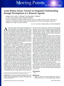
In this single-centre cohort study, we prospectively enrolled patients who were admitted to the eight-bed cardiac surgery ICU of Ghent University Hospital from May 2012 till February 2014. The inclusion and exclusion criteria of the study are incorporated in the flow diagram in Fig. 1. Primary endpoint
The primary endpoint of the current study was the development of AKI stage ≥1 within 48 h after t1. Reference SCr, representing baseline SCr, was defined as the lowest SCr value within the last 3 months (mo) prior to enrolment (lowest of history SCr value(s) and pre-operative SCr). In our cohort 30.0% of the patients had only the pre-operative SCr value available. In 49.3% the lowest history SCr was lower than or equal to the pre-operative SCr, while in 20.7% the pre-operative SCr was lower than the lowest history SCr. The details for calculation of UO are outlined by our group [21]. Note that UO was registered hourly in the ICU only. Secondary endpoint
The secondary endpoint was AKI stage ≥2 within 12 h after t1. Ethics approval and consent to participate
The Ethical Committee of Ghent University Hospital approved this study (Belgian Registration Number of the study: B670201213147). All patients or their legally authorized representatives provided written informed consent. We respected the Declaration of Helsinki and the Good Clinical Practice Guidelines. Prospective sample and data collection
The first collection of blood and urine was after the induction of anaesthesia and before the start of surgery [time 0 (t0) on the day of surgery (dsurgery)]. The rest of the specimens (n = 7) were collected post-operatively, starting at ICU admission (t1) and then at 2 hours (h) (t2), 4 h (t3), 6 h, 12 h, 24 h and 48 h after ICU admission. If the patient was discharged to the Midcare unit before 24 h or 48 h, those samples were collected there. Whenever possible, the routine collection times were followed [at 4 PM on the first post-operative day (d1post-op) and at 6 AM on the second post-operative day (d2post-op)]. The sample collection times for a fictional patient who underwent surgery in the morning are outlined on the timeline in Additional file 3: Fig. S3A, while those for a fictional patient who underwent surgery in the afternoon are outlined on the timeline in Additional file 4: Fig. S3B. These paired blood and urine samples were collected by standard methods and centrifuged by standard protocols,
Page 3 of 12
as described previously by our group [21]. Serum and urine supernatants were stored at −80 °C and thawed at room temperature immediately prior to analysis. Clinical data needed to complete the individual clinical research files (Additional file 5: Table S1) were extracted from the hospital records by study coordinators. Note that Additional file 5 contains Tables S1, S2, S3, S4A, B, C, D and the legends of Figures S1, S2, S3A, B, S4, S5, S6. Samples were anonymized as were clinical data. All technicians were blinded to clinical data. Biomarker analysis and single‑biomarker diagnostic test possibilities
The CHI3L1 analysis was performed in-house. We measured the concentration of CHI3L1 by a sandwich enzyme-linked immunosorbent assay (ELISA) technique (DC3L10, R&D Systems, Minneapolis, MN, USA). Analyses performed externally were Cr and UNGAL. The Cobas c502 measured the concentration of Cr by a kinetic rate-blanked Jaffé assay (commercial reagents, Roche Diagnostics, Basel, Switzerland), whereas the Modular P measured the concentration of UNGAL by a particle-enhanced turbidimetric immunoassay (ST0013CA, BioPorto, Hellerup, Denmark). All details were recently described [21], except for the standard sample dilution scheme used in the CHI3L1 ELISA, which is presented in Additional file 5: Table S2. For blood samples that were collected at routine collection times, a SCr concentration was already available in the hospital records. Based on the temporal relationship of the predictive value of UNGAL for CSA-AKI [19], we measured this biomarker at t1 and t3 only. Besides UCHI3L1 and UNGAL, we also evaluated UCHI3L1 and UNGAL corrected for urine dilution by using the ratio to UCr as diagnostic test. Besides SCr, we also evaluated ΔSCrtx-t0 as diagnostic test, representing the absolute change in SCr between SCrtx and SCrt0. The most recent SCr value recorded prior to surgery was considered as SCrt0. Defining acute tubular damage and subclinical AKI
Following the recommendations of de Geus et al., acute tubular damage was defined as a CSA-NGAL score of 2 or greater; either as UNGALt1 or UNGALt3 ≥ 150 ng/ml or as ∆UNGALt3-t1 > 100 ng/ml with UNGALt3 ≥ 125 ng/ ml [26]. Subclinical AKI was defined when there was acute tubular damage (according to the ‘de Geus criteria’) and absence of AKI according to the KDIGO definition. Defining good and excellent biomarkers
An area under the receiver operating characteristics curve (AUC-ROC) of 0.750 or greater was considered to represent a good biomarker, whereas an AUC-ROC of
De Loor et al. Ann. Intensive Care (2017) 7:24
Page 4 of 12
No. of adults (≥ 18 y) admitted to the post-operative cardiac surgery ICU from May 2012 till February 2014 N = 1064
Inclusion criteria ►Elective surgerya ►Written informed consent
Exclusion criteria ►AKI stage ≥ 1b at time of enrolment ►CKD stage 5c ►Recent kidney transplantd ►Surgery on Sat or Sun
AKI stage ≥ 1 at enrolment or at ICU admission N=8
Enrolled N = 211
Excluded Analysed N = 203
12-h window
24-h window
48-h window Primary endpoint
7-d window
No AKI N = 157 77.3 %
No AKI N = 118 58.1 %
No AKI N = 108 53.2 %
No AKI N = 103 50.7 %
AKI stage 1 N = 43 21.2 %
Within AKI 93.5 %
AKI stage 1 N = 72 35.5 %
Within AKI 84.7 %
AKI stage 1 N = 67 33.0 %
Within AKI 70.5 %
AKI stage 1 N = 65 32.0 %
Within AKI 65.0 %
AKI stage 2 N=3 1.5 %
Within AKI 6.5 %
AKI stage 2 N = 12 5.9 %
Within AKI 14.1 %
AKI stage 2 N = 19 9.4 %
Within AKI 20.0 %
AKI stage 2 N = 21 10.3 %
Within AKI 21.0 %
AKI stage 3 N=0 0.0 %
Within AKI 0.0 %
AKI stage 3 N=1 0.5 %
Within AKI 1.2 %
AKI stage 3 N=9 4.4 %
Within AKI 9.5 %
AKI stage 3 N = 14 6.9 %
Within AKI 14.0 %
Fig. 1 Flow diagram of patient enrolment and primary endpoint analysis. aPlanned ≥4 h in advance. bKDIGO definitions for the diagnosis and staging of AKI, which are based on SCr and UO [25]. cKDOQI definitions for the diagnosis and staging of CKD [40]. d≤3 mo before. AKI acute kidney injury, CKD chronic kidney disease, d day, h hour, ICU intensive care unit, KDIGO Kidney Disease|Improving Global Outcomes, KDOQI Kidney Disease Outcomes Quality Initiative, mo month, No. number, Sat Saturday, SCr serum creatinine, Sun Sunday, UO urine output, y year
De Loor et al. Ann. Intensive Care (2017) 7:24
0.900 or greater was considered to represent an excellent biomarker [27]. Statistical analysis
The principal statistical analysis was based on comparison of the AUC-ROCs of UCHI3L1 with those of UNGAL, more frequently assessed early measurements of SCr, and various two-biomarker panels for predicting both defined endpoints. It was performed in MedCalc 15.2.1 (MedCalc Software, Oostende, Belgium). The unpaired comparison of a variable between two independent samples was done in SPSS 22 (IBM, Armonk, NY, USA). Categorical variables were analysed with Fisher’s exact or the Chi-square test, and continuous variables with the nonparametric Mann–Whitney U test. Additionally, we calculated the 95% confidence interval (CI) for a proportion using the Wilson procedure without a correction for continuity [28, 29]. For all analyses, two-sided P values 100 ng/ml with UNGALt3 ≥ 125 ng/ml [26]. Subclinical AKI was defined when there was acute tubular damage (according to the ‘de Geus criteria’) and absence of AKI according to the KDIGO definition. In this way 84.6% of AKI in our specific cohort (i.e. 77/[77 + 14]) was classified as AKI without acute tubular damage. Subclinical AKI, which was missed by KDIGO, occurred in 5.1% of the patients. AKI acute kidney injury, CSA cardiac surgery-associated, d day, KDIGO Kidney Disease|Improving Global Outcomes, t1 time of intensive care unit admission, t3 4 h after intensive care unit admission, UNGAL urinary neutrophil gelatinase-associated lipocalin
to UCr. Table S4A reports the performances of the biomarkers measured at t0.
Discussion We found that in adult patients who underwent elective cardiac surgery, UCHI3L1 had inadequate predictive value for CSA-AKI. This was also true for the well-known tubular damage biomarker UNGAL. In contrast, more frequent assessment of the functional biomarker SCr in the early post-operative ICU period (first 4 h) had good to excellent predictive value for CSA-AKI.
Similar to others, our ICU routinely measures SCr at t1 and either around t4 (morning patient) or t5 (afternoon patient) in the early post-operative period. However, with ±50% of AKI diagnosed before t5, of which ±50% before t4, our study highlights the importance of more frequent SCr assessment in the first 4 h. This aids in early AKI diagnosis and could also reveal some cases of rapid reversal of AKI (i.e. ‘complete reversal of AKI by KDIGO criteria within 48 h of AKI onset’ [30]). These findings are in accordance with those of a small retrospective study (n = 29) by Maciel et al. [31].
De Loor et al. Ann. Intensive Care (2017) 7:24
Page 7 of 12
Table 1 Characteristics of the patients and procedures at baseline, as well as short-term patient outcomes All patients 203 (100) [98.1–100]
AKI stage ≥1a within 48 h 95 (46.8) [40.1–53.7]
No AKIa within 48 h 108 (53.2) [46.3–59.9]
P value
Male sex—no. (%) [95% CI]
133 (65.5) [58.7–71.7]
65 (68.4) [58.5–76.9]
68 (63.0) [53.6–71.5]
0.461
White race—no. (%) [95% CI]
202 (99.5) [97.3–99.9]
95 (100) [96.1–100]
107 (99.1) [94.9–99.8]
1.000
Ageb (IQR)—years
70.0 (61.0–76.0)
74.0 (65.0–80.0)
67.0 (58.0–75.0)