Elizabeth A. WOODCOCK, Jennifer K. TANNER, Meryl FULLERTON and Ivana J. ...... Berridge, M. J. & Irvine, R. F. (1989) Nature (London) 341, 197-205. 2.
Biochem. J. (1992) 281, 683-688 (Printed in Great Britain)
683
Different pathways of inositol phosphate metabolism in intact neonatal rat hearts and isolated cardiomyocytes Elizabeth A. WOODCOCK, Jennifer K. TANNER, Meryl FULLERTON and Ivana J. KURAJA Baker Medical Research Institute, Commercial Road, Prahran, Vic. 3181, Australia
In most tissues stimulation of the phosphatidylinositol turnover pathway causes release of inositol 1,4,5-trisphosphate [Ins(1,4,5)P3], which is subsequently metabolized to a wide range of inositol phosphate isomers deriving from both phosphorylation and dephosphorylation reactions. However, addition of noradrenaline to isolated intact neonatal-rat hearts generated only those inositol phosphates produced by dephosphorylation of Ins(1,4,5)P3. Products of the InsP3 kinase pathway were absent from the profiles, except after prolonged stimulation. In contrast, addition of noradrenaline to isolated cultured neonatal-rat cardiomyocytes caused the release of Ins(1,4,5)P3, which was metabolized by both phosphorylation and dephosphorylation pathways to yield a complex range of inositol phosphate isomers, as observed in many other cell types. These differences between the responses in intact tissues and in isolated cell preparations were not caused by the different conditions used for [3H]inositol labelling. Furthermore, results could not be explained by overgrowth of other cell types in the isolated cell preparations. Thus the results demonstrate that the isolation and culture of rat neonatal cardiomyocytes produces alterations in the nature of the phosphatidylinositol turnover pathway.
INTRODUCTION It is now generally recognized that the turnover of inositol phospholipids is an important mechanism by which cells recognize and respond to external stimuli. The pathway mediates such divergent responses as mitogenesis, contraction and secretion [1] and appears to be present in all mammalian cell types. Phosphatidylinositol (Ptdlns) turnover is a complex signaltransduction mechanism involving the generation of at least two sigalling molecules, Ins[ 1,4,5]P3 and sn- 1,2-diacylglycerol. Diacylglycerol activates the plasma-membrane-bound phospholipid-dependent Ca2l-dependent protein kinase C, and Ins(1,4,5)P3 releases Ca2+ from specific intracellular stores. These two arms of the pathway act in concert to initiate and sustain the observed cellular responses [2]. Both diacylglycerol and Ins(1,4,5)P3 are rapidly metabolized within the cell. For Ins(1,4,5)P3, metabolism is complex, involving both dephosphorylation via Ins(1,4)P2 to Ins4P and phosphosphorylation to Ins(1,3,4,5)P4, this latter compound being eventually degraded to InsIP and Ins3P [3]. The full significance of this complex metabolism is not yet clear, but Ins(1,3,4,5)P4 itself appears to have a role in maintaining intracellular Ca2+ stores via entry of intracellular Ca2+ in a number of different cell types [1,4]. Ins(1,3,4,5)P4 is also the starting point for metabolic pathways generating other isomers of InsP4 as well as higher inositol phosphates [3]. The generation of this compound thus may be important in determining cellular responses. Although quantitative differences in the profiles of inositol phosphates are observed in different tissues, the essential pattern of inositol phosphate metabolism appears to hold for most cell types. In adult heart tissue Ptdlns turnover is stimulated by aadrenergic agonists, cholinergic agonists, endothelin and histamine [5-8]. Although the importance of Ptdlns turnover in heart tissue is still to be established, it is likely to be involved in mediating some of the responses to stimulation of these receptors, including inotropic responses and cellular hypertrophy [9,10]. However, a number of features of the pathway in heart remain controversial. For instance, the role of protein kinase C in initiating contractile events is debated. Addition of exogenous diacylglycerols or other stimulators of protein kinase C has been variously reported to produce positive [11] or negative [12] inotropic effects, depending on the preparation used and the time Vol. 281
of stimulation [13]. The role of Ins(l,4,5)P3 also is controversial, and Ins(1,4,5)P3 has been reported to be both active [14] and inactive [15] and weak [16] in releasing Ca2` from cardiac sarcoplasmic reticulum. In addition, the nature of the Ptdlns turnover pathway itself has been the subject of debate. In 1987, we reported the absence of detectable levels of Ins(1,3,4,5)P4 and its dephosphorylation products in intact perfused adult rat heart stimulated with noradrenaline [17]. Since that time we have reported similar findings in isolated rat right and left atria [7], and others have reported similar findings in guinea-pig atria [8]. The absence of Ins(1,3,4,5)P4 and its metabolites in heart may explain the relative ineffectiveness of Ins(1,4,5)P3 in releasing Ca2+ from intracellular stores [16] and the lack of increase in Ca2+ after addition of endothelin [18], which stimulates Ptdlns turnover in heart [7]. However, in contrast with our findings, a number of reports have claimed to detect comparatively high concentrations of products of the Ins(1,4,5)P3 kinase pathway in heart tissue [19-21]. Some of these observations were made with isolated neonatal cardiomyocyte preparations [19]. Any differences in the Ptdlns turnover pathway between neonatal myocytes and adult intact tissue might reflect the different developmental stage of the animal and the degree of differentiation of the cardiomyocytes. Alternatively, cell isolation and culture may induce changes in the nature of the pathway. To investigate these possibilities, studies were performed to examine the profile of inositol phosphate release in intact neonatal hearts and in isolated neonatal cardiomyocytes when both are stimulated with noradrenaline. MATERIALS AND METHODS Inositol phosphate generation in neonatal myocytes Cardiomyocytes were prepared from 1-2-day-old rats by methods described previously [22]. This involved an initial preplating step to remove fibroblasts from the preparation. In addition, in some experiments cells were grown in serum-free medium (containing Ham's F12 medium and 5 % BSA) until 2 days before the experiment, to decrease further the possibility of fibroblast contamination. Cells were plated at a density of 5 x 10⁵ per well in 35 mm-diameter culture dishes and maintained in medium 199 containing 50% foetal-calf serum and 100 units of penicillin and strepomycin/ml each. At 2 days before the
E. A. Woodcock and others
684
experiment, medium was replaced with medium 199 containing 20% BSA and antibiotics. Inositol phospholipids were labelled by incubating with [3H]inositol (104,uCi/ml) either for 48 h in medium 199 or for 4 h in Hepes-buffered Krebs medium containing 2 mM-Ca2+, as indicated. After the labelling period, the cells were washed in medium containing non-radioactive inositol (5 mM) for 10 min and subsequently stimulated with noradrenaline in the presence of 10 mM-LiCl for the indicated time. Release of inositol phosphates was terminated by adding ice-cold 50% trichloroacetic acid containing 5 mM-EDTA and 1 mMphytic acid. Phytic acid was added to protect labelled inositol phosphates. After centrifugation to remove trichloroacetic acidinsoluble material, trichloroacetic acid was removed from the extracts by four ether extractions. Samples were treated with Dowex-50 cation-exchange resin and freeze-dried before analysis. This procedure removed u.v.-absorbing material and cations, but did not alter the profiles of inositol phosphates. Inositol phosphate generation in intact neonatal hearts Rats of age 1-2 days were killed by decapitation. Hearts were removed immediately and transferred to an organ bath containing Hepes-buffered Krebs medium containing 2 mM-Ca2 , constantly gassed with 02/C02 (19: 1) at 37 'C. After removal of blood, hearts were incubated in medium containing [3H]inositol (10 ,uCi/ml) for 4 h. After the labelling time, hearts were washed in medium containing non-radioactive inositol (5 mM) for 10 min. Noradrenaline was then added together with 10 mM-LiCl. Inositol phosphate release was terminated by dropping the hearts into liquid N2. Frozen tissue was extracted with ice-cold 5% trichloroacetic acid containing 5 mm-EDTA and I mM-phytic acid. Samples were homogenized, sonicated and centrifuged and the supernatants treated as described above.
Analysis of inositol phosphates For quantification of total inositol phosphates, samples were loaded on to 1 ml columns of Dowex-1 anion-exchange resin (formate form). Inositol phosphates were eluted with ammonium formate as described previously [23]. Analysis of the isomers of the inositol phosphates was carried out by using anion-exchange h.p.l.c. employing a Whatman Partisil SAX column in a Waters Radial Compression Unit. Inositol phosphates were eluted with a complex gradient of ammonium phosphate, pH 3.8, essentially as described by Dean & Moyer [24]. The gradient program involved the use of gradients of 0-0.08 M over 22 min, 0.2-0.28 M over 30 min and 0.5-0.56 M over 25 min for the separation of the isomers of InsPj, InsP2 and InsP3 respectively. Higher inositol phosphates were separated by isocratic elution with 2 M-ammonium phosphate. 3H-labelled compounds were detected and quantified by using an on-line /radiation counter (Radiomatic Instruments, model CR). This provided retention times and integrated peak values for each of the isomers of the inositol phosphates. The identities of the various isomers were established by using appropriate standards. Analysis of I3Hlinositol-labelled phospholipids Inositol phospholipids were extracted from the TCA pellets obtained during the extraction of inositol phosphates. These pellets were extracted with chloroform/methanol/HCl (200: 100: 1, by vol.) by sonication and vigorous vortex-mixing. Water-soluble material was removed by extraction with 5 mMEDTA. The chloroform phase was extracted twice more with methanol/EDTA (1:2, v/v) and evaporated under N2. The evaporated lipids were redissolved in chloroform and the soluble material was again dried under N2. The final evaporated material was resuspended in chloroform and applied to silica-gel
plates (Whatman 60A) preloaded with potassium oxalate (1% in methanol/water, 2:3, v/v) and air-dried for 1 h at 110 °C before use. Phospholipids were separated in a solvent system of chloroform/acetone/methanol/acetic acid/water (40:17:15:12:8, by vol.) under saturated conditions. Plates were sprayed with En3Hance (an autoradiographic enhancer) and autoradiographed on Kodak X-Omat film. Inositol phospholipids were identified according to the positions of authentic standards after iodine staining. Spots were scraped from the plates and counted for radioactivity in a liquid-scintillation counter.
Materials [3H]Inositol, [3H]Ins(1,4,5)P3 and [14C]InslP were obtained from The Radiochemical Centre, Amersham, Bucks., U.K. [3H]Ins4P, [3H]Ins(1 ,4)P2, [3H]Ins(l ,3,4)P3 and [3H]Ins( ,3,4,5)P4 were supplied by DuPont-New England Nuclear, Melbourne, Vic., Australia. En3Hance was supplied by DuPont-New England Nuclear. Cell-culture media were supplied by the Commonwealth Serum Laboratories, Parkville, Vic., Australia.
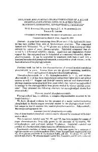
RESULTS Inositol phosphate response to noradrenaline stimulation in intact neonatal-rat hearts Experiments were performed to examine the nature of the Ptdlns turnover pathway in noradrenaline-stimulated neonatal rat hearts. Neonatal-rat hearts were labelled with [3H]inositol and subsequently stimulated with noradrenaline (0.1 mM) in the presence of propranolol (1 /SM) and LiCl (10 mM). The accumulations of the different isomers of the inositol phosphates were determined at different times after addition of noradrenaline. A typical profile of [3H]inositol-labelled products in a neonatal heart stimulated with noradrenaline for 20 min, in the presence of LiCl, is shown in Fig. 1. The major inositol phosphates identified in the profile were Ins(1,4,5)P3, Ins(1,4)P2, Ins4P and Ins3P or Ins lP. In the 20 min profiles there was also a peak of Ins(1,3,4)P3, which was small compared with the peak of Ins(1,4,5)P3 even after 20 min treatment with LiCl. There was no sign of Ins(1,3,4)P3 in profiles obtained at earlier time points.
E 6.
I x 0
40 60 Elution time (min) an intact neonatal-rat heart stimulated for 20 nin with noradrenaline in the presence of LiCI Anion-exchange h.p.l.c. was performed as described in the Materials and methods section. The experiment was performed four times with similar results. Average results are shown in Fig. 2.
Fig. 1. Profile of inositol phosphates in
1992
Inositol phosphate metabolism in intact hearts and cardiomyocytes V4
25 r
Ins(1,4,5)P3
Q
685 40
(1,4)P2
Ins4P
a CL . U, 0 m 0
KS\4
0
Ins(1,3,4)P3
InslP
x
00
.1
0
0
1
20
5
0
1 5 Time (min)
1_
20
0
1
5
20
Fig. 2. Time course of accumulation of inositol phosphate isomers in neonatal-rat hearts stimulated with noradrenailne (0.1 mM) in the presence of LiCI Inositol phosphates were separated and quantified as described in the Materials and methods section and as shown in Fig. 1. Results shown are radioactivity (c.p.m.) in the inositol phosphate isomer relative to the label in the inositol phospholipids. Total label in the inositol phospholipids was 5860 + 396 c.p.m./mg wet wt. of tissue (mean + S.E.M. of four experiments). Accumulation of inositol phosphates in the absence of noradrenaline expressed relative to the lipid label was as follows: Ins(1,4,5)P3, 1.8 + 0.2; Ins(1,4)P2, 7.4 + 1.6; Ins4P, 15.5 + 2.7; Insl/3P, 6 + 1.1 (all x 10-'; means + S.E.M., n = 4).
Ins4P
Ins°lP E
Ins(1,4)P2 x
u
l~~~~~~~~~ns(1,3,4,5)P4 Ins(1,4,5)P3
4
lns(1,3,4)P3 0
20
40
60
80
Elution time (min)
Fig. 3. Profile of inositol phosphates in isolated neonatal cardiomyocytes stimulated with noradrenafine for 20 min in the presence of LiCI The cells had been maintained in culture for 4 days and labelled with [3H]inositol for 4 h in Hepes-buffered Krebs medium. Anionexchange h.p.l.c. was performed as described in the Materials and methods section. The experiment was performed three times with similar results.
The time course of the accumulation of these different isomers is shown in Fig. 2, where each point represents the average of data obtained from four different hearts. To allow comparison of inositol phosphate release studied by different labelling protocols, data are expressed as a percentage of the labelled inositol phospholipids. Addition of noradrenaline caused a rapid and transient increase in Ins(1,4,5)P3, which was followed by increases in its dephosphorylation products Ins(1,4)P2 and Ins4P. In addition, there was a small but significant increase in Ins3P or Insl P over the 20 min period. Ins(1,3,4)P3 was detected at low levels relative to the 1,4,5-isomer in the hearts which had been stimulated with noradrenaline in the presence of LiCl for 20 min, but not at earlier time points. LiCl inhibits the breakdown of Ins(1,3,4)P3 [25] {at least in the presence of Ins(1,4)P2 [26]}, but not that of Ins( 1 ,4,5)P3. The profile of release of inositol phosphates is similar to that reported previously by us in intact adult-rat heart tissue, except for the detectable peak of Ins(1,3,4)P3 at 20 min [17,27,28]. Examination of the isomers of the inositol phosphates allows an evaluation of the relative activities of the phosphorylation and Vol. 281
dephosphorylation pathways of metabolism of Ins(1,4,5)P3. If it is assumed that LiCl has produced an effective blockade of inositol phosphate degradation and that all the InsI P is derived from Ins(1,4,5)P3, it is possible to calculate the proportional activities of the phosphorylation and dephosphorylation pathways after noradrenaline stimulation. Total accumulation from the dephosphorylation pathway equals the increase in Ins4P plus the increase in Ins(1,4)P,. Accumulation from the kinase pathway is the increase in Ins lP/Ins3P plus any increases in Ins(1,3)P2 and Ins(3,4)P2, in Ins(1,3,4)P3 and Ins(I,3,4,5)P4. When this calculation was performed by using the stimulated accumulation of inositol phosphates (i.e. accumulation with noradrenaline plus LiCl minus the accumulation with LiCl alone), the phosphorylation pathway was found to contribute no more than 6.20% of the total metabolism of Ins(1,4,5)P3. This value was calculated from data derived from the average of four hearts incubated with or without noradrenaline for 20 min. A separate experiment also using four hearts in each group provided a similar value (5.8 %). Most of this accumulation was due to the isomers of InsP, and if any direct breakdown of Ptdlns to InsI P occurs in heart then the contribution of the phosphorylation pathway is even less. The possibility that LiCl inhibits the Ins(1,4,5)P3 phosphorylation pathway in intact heart tissue was addressed by performing experiments in the absence of LiCl. The stimulation of inositol phosphate accumulation in heart is low, and in the absence of LiCl increases in inositol monophosphates only were detected. Over 20 min incubation with noradrenaline plus propranolol, InsIP increased from 17.5+5 to 37.4+4 c.p.m./mg of tissue (mean + S.E.M., n = 4). Ins4P increased from 10 + 3 to 136 + 13 c.p.m./mg. Calculation of the contribution of the phosphorylation pathway to this accumulation provided a value of 6.8 %. Thus inclusion of LiCl did not influence the direction of metabolism of Ins(1,4,5)P3. Inositol phosphate response to noradrenaline stimulation in cardiomyocytes As a comparison with the results obtained in intact neonatal hearts, an investigation was made of the inositol phosphate response to noradrenaline stimulation in isolated neonatal cardiomyocytes. Myocytes were maintained in culture for 4 days after isolation and then labelled with [3H]inositol for 4 h in Hepes-buffered Krebs medium. After excess radioactivity was washed out, cells were stimulated with noradrenaline (0.1 mM) in the presence of 1 ,#M-propranolol and 10 mM-LiCl, and the accumulations of the different isomers of the inositol phosphates
686
E. A. Woodcock and others 25 a
50 r
0.
600 r V
Ins(1,3,4)P
Ins4P Ins(1,4)P2
C',
0
-C
S00. Q
0
Ins(1,4,5)P3
0 x
0
0
20
5
1
InslP
...........
Ins(1,3)P2
YY
Ins(1,3,4,5)P
0
1 5 Time (min)
20
1 0
5
1
20
Fig. 4. Time course of accumulation of the isomers of the inositol phosphates in 4-day-cultured neonatal cardiomyocytes after addition of noradrenaline (0.1 mM) plus LiCi Cells were labelled for 4 h with [3H]inositol. Inositol phosphates were separated and quantified as described in the Materials and methods section and as shown in Fig. 3. Results shown are radioactivity (c.p.m.) in inositol phosphates relative to the label in the inositol phospholipids. The total label in the lipids averaged 14706 c.p.m./well of cells. In the absence of noradrenaline, there was no detectable increase in inositol phosphates over 20 min, except for Ins(1,4)P2 (0.004), Ins4P (0.163) and Insl/3P (0.015). The experiment was performed three times with similar results.
._-
3
Ins(1,3,4)P3
._
45
300
50
\
Ins(1,4,5)P3
cn 0
V
Ins4P
v V
/
Ins(1,4)P2
/
/:
a Q ._0
x
X / Ins(1,3,4,5)P4
rX
,&0,.,
o o
"-
llns(1,3)P2
X
1
0 0
1
5
20
0
1 5 Time (min)
20
0
1
5
20
Fig. 5. Inositol phosphate response in isolated neonatal myocytes maintained in culture for 2 days and then labelled with I'Hlinositol for 2 days in medium 199 containing 2 % BSA The response after addition of noradrenaline (0.1 mM) plus LiCI is shown. Separation and determination of the isomers of the inositol phosphates were performed as described in the Materials and methods section and as shown in Fig. 3. Results are expressed as radioactivity (c.p.m.) in the inositol phosphate relative to the label in the inositol phospholipids. The label in the inositol phospholipids averaged 146535 c.p.m./well. Increases in the inositol phosphates in the absence of noradrenaline were as follows: Ins(1,4)P2 (0.0014), Ins4P (0.02) and Insl/3P (0.01). The experiment was performed three times with similar results.
measured. A typical profile of [3H]inositol-labelled products accumulated in cardiomyocytes after 20 min stimulation with noradrenaline plus LiCl is shown in Fig. 3. Unlike the profiles observed in intact tissue, extracts of cultured cells contained a number of different inositol phosphate isomers, and chromatographic profiles were qualitatively similar to profiles obtained from a number of different cell types. Addition of noradrenaline caused a rapid accumulation of Ins(l,4,5)P3, which was maximal at 15 s and decreased thereafter. The increase in Ins(1,4,5)P3 was followed by increases in its metabolites Ins(1,4)P2 and Ins4P. In addition to these dephosphorylation products, Ins(1,3,4,5)P4 generated by phosphorylation of Ins(1,4,5)P3 also showed a transient increase. This was followed by increases in its breakdown products Ins(1,3,4)P3 and Ins3P or InslP (Fig. 4). These data demonstrate that in isolated neonatal-rat cardiomyocytes metabolism of the generated Ins(1,4,5)P3 occurs via both phosphorylation and dephosphorylation pathways. These profiles are similar to those reported by us and by others in a range of different cell types [3,23]. Total inositol phosphate accumulation from the phosphorylation and dephosphorylation pathways under noradrenaline stimulation over 20 min was calculated as described for the intact heart experiments. The phosphorylation pathway was found to contribute 23.2 + 5.7 (mean+S.E.M., n = 3) to the total inositol phosphate accumulation. were
Effect of the labelling conditions on the observed inositol phosphate response Experiments using isolated cells are usually performed after labelling with [3H]inositol for 24-48 h. In order to compare our results with those in the literature, experiments were performed with neonatal cardiomyocytes labelled for 48 h 2 days after isolation. As shown in Fig. 5, the profile of release of inositol phosphates was similar to that observed in cells labelled for 4 h. The contribution of the phosphorylation pathway calculated from these experiments was 25.1 + 6.7 % (mean + S.E.M., n = 3), similar to findings in cells labelled for 4 h. Additional experiments were performed using cells maintained in culture for 12-14 days. The inositol phosphate response to noradrenaline was similar to that observed in cells 4 days after isolation.
Specificity of the stimulation in neonatal cardiomyocytes One possible explanation for the observed difference between cultured cells and the intact tissue is the presence of other cell types, especially fibroblasts, in the cultured preparations. As explained in the Materials and methods section, steps were taken to remove fibroblasts from cardiomyocyte preparations, and microscopic examination indicated that most cells were
spontaneously beating cardiomyocytes. However, the possibility 1992
Inositol phosphate metabolism in intact hearts and cardiomyocytes Table 1. Specificity of the inositol phosphate response in 12-day-cultured neonatal cardiomyocytes
Cells were incubated with stimulatory factor for 20 min in the presence of LiCl, and total inositol phosphate accumulation was measured. Results shown are c.p.m./well of cells, as means + S.E.M. of three different experiments. Abbreviation: FGF, fibroblast growth factor. Treatment
LiCl Noradrenaline (0. 1 mM) Endothelin ( 1,M) Vasopressin (I gM) FGF (10 ng/ml) Bombesin (0.1 pM)
InsP,
InsP2
25686+ 3624 2030+280 61628 + 3420 4175 + 140 73458 +2130 5166+200 32682 +2124 2570+ 215 32505 + 1950 2315 +90 24252 +2610 2125 + 160
InsP3 966+210 2398 + 168 2747 + 119 1092 + 193 1106+98 1134+ 70
of a small number of fibroblasts contributing to the observed inositol phosphate response is possible. To examine the contribution of other cell types, 4-day-isolated myocytes were stimulated with a range of different agonists and the total inositol phosphate response was determined. Results are shown in Table 1. Stimulatoin of inositol phosphate accumulation was observed only with noradrenaline and endothelin, both of which are known activators of cardiac Ptdlns turnover. Vasopressin, fibroblast growth factor and bombesin were ineffective. These factors activate vascular smooth-muscle cells or fibroblasts [29,30]. Thus neither of these cell types appears to make a significant contribution to the observed inositol phosphate response. DISCUSSION The experiments reported here show that the profile of inositol phosphates generated in neonatal-rat hearts stimulated with noradrenaline is different from the reponse observed in isolated neonatal cardiomyocytes in culture. Inositol phosphate profiles in intact tissue were unusual in that only a small number of inositol phosphates were observed. The cultured cardiomyocyte, on the other hand, contained a large number of different inositol phosphates, and the profiles obtained were similar to those reported in many different cell types. The observed difference between intact tissue and myocyte preparations was not caused by the different labelling conditions used in the two types of experiment. Additionally, there was no evidence that responses in myocyte preparations reflect contaminating cell types. Thus the data indicate a change in the Ptdlns turnover pathway when cardiomyocytes are isolated and maintained in culture ex vivo. Addition of noradrenaline to isolated cardiomyocytes caused a rapid and transient release of Ins(1,4,5)PJ, which was metabolized via both dephosphorylation to Ins(1,4)P2 and by phosphorylation to Ins( 1,3,4,5)P4. These two metabolic pathways lead eventually to the accumulation of Ins4P and Insl P or Ins3P respectively. Although maximal release of Ins(1,4,5)PJ was observed within 15 s, accumulation of Ins(1,3,4)P3 continued to increase until I min after addition of noradrenaline. A secondary rise was observed after 5 min stimulation. The accumulation of the dephosphorylation product Ins(1,4)P2 was similarly slow, and substantial accumulations of InsIP and Ins4P were not observed during the first 5 min of stimulation. These findings contrast with results obtained in adrenal glomerulosa cells, where addition of angiotensin II, endothelin-1 or vasopressin caused a rapid and transient release of Ins(1,4,5)P3, which was immediatedly followed by increases in all of its metabolic products [23]. Vol. 281
687
The pathway in intact neonatal hearts was similar to that reported previously by us in adult heart [17], except for the generation of a small amount of Ins(1,3,4)P3 in neonatal hearts stimulated with noradrenaline for 20 min in the presence of LiCI. This indicates a small amount of phosphorylation of Ins(1,4,5)P3 in neonatal tissue. No Ins( 1 ,3,4)P3 was observed in noradrenalinestimulated adult heart [17], even in the presence of LiCl, which inhibits its breakdown [25,26]. Thus, although the pathway in neonatal tissue is similar to that observed in adult tissue, a small vestige of the Ins(1,4,5)P3 kinase pathway remains. This is probably a feature of the incomplete differentiation of these cells compared with the adult tissue. The pathways of inositol phosphate metabolism appear to be similar in a number of different heart preparations, isolated perfused rat heart [16,27,28], isolated guinea-pig atria [8], isolated right and left atria [7] and isolated neonatal-rat heart. This demonstrates that the absence of products of the Ins(1,4,5)P3 kinase pathway is not a feature of a particular preparation and is not related to factors such as ischaemia. In the present study, the apparent absence of detectable levels of Ins(1,3,4)P3 and Ins(1,3,4,5)P4 was further substantiated by measuring the total contribution of metabolites of the phosphorylation and dephosphorylation pathways. This calculation depends mainly on the accumulation of the isomers of inositol monophosphate, and is therefore relatively independent of the determination of Ins(1,3,4)P3. In intact neonatal hearts the phosphorylation pathway was found to contribute 5-6%0 to the metabolism of Ins(1,4,5)P3. When the same calculation was performed from data derived from isolated cardiomyocytes, the phosphorylation pathway was found to account for 25 % of this metabolism. In contrast with our findings and those of Sakuma et al. [8], some other laboratories have reported finding Ins(1,3,4)P3 in intact heart tissue [20,21]. One of these studies used an acidic chloroform/methanol extraction, which produces artifacts which chromatograph with ATP [31]. Co-chromatography with ATP was used by these workers to identify Ins( 1,3,4)P3, and it is possible that the material identified as Ins(1,3,4)P3 in those studies is actually methyl-phosphoryl-inositol 1,4,5-trisphosphate [31]. The other study claimed to detect Ins(1,3,4)P3 but not Ins(1,3,4,5)P4, and used a HC104-extraction procedure [21]. In our hands, HC04 can also cause artifacts in the preparation of heart extracts which require prolonged exposure to the acid. Trichloroacetic acid extraction does not generate unidentified compounds over the extended extraction times used in our studies. Unfortunately, the chromatographic procedures used in these two studies [20,21] did not separate the isomers of inositol monophosphate, and so it is not possible to evaluate the relative contributions of the phosphorylation and dephosphorylation pathways. Thus it is possible thatextraction procedures together with chromatographic differences explain the different reports concerning the presence of InsP3 kinase products in intact heart tissue. Alternatively, if the heart can redevelop the InsP3 kinase pathway when cells are isolated, then it is also possible that the heart in situ can, under some conditions, also regain this part of the pathway. Ins(1,3,4,5)P4 has a role in controlling the influx and distribution of Ca21 in a number of different cell types [1], and the absence of this pathway might be expected to influence Ca2+ handling. Absence of the Ins(1,4,5)P3 kinase pathway has been reported in a number of other cell types, including BC3H-1 muscle cells [32]. This finding is of interest because, in the differentiated state, these cells do not show a Ca2' response to Ins(1,4,5)P3 [33]. Synaptosomes [34] and ram spermatozoa [35] also metabolize Ins(1,4,5)P3 solely by dephosphorylation when activated by Ca2+ plus ionophore. Under these conditions there is no requirement for mobilization of Ca2` from either intracellular or extracellular sources. However, pancreatoma cells
E. A. Woodcock and others
688
stimulated with substance P were able to increase intracellular Ca2+ from both intracellular and extracellular sources in the apparent absence of Ins(1,3,4,5)P4 generation [36]. Thus other methods of control of entry of extracellular Ca2+ associated with activation of Ptdlns turnover must be possible. Studies of Ptdlns turnover using intact tissue have the obvious disadvantage that labelling with [3H]inositol is restricted to 4-5 h at maximum. This may mean that equilibrium has not been reached, and therefore some change in specific radioactivity of the inositol phosphates may occur during stimulation. This is clearly a problem if the level of radioactivity is to be related to absolute mass of the compound. However, even when cells have been labelled for an extended period, the label in the inositol phosphates does not relate well to absolute mass measurements [37,38]. In any case, change in specific radioactivity cannot readily explain the lack of appearance of products of the Ins(1,4,5)P3 kinase pathway. If dephosphorylation products are clearly detectable, then products of the phosphorylation pathway should also be obvious, if they are present in comparable amounts. Although methods for the mass measurement of Ins(1,4,5)P3 are available [39], these do not provide any information about the metabolic fate of the Ins(1,4,5)P3. Furthermore, Ins(1,4,5)P3 is Very rapidly degraded in most cell types, and the continued generation of metabolites demonstrates its continued release. For these reasons there are considerable advantages in examining the complete profile of inositol phosphate isomers, even if isotopic equilibrium has not been established. The heart has the potential to metabolize Ins(1,4,5)P3 to Ins(1,3,4,5)P4, because InsP3 kinase activity has been demonstrated in heart homogenates [21,40], albeit at a lower activity than in other tissues (J. Cox, personal communication), although it is possible that mast cells, which are present in large numbers in heart [41], contribute to the activity measured in homogenates. Mast cells do not contribute significantly to the inositol phosphate response measured in intact heart preparations [6]. Given the proposed importance of Ins(1,3,4,5)P4 in maintaining Ca2+ responses, it would seem unlikely that this enzyme in situ would not be under tight control. The finding that neonatal cardiomyocytes in culture develop the Ins(1,4,5)P3 kinase pathway suggests a loss of cellular differentiation and implies that this part of the pathway is selectively deleted during maturation and differentiation of cardiac myocytes in situ. Studies of Ptdlns turnover are conventiently performed with isolated neonatal cardiomyocytes, and many reports in the literature involve the use of this model system. The findings reported here show that observations made concerning inositol phosphate metabolism in isolated neonatal cardiomyocytes cannot be extrapolated to intact cardiac tissue, and therefore caution must be observed in relating any such results to cardiac function. This work was supported by grants from the National Heart Foundation of Australia, the Australian National Health and Medical Research Council and the Ramaciotti Foundation.
REFERENCES 1. Berridge, M. J. & Irvine, R. F. (1989) Nature (London) 341, 197-205 2. Berridge, M. J. (1987) Annu. Rev. Biochem. 56, 159-193
3. Shears, S. B. (1989) Biochem. J. 260, 313-324 4. Irvine, R. F. & Moor, R. M. Biochem. J. 240, 917-920 5. Brown, J. H., Buxton, I. L. & Brunton, L. L. (1985) Circ. Res. 57,
(1986)
532-537 6. Woodcock, E. A., White, L. B. S., Smith, A.I. & Tanner, J. K. (1987) Circ. Res. 61, 625-631 7. Kuraja, I. J., Tanner, J. K. & Woodcock, E. A. (1990) Eur. J. Pharmacol. Mol. Pharmacol. Sect. 189, 299-306 8. Sakuma, I., Gross, S. S. & Levi, R. (1988) J. Pharmacol. Exp. Ther. 247, 466-472 9. Scholz, J., Schaeffer, B., Schmitz, W., Steinfath, M., Lohse, M., Schwabe, U. & Purunen, J. (1988) J. Pharmacol. Exp. Ther. 245, 327-335 10. Simpson, P. (1985) Circ. Res. 56, 884-894 11. Otani, H., Otani, J. & Das, D. K. (1987) Circ. Res. 62, 8-17 12. Yuan, S., Sunhara, F. A. & Sen, A. K. (1987) Circ. Res. 61, 372-378 13. Lacerada, A.E., Rampe, D. & Brown, A. M. (1988) Nature (London) 335, 249-251 14. Nosek, T. M., Williams, M. F., Zeighter, S. T. & Godt, R. E. (1986) Am. J. Physiol. 250, C807-C811 15. Movsesian, M. A., Thomas, A. A., Selak, M. & Williamson, J. R. (1985) FEBS Lett. 185, 328-332 16. Kentish, J. C., Barsotti, R. J., Lea, T. J., Mulligan, I. P., Patel, J. R. & Ferenczi, M. A. (1991) Am. J. Physiol. 258, H610-H615
17. Woodcock, E. A., Smith, A. I., Wallace, C. A. & White, L. B. S.
(1987) Biochem. Biophys. Res. Commun. 148, 68-77 18. Kelly, R. A., Eid, H., Kramer, B. K., O'Neil, M., Liang, B. T., Reers, M. & Smith, T. W. (1990) J. Clin. Invest. 86, 1164-1171
19. Steinberg, S. F., Kaplan, L. M., Inouye, T., Zhang, J. F. & Robinson, R. B. (1989) J. Pharmacol. Exp. Ther. 250, 1141-1148 20. Kohl, C., Schmitz, W. & Scholz, H. (1990) Circ. Res. 66, 580-583 21. Renard, D. & Poggioli, J. (1987) FEBS Lett. 217, 117-123 22. Weissberg, P. L., Little, P. J., Cragoe, E. J. & Bobik, A. (1989) Circ. Res. 64, 676-685 23. Woodcock, E. A., Little, P. J. & Tanner, J. K. (1990) Biochem. J. 271, 791-796 24. Dean, N. M. & Moyer, J. D. (1987) Biochem. J. 242, 361-366 25. Hughes, P. J. & Drummond, A. H. (1987) Biochem. J. 248, 463-470
26. Shears, S. B., Storey, D. J., Morris, A. J., Cubitt, A. B., Parry, J. B., Michell, R. H. & Kirk, C. J. (1987) Biochem. J. 242, 393-402
27. von Harsdorf, R., Lang, R., Fullerton, M., Smith, A. I. & Woodcock, E. A. (1988) FEBS Lett. 233, 201-205
28. von Harsdorf, R., Lang, R. E., Fullerton, M. & Woodcock, E. A. (1989) Circ. Res. 65, 495-501 29. Aiyar, N., Nambi, P., Stassen, F. L. & Crooke, S. T. (1986) Life Sci. 39, 37-45
30. Takuwa, N., Takuwa, Y., Yanagisawa, M., Yamashita, K. & Masaki, T. (1989) J. Biol. Chem. 264, 7856-7861 31. Brown, J. E., Rudnick, M., Letcher, A. J. & Irvine, R. F. (1988)
Biochem. J. 253, 703-710 32. Ambler, S. K., Thompson, B., Solski, P. A., Brown, J. H. & Taylor, P. (1987) J. Pharmacol. Exp. Ther. 32, 376-383 33. De Smedt, H., Parys, J. B., Himpens, B., Missiaen, L. & Borghgraef, R. (1991) Biochem. J. 273, 219-224 34. Weaver, K. & Brammer, M. (1989) Biochem. Soc. Trans. 17, 711-712
35. Harrison, R. A. P., Roldan, E. R. S., Lander, D. J. & Irvine, R. F. (1989) Cell. Signalling 2, 277-284 36. Horstman, D. A., Takemura, H. & Putney, J. W., Jr. (1988) J. Biol.
Chem. 263, 15297-15303 37. Bird, G. St. J., Oliver, K. G., Horstman, D. A., Obie, J. & Putney, J. W., Jr. (1991) Biochem. J. 273, 541-546 38. Lambert, D. G., Challis, R. A. & Nahorski, S. R. (1991) Biochem.
J. 273, 791-794 39. Kawaguchi, H., lizuka, K., Takahashi, H. & Yasuda, H. (1990) Biochem. Med. Metab. Biol. 44, 42-50 40. McKee, E. E., Clark, M. G., Beinlich, C. J., Lins, J. A. & Morgan,
H. E. (1979) J. Mol. Cell. Cardiol. 11, 1033-1051 41. Keller, A. M., Clancy, R. M., Barr, M. L., Marboe, C. C. & Cannon,
P. J. (1988) Circ. Res. 63, 1044-1052
Received 18 April 1991/28 August 1991; accepted 12 September 1991
1992