0013-7227/98/$03.00/0 Endocrinology Copyright © 1998 by The Endocrine Society
Vol. 139, No. 5 Printed in U.S.A.
Differential Dose-Dependent Effects of Epidermal Growth Factor on Gene Expression in A431 Cells: Evidence for a Signal Transduction Pathway That Can Bypass Raf-1 Activation* MONIQUE SILVY, PIERRE-MARIE MARTIN, NAIMA CHAJRY, YOLANDE BERTHOIS
AND
Laboratoire Interactions Cellulaires Intratumorales, CJF INSERM 9311, IFR Jean Roche, Faculte´ de Me´decine Secteur Nord, 13916 Marseille Cedex 20, France ABSTRACT Epidermal growth factor (EGF), which plays an important role in normal and tumoral cell growth regulation, displays an ambivalent dose-dependent effect on the proliferation of epithelial cells overexpressing EGF receptor. However, the underlying molecular mechanisms remain obscure. In this study we have examined the regulation of amphiregulin (AR) gene expression by growth inhibitory (1029 M) and stimulatory (10212 M) EGF concentrations in A431 cells. The time course of AR messenger RNA (mRNA) accumulation was different with 10212 and 1029 M EGF; AR induction by 1029 M EGF peaked between 1 and 1.5 h, then decreased to the basal level within 2 h. Conversely, the induction by 10212 M EGF was slightly delayed, but persisted for 4 h. The involvement of tyrosine phosphorylation in AR induction by EGF was suggested by the ability of the tyrosine phosphatase inhibitor sodium orthovanadate to prolong AR expression induced by 10212 or 1029 M EGF. In the presence of the protein phosphatase 2A inhibitor, okadaic acid, 1029 M EGF induced a persistent accumulation of AR mRNA. On the contrary, okadaic acid abrogated the stimulation of AR mRNA level induced by a low EGF concentration, suggesting that both EGF concentrations activated
E
PIDERMAL growth factor (EGF) elicits a number of biological responses, including profound alterations in differentiation and growth, in a variety of normal and tumoral cell types (1, 2). These responses are mediated by ligand activation of the EGF receptor (EGFR) tyrosine kinase, resulting in the phosphorylation of several protein substrates, including Grb/Sos (which complex with and activate Ras), phospholipase Cg1, phosphatidylinositol 3-kinase, and STAT (signal transducers and activators of transcription) proteins (1– 4). These EGFR tyrosine kinase substrates then transmit the EGF signal to various intracellular compartments via phosphorylation of downstream serine/threonine kinases or by other protein-protein or protein-DNA interactions. Until now, much attention has been paid to the mitogenic Received August 21, 1997. Address all correspondence and requests for reprints to: Dr. Yolande Berthois, Laboratoire Interactions Cellulaires Intratumorales, CJF INSERM 9311, IFR Jean Roche, Faculte´ de Me´decine Secteur Nord, boulevard Pierre Dramard, 13916 Marseille Cedex 20, France. E-mail:
[email protected]. * This work was supported by grants from the Fe´de´ration Nationale des Centres de Lutte contre le Cancer.
distinct regulatory mechanisms. The signaling components involved in the differential activities of EGF in A431 cells were then examined. We previously reported a relationship between the ambivalent activity of EGF and the p42-mitogen-activated protein (MAP) kinase activity. Thus, 10212 M EGF induced a sustained MAP kinase activation, whereas 1029 M EGF led to a sharp, but transitory, activation. The MAP kinases are activated by MAP kinase kinases (MEK1 and MEK2). Whereas no significant effect of 10212 M EGF could be detected, 1029 M EGF was shown to activate MEK1 and, to a lesser extent, MEK2. Also, both MAP kinase activation and AR induction by 1029 M, but not by 10212 M, EGF were inhibited by the MEK1 inhibitor PD98059. Moreover, the involvement of c-Raf-1 in the signaling pathway induced by EGF was verified. A concentration of 1029 M EGF induced stimulation of c-Raf-1 kinase activity, whereas 10212 M EGF not only failed to activate c-Raf-1, but led to a moderate decrease in its kinase activity. These results demonstrate that in EGF receptoroverexpressing cells, EGF may differently affect gene expression and cell proliferation through distinct mechanisms of regulation. (Endocrinology 139: 2382–2391, 1998)
action of EGF on various tissues and cell lines in culture. Nevertheless, several pieces of evidence have emerged describing an ambivalent activity of EGF on the proliferation of epithelial cells overexpressing the EGFR (5). Whereas this phenomenon has been clearly related to receptor frequency and occupation (6), the molecular mechanism involved has not been elucidated. In a study aimed at dissecting the biochemical events leading to the dual actions of EGF on cell proliferation (7, 8), we previously described in the human epidermoid carcinoma A431 cell line, a relationship between the dual stimulator/inhibitor effects of EGF and the activity of a serine/threonine protein kinase [p42 mitogen-activated protein (MAP) kinase] that occupies a pivotal position in the signal transduction triggered by a large variety of growth effectors. Thus, MAP kinase was shown to be activated in response to growth stimulatory doses of EGF, and this activation persisted throughout cell treatment. Conversely, under conditions of EGF growth inhibition, a sharp, but transitory, activation of MAP kinase was followed by its rapid decrease to basal levels. However, the functional significance of various signal transduction pathways and the mechanisms by which they regulate cell growth or differentiation remain unclear. There-
2382
DOSE EFFECTS OF EGF ON GENE EXPRESSION
fore, investigation of the regulatory mechanisms should help to unravel cellular responses to extracellular signals at the molecular level. In many cases, cellular responses to extracellular stimuli accompany the induction of various genes. Among the genes induced by EGF, amphiregulin (AR) is of interest because a number of studies suggest that AR is important as an autocrine growth and/or differentiation factor that might be involved in the pathogenesis of a variety of human cancers. AR is a heparin-binding EGF-related growth factor originally isolated from serum-free conditioned medium of the human breast carcinoma cell line MCF-7, that had been treated with the phorbol ester, tumor promoter 12-O-tetradecanoyphorbol-13-acetate (TPA) (9). AR is a multifunctional growth regulatory factor. Depending on the concentration, target cell, and presence of other growth factors, AR is able to either inhibit or stimulate cell proliferation (9 –12). AR binds to the EGFR, but with a lower affinity than EGF or transforming growth factor-a (13). However, AR appears to be a more specific and natural regulator of normal epithelial cells than the other factors, as evidenced by its discovery as an autocrine mitogen for cultured human keratinocytes, mammary epithelial cells, and colorectal cells and by its reduced activity on mesenchymal cells (11–14). Moreover, a nuclear localization of AR has been demonstrated by immunocytochemistry in normal mucosa epithelial cells and ovarian surface epithelial cells, indicating that AR could play a role in the differentiation of cells through a direct effect in the nucleus (10, 11). More important, the frequency and intensity of AR expression and nuclear localization were enhanced in several human cancer cell lines and primary carcinomas (12, 15–19). These data in conjunction with the presence of specific nuclear acceptor proteins for this peptide suggest that AR has an additional intracellular site of action. This may be the case, as mutation of the nuclear targeting sequences from the rat homolog of AR, schwannoma-derived growth factor, impairs AR’s ability to function as a mitogen (20). The mechanism by which gene activation is produced after EGF binding to its receptor remains unclear. Although AR gene regulation by estradiol and the phorbol ester TPA has been previously examined by Martinez-Lacaci et al. (21) and demonstrated to employ separate mechanisms, the possibility that growth inhibitory and stimulatory doses of EGF could regulate gene expression in EGFR-overexpressing cells through distinct signal transduction pathways has not been examined. In the study described here, we have investigated the interaction between EGF and c-Raf-1 in regulating the AR messenger RNA (mRNA) level in A431 cells and the roles of c-Raf-1 and MAP kinase in mediating the EGF response. The results demonstrate that EGF, depending on its concentration, is able to activate MAP kinase cascade through distinct mechanisms of regulation that allow to differential induction of AR gene expression. Materials and Methods Materials The AR complementary DNA (cDNA) probe was a gift from Dr. Plowman (Bristol-Myers Squibb, Seattle, WA). Rabbit polyclonal antiRaf-1 antibody and enzyme-inactive p42 MAP kinase were provided by Santa Cruz Biotechnology (Santa Cruz, CA). Rabbit anti-p42 MAP kinase
2383
serum was obtained as previously described (8). Rabbit antiphosphorylated p42 MAP kinase antibody and MAP kinase kinase (MEK1) inhibitor (PD98059) were obtained from New England Biolabs (Beverly, MA). c-Raf-1 kinase peptide substrate was provided by Promega (Madison, WI). MAP kinase substrate (MBP) was purchased from Sigma Chemical Co. (St. Louis, MO).
Cell culture A431 cells were cultured in 50% DMEM-50% Ham’s F-12 (Life Technologies, Grand Island, NY) supplemented with 10% FBS and 2 mm l-glutamine and maintained at 37 C in a humid atmosphere of 5% CO2 in air. Two days before the experiments, complete medium was replaced by serum-free medium supplemented with 10 mm HEPES for starvation of the cells. Cell treatment in the presence of EGF was performed in serum-free culture conditions.
Isolation of cellular RNA A431 cells, plated into 25-cm2 T-flasks were incubated at 37 C for various periods of time (0.5–12 h) in the presence of 10212 or 1029 m EGF. At the end of the treatment, RNA extraction was carried out using RNazol (Cinna-Biotex, Friendswood, TX). After the addition of equal volumes of chloroform, extracted RNA was precipitated with 1 vol isopropanol. After washing, pellet was resuspended in 20 – 40 ml diethylpyrocarbonate-water.
RT-PCR First strand cDNA was synthesized using Moloney mouse leukemia virus reverse transcriptase. Five micrograms of total RNA, extracted as previously described, were incubated with oligo(deoxythymidine) (1 mg/reaction) and diethylpyrocarbonate-water (total volume 6 ml) for 10 min at 65 C, then for 5 min at 0 C. Reverse transcription was initiated in the presence of 2 ml Moloney mouse leukemia virus (400 U), 5 ml of 5 3 reaction buffer [250 mm Tris-HCl (pH 8.3), 375 mm KCl, and 15 mm MgCl2], 1 ml 10 mm deoxy (d)-NTP, 2 ml 100 mm dithiothreitol (DTT), and 0.5 ml ribonuclease inhibitor (40 U/ml). Reaction was performed for 1 h at 37 C, then for 30 min at 52 C. Coamplification of the cDNA was carried out in a reaction buffer containing 1.5 mm MgCl2, 50 mm KCl, 10 mm Tris-HCl (pH 8.3), four deoxynucleotide triphosphates (0.2 mm each), 2.5 U Taq DNA polymerase, 1 pmol/ml of the specific amphiregulin primers, and 0.5 pmol/ml of the specific b2-microglobulin primers. The primers used correspond to human AR cDNA sequences 403– 424 (primer sense) and 540 –521 (primer antisense) and to human b2microglobulin cDNA sequences 97–116 (primer sense) and 242–261 (primer antisense). Four microliters of cDNA were added to 46 ml amplification reaction buffer. The samples were overlaid with mineral oil, and PCR was performed for 20 cycles, consisting of 50 sec at 94 C, 50 sec at 57 C, and 20 sec at 72 C. The samples were heated for 10 min at 94 C before the first cycle, and the extension time was lengthened to 10 min during the last cycle.
Southern blot analysis Southern analysis of the PCR products was performed as reported previously (22). Briefly, 30 ml of the PCR products were electrophoresed in 2% agarose gel. The gel was stained with ethidium bromide to allow visualization of the DNA, which was then denatured and transferred to a Hybond N membrane (Amersham, Arlington Heights, IL). The AR cDNA probe was labeled with [a-32P]dCTP using the random priming method (23). The b2-microglobulin probe corresponding to the human b2-microglobulin sequence 121–150 was labeled with [g-32P]dCTP using the T4 kinase enzyme.
Preparation of cytosolic cell extracts and immunoprecipitation After serum starvation for 48 h at 37 C, A431 cells seeded in six-well culture plates were incubated at 37 C for various periods of time in the presence of 10212 or 1029 m EGF. Control cells were not exposed to EGF. Cells were then washed with ice-cold PBS (NaCl-phosphate) and incubated in lysis buffer [50 mm Tris-HCl (pH 7.5), 100 mm NaCl, 5 mm
2384
DOSE EFFECTS OF EGF ON GENE EXPRESSION
EDTA, 50 mm sodium fluoride, 200 mm sodium orthovanadate, 40 mm b-glycerophosphate, 10 mg/ml leupeptin, 10 mg/ml aprotinin, 1 mm phenylmethylsulfonylfluoride, and 0.5% Triton X-100]. Insoluble material was removed by centrifugation at 4 C for 10 min at 12,000 3 g. Cell lysates (500 ml) containing equivalent amounts of protein were incubated for 2 h at 4 C with primary antibodies preadsorbed to protein A-Sepharose overnight at 4 C. For the immunoprecipitation of MEK1, preadsorption of monoclonal mouse anti-MEK1 antibody was performed in the presence of rabbit antimouse Igs. Controls were performed in which primary antibodies were omitted.
Endo • 1998 Vol 139 • No 5
cell growth stimulation that is induced in the presence of low EGF concentrations (10212–10211 m) was totally abolished with increasing EGF doses (data shown in Ref. 7). Moreover, a dramatic inhibition of cell proliferation was described when the growth factor concentration attained 1029 m. As AR may be induced by EGF in a variety of cancer cell lines in culture, we have examined the induction of AR under conditions of growth stimulation or inhibition by EGF. Figure 1
Immunocomplex kinase assay Immunoprecipitates were washed three times with cold RIPA buffer (150 mm NaCl, 1% NP-40, 0.5% sodium deoxycholate, 0.1% SDS, 50 mm Tris-HCl, pH 8.0) and twice with kinase buffer [50 mm Tris-HCl (pH 7.3), 150 mm NaCl, 12.5 mm MnCl2, 1 mm DTT, and 0.2% Tween-20]. Raf-1 kinase activity was determined according to the procedure previously described (24). Raf-1 immunoprecipitates were incubated in 80 ml kinase buffer with 20 mCi [g-32P]ATP and 100 mg Raf-1 substrate peptide for 20 min at 25 C. The essay was verified to be linear for at least 30 min. The phosphorylation reaction was terminated by the addition of 80 ml of 2-fold concentrated Laemmli buffer. Samples were heated at 95 C for 5 min and submitted to SDS-electrophoresis on a 20% acrylamide resolving gel. The gels were dried and submitted to autoradiography. Phosphate incorporation was measured by quantitation of the substrate bands by image analysis. A similar procedure was used for measuring MEK1 and MEK2 activities, except that enzyme-inactive p42 MAP kinase (5 mg) was used as substrate. p42-MAP kinase activity was determined as previously described (7). MAP kinase immunoprecipitates were washed four times in lysis buffer, then once with kinase buffer [25 mm HEPES (pH 7.4), 10 mm MgCl2, 1 mm DTT, and 10 mm p-nitrophenyl phosphate]. The kinase reaction was performed by resuspending the immunoprecipitate in 40 ml kinase buffer containing 250 mm MBP as substrate, 25–50 mm ATP, and 3–5 mCi [g-32P]ATP (Amersham; SA, 3000 Ci/mmol). The reaction was continued for 10 min at 37 C; it was stopped by the addition of 40 ml of 2-fold concentrated Laemmli buffer. Samples were heated at 95 C for 5 min and submitted to SDS-electrophoresis on a 12% acrylamide resolving gel. The level of phosphorylation of MBP was evaluated as described above.
Western immunoblotting Cell extracts prepared from EGF-treated A431 cells were resolved by SDS-PAGE. Separated proteins were then electroblotted for 1 h on a nitrocellulose membrane (Amersham) using a semidry blotter (Bio-Rad, Richmond, CA). Raf-1 was detected using a rabbit polyclonal anti-cRaf-1 antibody. Phosphorylated p42 MAP kinase was visualized with a antibody that reacts with tyrosine-phosphorylated and doubly threonine/tyrosine-phosphorylated MAP kinase. Revelation was performed using the chemiluminescence procedure (Amersham). Quantitation of the phosphorylated MAP kinase was performed by image analysis.
Image acquisition The autoradiographies were scanned with an Agfa ARCUS scanner (Agfa-Geavaert, Morstel, Belgium) in the transparency mode. The resolution of the scanner is adjustable from 1–1200 pixels/in.
Image analysis The software used in the analysis was the Macintosh-based public domain program Image, written by Rasband at the NIH. The program contains a built-in macrolanguage permitting complicated repetitive procedures to be converted to single commands, as we use here. The integrated optical density of each band was measured after background subtraction.
Results Dose-dependent induction of AR gene expression by EGF in A431 cells
EGF is known to display ambivalent activity on the proliferation of the A431 cells that overexpress EGFR. Thus, the
FIG. 1. Effect of EGF on AR mRNA level in A431 cells. A431 cells were grown in DMEM-Ham’s F-12 supplemented with 10% FCS. After 2–3 days, medium was replaced with serum-free medium containing 10 mM HEPES. Two days later, cells received EGF for different periods of time. AR mRNA regulation was analyzed by RT-PCR as described in Materials and Methods. A and B, Southern blotting was performed on RT-PCR products prepared from cells treated with 10212 M (A) or 1029 M EGF (B). After separation on 2% agarose gel and transfer to a Hybond N membrane, RT-PCR products were hybridized with 32P-labeled probes for amphiregulin (bands at 136 bp) and b2microglobulin (bands at 165 bp). After visualization by autoradiography, the integrated optical density (IOD) of each band was measured after background subtraction, as indicated in Materials and Methods. C, Graphic representation of four time-course experiments concerning the effects of 10212 M (M) and 1029 M (E) EGF on AR mRNA levels. Values obtained for each sample were corrected for the corresponding b2-microglobulin mRNA level. Data are the mean 6 SD of values obtained from four separate experiments and are expressed as a percentage of the control value.
DOSE EFFECTS OF EGF ON GENE EXPRESSION
shows that both growth stimulatory and inhibitory concentrations of EGF (10212 and 1029 m, respectively) induced an increase in AR mRNA. Time-course analysis demonstrated that 1029 m EGF allowed to a sharp and transitory stimulation of AR expression that reached a maximum at 1 h, then rapidly decreased to the basal level at 2 h. AR induction by 10212 m EGF was slightly delayed, but persisted longer than induction by 1029 m, as the return to the basal level was shown to occur between 4 – 8 h. To test whether the AR gene was the primary target in 10212 and 1029 m EGF induction, cells were pretreated with the protein synthesis inhibitor cycloheximide (not shown). Cycloheximide inhibited AR mRNA induction by 10212 and 1029 m EGF, indicating that both EGF doses required de novo protein synthesis to activate signal transduction pathways and induce AR gene expression. Equally, to examine possible modifications of the stability of AR mRNA under 10212 and 1029 m EGF, cells were pretreated with the transcriptional inhibitor actinomycin D (not shown). In all cases, AR transcripts revealed similar half-lives. Effect of protein phosphatase inhibitors on AR gene induction by EGF in A431 cells
The activity of intracytoplasmic molecules involved in the signal transduction induced by extracellular stimuli is regulated by mechanisms of phosphorylation/dephosphorylation that are under the control of a variety of tyrosine and serine/threonine phosphatases. To examine the requirement for tyrosine phosphorylation in the induction of AR gene transcription by EGF, the tyrosine phosphatase inhibitor NaVO4 was used. Figure 2 demonstrates that the return to the mRNA control level that follows the AR gene induction by 10212 and 1029 m EGF was totally abolished in the presence of NaVO4. In both cases, NaVO4 was able to maintain the AR mRNA level at 250 –280% of the control value throughout EGF treatment. Although NaVO4 alone also induced a stimulation of AR expression, this stimulation did not exceed 150% of the control value. The tumor promotor okadaic acid (OA) is known as a powerful inhibitor of serine/threonine protein phosphatases 1 and 2A (PP1 and PP2A). PP2A, which plays an essential role in the regulation of MAP kinase activity, has been described to be specifically inhibited by 2 nm OA. When AR gene induction by 1029 m EGF was examined in the presence of 2 nm OA, a sustained and prolonged stimulation of AR gene expression was observed (Fig. 2). In parallel, it was verified that OA abolished the dephosphorylation of MAP kinase observed under long term cell treatment with 1029 m EGF (data not shown) (8), suggesting that PP2A is involved in AR gene regulation induced by growth inhibitory EGF concentrations. On the contrary, whereas OA per se induced a moderate increase in AR mRNA (150% of the control value), it was shown to antagonize AR induction by 10212 m EGF, supporting the hypothesis that 10212 and 1029 m EGF modulate AR gene expression through separate regulatory mechanisms.
2385
FIG. 2. Effects of sodium orthovanadate (A and C) and OA (B and D) on EGF induction of AR mRNA. A431 cells, grown as described in Fig. 1, were treated with EGF in the absence or presence of inhibitor for various periods of time. A and B, Cells were treated with 10212 M EGF in the absence (M) or presence (f) of 400 mM sodium orthovanadate (A) or 2 nM okadaic acid (B). C and D, Cells were treated with 1029 M EGF in the absence (E) or presence (F) of 400 mM sodium orthovanadate (C) or 2 nM OA (D). In parallel, the effects of sodium orthovanadate and okadaic acid alone were verified (‚). AR mRNA levels were analyzed as described in Fig. 1. The AR bands were corrected with the b2-microglobulin bands, and values, expressed as a percentage of the control value, are the mean 6 SD obtained from three separate experiments.
MAP kinase activation pathways induced by EGF in A431 cells
Activation of a signaling pathway involves the activation by phosphorylation of several proteins making up the pathway. Various growth factors are known to activate a signal transduction pathway involving Ras/Raf/MAP kinase. In a previous study, aimed to analyze differential mechanisms of signal transduction that determine positive or negative regulation of A431 cell proliferation, we investigated the effect of EGF on MAP kinase activity (7, 8). As reported in Fig. 3, we demonstrated a relationship between the p42 MAP kinase activity and the dual growth effect of EGF, as 10212 m EGF led to a moderate, but persistent, activation of MAP kinase, whereas a high, but transitory, activation was induced by 1029 m. MAP kinase is activated by phosphorylation on specific threonine and tyrosine residues. The level of activated p42 MAP kinase, determined in parallel by Western blot using an antibody raised to tyrosine-phosphorylated and doubly tyrosine/threonine-phosphorylated MAP kinase, was shown to fit with its kinase activity (Fig. 3). Moreover, it was verified that EGF treatment did not affect the total amount of MAP kinase (not shown). MAP kinase activation that occurs after cell stimulation with growth factors and hormones may be triggered by two dual specificity kinases, MEK1 and MEK2, that phosphorylate MAP kinase on threonine and tyrosine. To analyze the molecular mechanism that leads to the activation of p42 MAP kinase under growth stimulatory and inhibitory actions of
2386
DOSE EFFECTS OF EGF ON GENE EXPRESSION
Endo • 1998 Vol 139 • No 5
EGF, the effects of 10212 and 1029 m EGF on both MEK1 and MEK2 activities were examined (Fig. 4). Whereas 10212 m EGF was unable to activate MEK1 and MEK2, cell treatment with 1029 m EGF for 5 and 15 min stimulated the kinase activity of MEK1. Also, a moderate MEK2 activation could be observed in the short term presence (5 min) of high doses of EGF. Additionally, the effect of the MEK1 inhibitor PD098059 on MAP kinase activation was analyzed. Whereas PD098059 had no effect on MAP kinase phosphorylation induced by 10212 m EGF, the MEK1 inhibitor was shown to reverse in large part MAP kinase activation under 1029 m EGF. The inhibitor displayed similar activity on the EGF-induced AR gene expression. Indeed, PD098059 was observed to partially abolish AR mRNA stimulation induced by 1029 m EGF, but not by 10212 m EGF, confirming that, depending on its concentration, EGF may regulate MAP kinase activation and AR expression through different mechanisms. Involvement of c-Raf-1 in signaling pathway induced by EGF in A431 cells
Multiple lines of evidence have demonstrated that stimulation by growth factors activates MAP kinase through activation of endogenous c-Raf-1 (24 –27). Activation of cRaf-1 involves multiple phosphorylations of Raf-1 on serine and, in some cases, on tyrosine residues, and these phosphorylations can induce a shift of Raf mobility in SDS-PAGE (26 –28). We have analyzed and compared Raf-1 activation and mobility shift in A431 cells treated with 10212 and 1029 m EGF. We found that 1029 m EGF induced a reduced electrophoretic mobility of Raf-1 that was observable by 5 min of EGF treatment (Fig. 5). Inversely, 10212 m EGF was unable to induce hyperphosphorylation of Raf-1, as indicated by the absence of reduced mobility forms. In parallel, the regulation of kinase activity of c-Raf-1 by EGF was determined by measuring the ability of immunoprecipitated c-Raf-1 to phosphorylate a specific substrate. The results presented in Fig. 5 demonstrated an early and transitory increase in cRaf-1 kinase activity that peaked between 15–30 sec when cells were treated in the presence of 1029 m EGF. On the contrary, 10212 m EGF not only failed to activate c-Raf-1 kinase, but led to a moderate, but reproducible, decrease in the kinase activity within 15 and 45 sec of EGF cell treatment. FIG. 3. Time course of p42MAP kinase activation by EGF. Cells grown as described in Fig. 1 were incubated in the presence of 10212 or 1029 M EGF for various periods of time. At the end of the incubation, cell extracts were prepared as described in Materials and Methods. A, Equivalent amounts of proteins were separated by electrophoresis on a 10% SDS-polyacrylamide gel, transferred on nitrocellulose membrane, then probed with an antibody raised to tyrosine- and tyrosine/ threonine-phosphorylated p42 MAP kinase. Immunoreactive proteins were revealed using chemiluminescence detection. B, p42 MAP kinase activity was measured in cells treated with 10212 M EGF (M) or 1029 M EGF (E). At the end of the treatment, cells were lysed, and p42 MAP kinase was immunoprecipitated. The kinase activity was then measured, as described in Materials and Methods, by the ability of the immunoprecipitate to phosphorylate exogenous MBP substrate. After separation on SDS-polyacrylamide gel and autoradiography, the bands illustrating the phosphorylation of MBP were quantified by image analysis. The values obtained were plotted as a percentage of those in unstimulated control cells. Each value represents the mean 6 SD of three separate determinations and is representative of data from three separate experiments.
Discussion
We have shown in EGFR-overexpressing A431 cells that EGF, depending on its concentration, may differentially activate the MAP kinase signaling cascade, allowing differential regulation of gene expression and, ultimately, of cell proliferation. The A431 cells have been frequently used to examine the molecular mechanisms by which EGF acts on the proliferation of tumoral epithelial cells. However, the paradoxical ambivalent growth response of A431 cells to EGF has been little considered, as most reports have studied the signal transduction pathway induced by EGF under growth inhibitory conditions. To our knowledge, only a few studies have examined the regulation of gene expression by different doses of EGF in A431 cells (29, 30). Thus, the recent work by Gulli et al. (30) described differential induction of c-myc by
DOSE EFFECTS OF EGF ON GENE EXPRESSION
2387
FIG. 4. Involvement of MEK1 and MEK2 in the signaling pathway induced by EGF. Cells grown as described in Fig. 1 were treated with 10212 or 1029 M EGF for 5 min or 15 min. At the end of the treatment, cell lysate was prepared, and MEK1 and MEK2 were immunoprecipitated. The kinase activities of MEK1 (A) and MEK2 (B) were measured, as described in Materials and Methods, by the ability of the immunoprecipitates to phosphorylate exogenous enzyme-inactive p42 MAP kinase. After separation on SDS-polyacrylamide gel, the bands illustrating the phosphorylation of the substrate were visualized by autoradiography. C, Effect of PD98059 on MAP kinase phosphorylation induced by EGF. Cells were treated with 10212 M EGF for 15 min or with 1029 M EGF for 5 min in the absence or presence of the inhibitor. After preparation of cell extracts as described in Materials and Methods, phosphoMAP kinase was immunodetected as described in Fig. 3. The intensity of the bands was quantified by image analysis. The values obtained were plotted as a percentage of those in unstimulated control cells. Each value represents the mean 6 SD of four separate experiments. D, Effect of PD98059 on AR mRNA induction by EGF. Cells were treated with 10212 M EGF for 1.5 h or with 1029 M for 1 h in the absence or presence of the inhibitor. The AR mRNA level was analyzed as described in Fig. 1. The AR bands were corrected with the b2-microglobulin bands, and values, expressed as a percentage of the control value, are the mean 6SD obtained from four separate experiments. **, Significantly different from 1029 M EGF alone with P , 0.01, by Student’s test.
low and high EGF concentrations, but the underlying mechanisms remained to be clarified. The activation of signal transduction pathways by extracellular stimuli, such as growth factors or hormones, ultimately results in changes in the expression of specific genes. The altered pattern of expression eventually determines the resulting cellular consequences, cell growth, division, or differentiation. We were then interested in comparing the regulation of the EGF-inducible AR gene under conditions of growth inhibition and stimulation by EGF in A431 cells. It was shown that the time course of AR mRNA accumulation was different with 10212 and 1029 m EGF; the induction of AR by 1029 m EGF peaked between 1 and 1.5 h, then decreased to the basal level within 2 h of treatment, whereas 10212 m EGF induction showed an initial increase that was slightly delayed but persisted for 4 h. However, it has to be noted that
when cell treatment with 1029 m EGF was prolonged, the AR mRNA level newly increased by 18 h and was maintained to 150 –180% of the control value for at least 72 h. On the contrary, similar long term treatment with 10212 m EGF was unable to affect AR mRNA expression (not shown). The induction by both EGF concentrations did require ongoing protein synthesis, indicating that 10212 and 1029 m EGF needed the de novo synthesis of molecules necessary for the induction of AR gene expression. Moreover, whatever its concentration, EGF did not affect AR mRNA half-life, indicating that the different patterns of AR induction observed with 10212 and 1029 m EGF were not due to modifications of the stability of the transcripts (not shown). All of these data led us to investigate the molecular mechanisms that allowed different doses of EGF to differentially affect AR gene expression.
2388
DOSE EFFECTS OF EGF ON GENE EXPRESSION
FIG. 5. Time course of Raf-1 mobility shift upon EGF treatment. Cells grown as described in Fig. 1 were treated with 10212 or 1029 M EGF for various periods of time. At the end of the incubation, cell extracts were prepared as described in Materials and Methods. Equivalent amounts of proteins were then separated by electrophoresis on an 8% SDS-polyacrylamide gel, transferred to a nitrocellulose membrane, then probed with an antibody raised to c-Raf-1. Immunoreactive proteins were revealed using chemiluminescence detection. Extracts from breast cancer epithelial MCF-7 cells incubated in the absence (a) or presence of 50 ng/ml TPA (b) for 3 h were included as a control to verify the mobility shift of c-raf-1 (70). The experiment was performed three times with similar results.
It has been well established that the reversible phosphorylation of proteins is a major regulatory mechanism in the processes that lead to gene transcription. The molecular events that drive the signal transduction are thus under the control of a variety of protein kinases and phosphatases. The tyrosine phosphatase inhibitor, sodium orthovanadate, was used to examine the involvement of tyrosine phosphorylation in the induction of AR gene expression by EGF. As expected, NaVO4 was able to prolong the stimulation of AR gene expression induced by EGF regardless of its concentration. We have previously demonstrated that NaVO4 prevented the inactivating tyrosine dephosphorylation of p42 MAP kinase induced by long term treatment with high doses of EGF (7). Moreover, the stimulation of MAP kinase tyrosine phosphorylation by low EGF concentrations was amplified in the presence of NaVO4 (not shown). Although NaVO4 has been reported to affect various metabolic events (31, 32), our data suggest the involvement of tyrosine phosphorylation in the induction of AR gene induction by both low and high doses of EGF. Among the protein phosphatases involved in the regulatory mechanisms of the signaling pathways activated by growth factors, serine/threonine PP2A plays an important role, especially in the down-regulation of MAP kinase activity (7, 8). The presence of OA at a concentration reported to inhibit PP2A was shown to prolong the stimulation of AR gene expression induced by 1029 m EGF. This prolongation of EGF-AR induction was associated with a partial reversion of the inactivating dephosphorylation of MAP kinase that was observed during long term treatment
Endo • 1998 Vol 139 • No 5
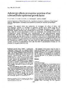
with 1029 m EGF (results not shown) (8), suggesting that PP2A might interfere in the signaling pathway that leads to the regulation of AR by elevated EGF doses. Surprisingly, an inverse effect of OA on AR gene induction by 10212 m EGF was observed, as OA was shown to abrogate the stimulation of the AR mRNA level induced by a low EGF concentration. A number of studies have previously described a similar inhibitory effect of OA on gene expression induced by a variety of effectors (33–35). It seems that OA could act through both transcriptional and posttranscriptional mechanisms. In particular, OA has been shown to be able to induce the expression and to stabilize via the inhibition of PP2A activity, some transcriptional factors, including c-Jun, c-Fos, JunB (36, 37), and Egr-1 (38, 39), whereas other factors, such as JunD, were not affected (40). However, the precise mechanisms by which OA acts on gene expression are unclear. Although OA at 2 nm has been shown to inhibit PP2A, additional effects on a number of intracellular events can occur, and the involvement and precise role of PP2A in the regulation of AR gene expression by EGF need to be confirmed. Nevertheless, the opposite effects of OA on the induction of AR by 10212 vs. 1029 m EGF reflect the existence of distinct regulatory mechanisms, differentially activated by high and low doses of EGF. Our preliminary observations, indicating that 10212 and 1029 m EGF differently affect the expression and activation of Jun family transcription factors, constitute additional evidence (not shown). Like a number of growth factors acting via tyrosine kinase transmembrane receptors, EGF is known to act through the activation of the Ras/Raf/MAP kinase pathway. In a study aimed at comparing the molecular mechanisms activated by growth stimulatory or inhibitory doses of EGF, we have previously described different patterns of p42 MAP kinase activation depending on the EGF concentration (Fig. 6) (7). Thus, it was shown that under conditions of growth stimulation, EGF induced a sustained and persistent activation of p42 MAP kinase, whereas an early, but transitory, peak of activation that rapidly fell under the basal level was observed with a growth inhibitory dose of EGF (1029 m). To assess the mechanisms through which various doses of EGF may differentially regulate MAP kinase activity in A431 cells, the potential activation of signaling components known to be involved in the MAP kinase cascade was then investigated. The MAP kinases are dually phosphorylated on threonine and tyrosine by MEK1 and MEK2 (41– 45), which are themselves activated by phosphorylation at serine/threonine by MEK kinase. Many MEK kinases have been identified; among them, c-Raf-1 has been described to play an important role in the signaling pathway activated by growth factors (46 – 49). To verify the involvement of c-Raf-1 in the signal transduction induced by EGF in A431 cells, both the activation and kinase activity of c-Raf-1 were evaluated during treatment with 10212 and 1029 m EGF. In agreement with a number of studies (50), 1029 m EGF induced an early stimulation of the c-Raf-1 kinase activity that was associated with modifications of the protein, as evaluated by electrophoretic mobility shift. On the contrary, the electrophoretic migration of c-Raf-1 was not affected by 10212 m EGF, indicating no or little modifications of the protein. Moreover, 10212 m EGF not only failed to activate c-Raf-1, but led to a moderate, but
DOSE EFFECTS OF EGF ON GENE EXPRESSION
FIG. 6. Effect of EGF on Raf-1 kinase activity. Cells grown as described in Fig. 1 were incubated in the presence of EGF for various periods of time. At the end of the treatment, cells were lysed, and c-Raf-1 was immunoprecipitated. The kinase activity was then measured as described in Materials and Methods by the ability of the immunoprecipitate to phosphorylate a specific c-Raf-1 kinase substrate. After separation on SDS-polyacrylamide gel and autoradiography, the bands illustrating the phosphorylation of the substrate were quantified by image analysis. A and B, Representative autoradiograms of substrate phosphorylation by c-Raf-1 during treatment with 10212 and 1029 M EGF, respectively. C, Graphic representation of three time-course experiments concerning the effects of 10212 M (M) and 1029 M EGF (E) on c-Raf-1 kinase activity. The values obtained were plotted as a percentage of those in unstimulated control cells. Each value represents the mean 6 SD of three separate experiments.
reproducible, decrease in its kinase activity. In a number of cases, activated c-Raf-1 has been reported to form Triton X-100-insoluble complexes with caveolae membranes and cytoskeletal elements (51–53). However, it was verified that under our experimental conditions, a large proportion of Raf could be extracted from EGF-treated and -untreated cells. Moreover, low and equivalent amounts of Triton X-100-insoluble Raf were detected in 10212 and 1029 m EGF-treated cells, indicating that differences in the cellular location and/or extractability of the protein could not account for the different patterns of c-Raf-1 activity. The processes that lead to differential regulation of c-Raf-1 kinase activity by EGF in A431 cells are as yet unclear. The activation by phosphory-
2389
lation of a tyrosine kinase activity intrinsic to the EGFR is the primary event induced by binding of the ligand. As described by Gulli et al. (30), we observed hyperphosphorylation of EGFR by 1029 m EGF, whereas lower growth factor concentrations led to a moderate phosphorylation of the receptor (data not shown). These data along with the demonstration by App et al. (24) that elevated doses of EGF stimulated kinase activity of Raf-1 by facilitating its association with EGFR lead us to suggest that the different patterns of c-Raf regulation by 1029 vs. 10212 m EGF could be conditioned by the extent of receptor activation. Thus, it is suggested that the inability of 10212 m EGF to activate c-Raf-1 kinase might be due to insufficient or inadequate EGFR activation. However, the decrease in Raf kinase activity observed during 10212 m EGF treatment indicates an alternative mechanism of regulation. A number of studies have previously demonstrated that the activation of cAMP-dependent protein kinase A may down-regulate c-raf-1 kinase activity in a variety of cell lines (54 –56). Thus, the involvement of protein kinase A in the signaling pathway activated by 10212 m, but not 1029 m, EGF might be envisaged. Activated Raf family members are able to catalyze the phosphorylation of MAP kinase kinases MEK1 and MEK2. In this study we could detect a sustained activation of MEK1 and to a lesser extent of MEK2 by 1029 m, but not by 10212, m EGF. Moreover, when a specific inhibitor of MEK1 was applied to A431 cells, the activation of p42 MAP kinase as well as the induction of AR gene expression by 1029 m EGF were in large part abrogated. The activating phosphorylation of MEKs require their association with activated Ras-Raf complexes. However, in a number of cases, differential activation of MEK1 and MEK2 by various Raf family members has been described (57). Thus, whereas the study by Catling et al. (58) indicated that both MEK1 and MEK2 were able to interact with Ras-bound Raf-1, Jelenik et al. (59) reported that in NIH-3T3 cells, immobilized Ras-Raf-1 complexes preferentially bound MEK1. These late data are in agreement with our observations showing that the activation of c-Raf-1 by 1029 m EGF in A431 cells is associated with a preferential activation of MEK1. Taken together, our results support the hypothesis that MEK1 is required for the activation of MAP kinase and the consequent induction of AR by growth inhibitory doses of EGF. Conversely, MEK1 and MEK2 do not seem to interfere in the signaling pathway that leads to the induction of the MAP kinase cascade by low EGF concentrations. Although low and undetectable MEK1 and/or MEK2 activation by 10212 m EGF cannot be excluded, all of our data argue in favor of a MAP kinase pathway that drives the mitogenic signal of EGF without the intervention of c-Raf-1, MEK1, or MEK2. In this case, the activation of MAP kinase by alternative signaling routes (60–63) could be envisaged. In conclusion, our data imply that in A431 cells, EGF, depending on its concentration, may act on the MAP kinase cascade through different mechanisms of regulation, allowing differential regulation of gene expression and cell proliferation (Fig. 7). Based on our data, the signaling pathways that are activated by high and low EGF doses are not necessary distinct. In A431 cells, high concentrations of EGF have been shown to induce various metabolic events, including calcium fluxes, phospholipase Cg activation, and cytoskeleton reorganization (64 – 67). Thus, additional events
2390
DOSE EFFECTS OF EGF ON GENE EXPRESSION
Endo • 1998 Vol 139 • No 5
which is described to play a central role in mediating the mitogenic action of growth factors, was demonstrated herein to be activated under conditions of growth inhibition. Defining the precise contribution of the signaling components in the differential regulation of gene expression and cell proliferation might help to control the growth of tumors that contain high levels of receptor. Acknowledgments The authors thank Jeanne Carbone for excellent technical assistance. We also gratefully acknowledge Dr. Plowman for providing amphiregulin cDNA probe.
References
FIG. 7. Proposal mechanism of signal transduction induced by low and high EGF concentrations in A431 cells. Low and high EGF doses activate p42 MAP kinase (MAPK) through different mechanisms of regulation. In particular, MAPK activation induced by mitogenic doses of EGF does not require c-Raf-1, MEK1, and MEK2 activation. Under these conditions the intermediate signaling components between EGFR and MAPK are presently unknown. The sustained and persistent activation of MAPK by low concentrations of EGF is accompanied by its translocation into the nucleus (our data not shown), where MAPK may thus activate transcriptional factors. On the contrary, in the presence of high doses of EGF, MAPK remains in the cytosolic compartment (our data not shown). In this case, the intervention of an unidentified component(s) (X) that drives the signal from MAPK to gene transcription machinery is proposed. Finally, both MAPK pathways result in differential regulation of the expression of genes involved in cell proliferation or differentiation. Question marks represent unknown steps or possible activation processes.
activated by high doses of EGF might act by interfering with the signaling cascade induced by low EGF concentrations. Alternatively, turnover and degradation of EGFR may interfere in the signal transduction induced by EGF. Indeed, when administered to A431 cells, low doses of EGF are less efficient than high concentrations in down-regulating EGFR (68). Therefore, in the presence of low EGF doses, a significant level of functional receptor is available for a longer period of time and, thus, might drive the EGF signal in a more sustained manner. Nevertheless, a better knowledge of the mechanisms involved in the ambivalent activity of EGF should help in understanding how a given growth factor can alternatively produce differentiation or tumorigenesis. Thus, the overexpression of EGFR is described in a variety of human malignancies, including cancers of the lung, brain, and breast (69), and like A431 cells, a number of EGFR-overexpressing tumor cells display an in vitro ambivalent growth response to EGF. High levels of EGFR in cancers might give a growth advantage or disadvantage to the cells depending on the level of disposable EGF. Thus, under certain circumstances, blocking the signaling pathway activated by EGF could interfere with the growth inhibitory mechanisms. In this context, our data bring into question the role of Raf in the growth regulation of EGFR-overexpressing cells, as c-Raf-1,
1. Hernandez-Sotomayor S, Carpenter G 1992 Epidermal growth factor receptor: elements of intracellular communication. J Membr Biol 128:81– 89 2. Ullrich A, Schlessinger J 1990 Signal transduction by receptors with tyrosine kinase activity. Cell 61:203–212 3. Ruff-Jamison S, Chen K, Cohen S 1993 Induction by EGF and interferon-g of tyrosine phosphorylated DNA binding proteins in mouse liver nuclei. Science 261:1733–1736 4. Ruff-Jamison S, Chen K, Cohen S 1995 Epidermal growth factor induces the tyrosine phosphorylation and nuclear translocation of Stat 5 in mouse liver. Proc Natl Acad Sci USA 92:4215– 4218 5. Kawamoto T, Sato JT 1983 Growth stimulation of A431 cells by epidermal growth factor: identification of a high-affinity receptor for epidermal growth factor by an anti-receptor monoclonal antibody. Proc Natl Acad Sci USA 80:1337–1341 6. Dong XF, Berthois Y, Martin PM 1991 Effect of epidermal growth factor on the proliferation of human epithelial cancer cell lines: correlation with the level of occupied EGF receptor. Anticancer Res 11:737–744 7. Chajry N, Martin PM, Pages G, Cochet C, Afdel K, Berthois Y 1994 Relationship between the MAP kinase activity and the dual effect of EGF on A431 cell proliferation. Biochem Biophys Res Commun 203:984 –990 8. Chajry N, Martin PM, Cochet C, Berthois Y 1996 Regulation of p42 mitogenactivated-protein kinase activity by protein phosphatase 2A under conditions of growth inhibition by epidermal growth factor in A431 cells. Eur J Biochem 235:97–102 9. Shoyab M, McDonald VL, Bradley JG, Todaro JG 1988 Amphiregulin: a bifunctional growth-modulating glycoprotein produced by the phorbol 12myristate 13-acetate-treated human breast adenocarcinoma cell line MCF-7. Proc Natl Acad Sci USA 85:6528 – 6532 10. Johnson GR, Seaki T, Auersperg N, Gordon AW, Shoyab M, Salomon DS, Stromberg K 1991 Response to and expression of amphiregulin by ovarian carcinoma and normal ovarian surface epithelial cells: nuclear localization of endogenous amphiregulin. Biochem Biophys Res Commun 180:481– 488 11. Johnson GR, Seaki T, Gordon AW, Shoyab M, Salomon DS, Stromberg K 1992 Autocrine action of amphiregulin in a colon carcinoma cell line and immunocytochemical localization of amphiregulin in human colon. J Cell Biol 118:741–751 12. Cook PW, Mattox PA, Keeble WW, Pittelkow MR, Plowman GD, Shoyab M, Adelman JP, Shipley GD 1991 A heparin sulfate-regulated human keratinocyte autocrine growth factor is similar or identical to amphiregulin. Mol Cell Biol 11:2547–2557 13. Shoyab M, Plowman GD, McDonald VL, Bradley JG, Todaro JG 1989 Structure and function of human amphiregulin: a member of the epidermal growth factor family. Science 243:1074 –1076 14. Li S, Plowman GD, Buckley SD, Shipley GD 1992 Heparin inhibition of autonomaous growth implicates amphiregulin as an autocrine growth factor for normal human mammary epithelial cells. J Cell Physiol 153:103–111 15. Normanno N, Qi CK, Gullick WJ, Persico G, Yarden Y, Wen D, PLowman GD, Kenney N, Johnson GR, Kim N, Brandt R, Martinez-Lacaci I, Dickson RB, Salomon DS 1993 Expression of amphiregulin, cripto-1, and heregulin in human breast cancer cells. Int J Oncol 2:903–911 16. Ciardiello F, Kim N, Saeki T, Dono R, Persico MG, Plowman GD, Garrigues J, Radke S, Todaro GJ, Salomon DS 1991 Differential expression of epidermal growth factor-related proteins in human colorectal tumors. Proc Natl Acad Sci USA 88:7792–7796 17. Kitadai Y, Yasui W, Yokozaki H, Kuniyasu H, Ayhan A, Haruma K, Kajiyama G, Johnson GR, Tahara E 1993 Expression of amphiregulin, a novel gene of the epidermal growth factor family, in human gastric carcinomas. Jpn J Cancer Res 84:879 – 884 18. LeJeune S, Leek R, Horak E, Plowman G, Grenall M, Harris AL 1993 Amphiregulin, epidermal growth factor receptor, and estrogen receptor expression in human primary breast cancer. Cancer Res 53:3597–3602 19. Panico L, D’Antonio A, Salvatore G, Mezza E, Tortora G, De Laurentiis M,
DOSE EFFECTS OF EGF ON GENE EXPRESSION
20. 21.
22. 23. 24. 25. 26. 27.
28. 29. 30. 31.
32.
33. 34. 35. 36.
37. 38. 39. 40. 41. 42. 43.
44. 45.
De Placido S, Giordano T, Merino M, Salomon DS, Gullick WJ, Pettinato G, Schnitt SJ, Bianco AR, Ciardiello F 1996 Differential immunohistochemical detection of transforming growth factor a, amphiregulin and cripto in human normal and malignant breast tissues. Int J Cancer 65:51–56 Kimura H 1993 Schwannoma-derived growth factor must be transported into the nucleus to exert its mitogenic activity. Proc Natl Acad Sci USA 90:2165–2169 Martinez-Lacaci I, Saceda M, Plowman GD, Johnson GR, Normanno N, Salomon DS, Dickson RB 1995 Estrogen and phorbol esters regulate amphiregulin expression by two separate mechanisms in human breast cancer cell lines. Endocrinology 136:3983–3992 Southern E 1975 Detection of specific sequences among DNA fragments separated by gel electrophoresis. J Mol Biol 98:503–510 Feinberg AP, Vogelstein BA 1983 Technique for radiolabelling DNA restriction endonuclease fragments to high specific activity. Anal Biochem 132:6 –13 App H, Hazan R, Zilberstein A, Ullrich A, Schlessinger J, Rapp U 1991 Epidermal growth factor (EGF) stimulates association and kinase activity of Raf-1 with the EGF receptor. Mol Cell Biol 11:913–919 Howe LR, Leevers SJ, Gomez N, Nakielny S, Cohen P, Marshall CJ 1992 Activation of the MAP kinase pathway by the protein kinase Raf. Cell 71:335–342 Kyriakis TM, Force TL, Rapp UR, Bonventre JV, Avruch J 1993 Mitogen regulation of c-Raf-1 protein kinase activity toward mitogen-activated protein kinase-kinase. J Biol Chem 268:16009 –16019 Morrison DK, Kaplan DR, Rapp U, Roberts TM 1988 Signal transduction from membrane to cytoplasm: growth factors and membrane-bound oncogenes products increase Raf-1 phosphorylation and associated protein kinase activity. Proc Natl Acad Sci USA 85:8855– 8859 Morrison DK, Kaplan DR, Escobedo JA, Rapp U, Roberts TM, Williams UT 1989 Direct activation of the serine/threonine kinase activity of Raf-1 through tyrosine phosphorylation by the PDGF B-receptor. Cell 58:649 – 657 Bravo R, Burckhardt J, Curran T, Muller R 1985 Stimulation and inhibition of growth by EGF in different A431 cell clones is accompanied by the rapid induction of c-fos and c-myc proto oncogenes. EMBO J 4:1193–1197 Gulli LF, Palmer KC, Chen YQ, Reddy KB 1996 Epidermal growth factorinduced apoptosis in A431 cells can be reversed by reducing the tyrosine kinase activity. Cell Growth Diff 7:173–178 Ueki H, Okuhama R, Sera M, Inoue T, Tominaga M, Morita T 1992 Stimulatory effect of vanadate on 39,59-cyclic guanosine monophosphate-inhibited low Michaelis-Menten constant 39,59-cyclic adenosine monophosphate phosphodiesterase activity in isolated rat fat pads. Endocrinology 131:441– 446 Ueki H, Mitsugi S, Kawashima Y, Motoyashiki T, Morita T 1997 Orthovanadate stimulates cyclic guanosine monophosphate-inhibited cyclic adenosine monophosphate phosphodiesterase activity in isolated rat fat pads through activation of particulate myelin basic protein kinase by protein tyrosine kinase. Endocrinology 138:2784 –2789 Westermarck J, Ilvonen E, Uitto J, Kahari VM 1995 Suppression of elastin gene expression in dermal fibroblasts by protein phosphatase inhibitor okadaic acid. Biochem Biophys Res Commun 209:175–181 Wang Q, Raghow R 1996 Okadaic acid-induced transcriptional downregulation of type I collagen gene expression is mediated by protein phosphatase 2A. Mol Cell Biochem 158:33– 42 O’Brien RM, Noisin EL, Granner DK 1994 Comparison of the effects of insulin and okadaic acid on phosphoenolpyruvate carboxykinase gene expression. Biochem J 303:737–742 Lee JS, Fabre B, Hemmings BA, Kiefer B, Nagamine Y 1994 Okadaic aciddependent induction of the urokinase-type plasminogen activator gene associated with stabilization and autoregulation of c-Jun. J Biol Chem 269:2887–2894 Kharbanda S, Datta R, Rubin E, Nakamura T, Hass R, Kufe D 1992 Regulation of c-jun expression during induction of monocytic differentiation by okadaic acid. Cell Growth Differ 3:391–399 Guy GR, Cao X, Chua SP, Tan YH 1992 Okadaic acid mimics multiple changes in early protein phosphorylation and gene expression induced by tumor necrosis factor or interleukin-1. J Biol Chem 267:1846 –1852 Hyun SW, Park K, Lee YS, Lee YI, Kim SJ 1994 Inhibition of protein phosphatases activates P4 promoter of the human insulin-like growth factor II gene through the specific promoter element. J Biol Chem 269:364 –368 Schonthal A, Alberts AS, Frost JA, Feramisco JR 1991 Differential regulation of jun family gene expression by the tumor promoter okadaic acid New Biol 3:977–986 Ahn NG, Seger R, Krebs EG 1992 The mitogen-activated protein kinase activator Curr Opin Cell Biol 4:992–999 Crews CM, Alessandrini A, Erikson RL 1992 The primary structure of MEK, a protein kinase that phosphorylates the ERK gene product. Science 258:478 – 480 Seger R, Ahn NGJ, Posada ES, Munar AM, Jensen JA, Cooper MH, Cobb MH, Krebs EG 1992 Purification and characterization of mitogen-activated protein kinase activator(s) from epidermal growth factor-stimulated A431 cells. J Biol Chem 267:14373–14381 Wu J, Harrison JK, Dent P, Lynch KR, Weber MJ, Sturgill TW 1993 Identification and characterization of a new mammalian mitogen-activated protein kinase kinase, MKK2. Mol Cell Biol 13:4539 – 4548 Zheng CF, Guan KL 1993 Cloning and characterization of two distinct human
46. 47. 48. 49. 50. 51.
52. 53. 54.
55. 56. 57. 58. 59. 60. 61. 62. 63.
64.
65.
66. 67.
68. 69. 70.
2391
extracellular signal-regulated kinase activator kinases, MEK1 and MEK2. J Biol Chem 268:11435–11439 Dent P, Hase W, Haystead TA, Vincent LA, Roberts TM, Sturgill TW 1992 Activation of mitogen-activated protein kinase kinase by v-RAF in NIH 3T3 cells and in vitro. Science 237:1404 –1407 Howe LR, Leevers SJ, Gomez N, Nakielny S, Cohen P, Marshall CJ 1992 Activation of the MAP kinase pathways by the protein kinase RAF. Cell 71:335–342 Kyriakis JM, App H, Zhang XF, Banerjee P, Brautigan DL, Rapp UR, Avruch J 1992 RAF-1 activates MAP kinase-kinase. Nature 358:417– 421 Macdonald SG, Crews CM, Wu L, Driller J, Clark R, Erikson RL, McCormick F 1993 Reconstitution of the RAF-1-MEK-ERK signal transduction pathway in vitro. Mol Cell Biol 13:6615– 6620 Chao TS, Foster DA, Rapp UR, Rosner MR 1994 Differential Raf requirement for activation of mitogen-activated protein kinase by growth factors, phorbol esters, and calcium. J Biol Chem 269:7337–7341 Gronowski AM, Bertics PJ 1993 Evidence for the potentiation of epidermal growth factor receptor tyrosine kinase activity by association with the detergent-insoluble cellular cytoskeleton: analysis of intact and carboxy-terminally truncated receptors. Endocrinology 133:2838 –2846 Gronowski AM, Bertics PJ 1995 Modulation of epidermal growth factor receptor interaction with the detergent-insoluble cytoskeleton and its effects on receptor tyrosine kinase activity. Endocrinology 136:2198 –2205 Mineo C, James GL, Smart EJ, Anderson RGW 1996 Localization of epidermal growth factor-stimulated Ras/Raf-1 interaction to caveolae membrane. J Biol Chem 271:11930 –11935 Graves LM, Bornfeldt KE, Raines EW, Potts BC, MacDonald SG, Ross R, Krebs EG 1993 Protein kinase A antagonizes platelet-derived growth factorinduced signaling by mitogen-activated protein kinase in human arterial smooth muscle cells. Proc Natl Acad Sci USA 90:10300 –10304 Cook SJ, McCormick F 1993 Inhibition by cAMP of Ras-dependent activation of Raf. Science 262:1069 –1072 Schramm K, Niehof M, Radziwill G, Romme C, Moelling K 1994 Phosphorylation of c-RAF-1 by protein kinase A interferes with activation. Biochem Biophys Res Commun 201:740 –747 Wu X, Noh SJ, Zhou G, Dixon JE, Guan KL 1996 Selective activation of MEK1 but not MEK2 by A-Raf from epidermal growth factor-stimulated Hela cells. J Biol Chem 271:3265–3271 Catling AD, Schaeffer HJ, Reuter CWM, Moodie SA, Wolfman A, Weber MJ 1995 A proline-rich sequence unique to MEK1 and MEK2 is required for Raf binding and regulates MEK function. Mol Cell Biol 15:5214 –5225 Jelenik T, Catling AD, Reuter CWM, Moodie SA, Wolfman A, Weber MJ 1994 RAS and RAF-1 form a signalling complex with MEK-1 but not MEK-2. Mol Cell Biol 14:8212– 8218 Huleihel M, Goldsborough M, Cleveland J, Gunnell M, Bonner T, Rapp UR 1986 Characterization of murine A-raf, a new oncogene related to the v-raf oncogene. Mol Cell Biol 6:2655–2662 Stephens RM, Sithanadam G, Copeland TD, Kaplan DR, Rapp UR, Morrison DK 1992 95-Kilodalton B-raf serine/threonine kinase: identification of the protein and its major auto-phosphorylation site. Mol Cell Biol 12:3733–3742 Lange-Carter CA, Pleiman CM, Gardner AM, Blumer KJ, Johnson GL 1993 A divergence in the MAP kinase regulatory network defined by MEK kinase and Raf. Science 260:315–319 Yamamori B, Kuroda S, Shimizu K, Fukui K, Ohtsuka T, Takai Y 1995 Purification of a Ras-dependent mitogen-activated protein kinase kinase kinase from bovine brain cytosol and its identification as a complex of B-Raf and 14 –3-3 proteins. J Biol Chem 270:11723–11726 Hepler JR, Nakahata N, Lvenberg TW, DiGuiseppi J, Herman B, Earp HS, Harden TK 1987 Epidermal growth factor stimulates the rapid accumulation of inositol (1,4,5)-triphosphate and a rise in cytosolic calcium mobilized from intracellular stores in A431 cells. J Biol Chem 262:2951–2956 Payrastre B, van Bergen en Henegouwen PMP, Breton M, den Hartigh JC, Plantavid M, Verkleij AJ, Boonstra J 1991 Phosphoinositide kinase, diacylglycerol kinase, and phospholipase C activities associated to the cytoskeleton: effect of epidermal growth factor. J Cell Biol 115:121–128 Elliget KA, Phelps PC, Smith MW 1996 Transforming growth factor b modulation of the epidermal growth factor Ca21 signal and c-Fos oncoprotein levels in A431 human epidermoid carcinoma cells. Cell Growth Differ 7:461–468 Tinhofer I, Maly K, Dietl P, Hochholdinger F, Mayr S, Obermeier A, Grunicke HH 1996 Differential Ca21 signaling induced by activation of the epidermal growth factor and nerve growth factor receptors. J Biol Chem 271:30505–30509 DePalo L, Das M 1988 Epidermal growth factor-induced stimulation of epidermal growth factor-receptor synthesis in human cytotrophoblasts and A431 carcinoma cells. Cancer Res 48:1105–1109 Gullick WJ 1991 Prevalence of aberrant expression of the epidermal growth factor receptor in human cancers. Br Med Bull 47:87–98 Blagosklonny MV, Schultle T, Nguyen P, Trepel J, Neckers L 1996 Taxolinduced apoptosis and phosphorylation of Bcl-2 protein involves c-Raf-1 and represents a novel c-Raf-1 signal transduction pathway. Cancer Res 56:1851– 1854