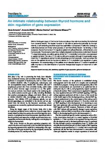
MOLECULAR AND CELLULAR BIOLOGY, June 2009, p. 3435–3450 0270-7306/09/$08.00⫹0 doi:10.1128/MCB.01805-08 Copyright © 2009, American Society for Microbiology. All Rights Reserved.
Vol. 29, No. 12
Differential Regulation of Human Interferon A Gene Expression by Interferon Regulatory Factors 3 and 7䌤§ Pierre Ge´nin,1 Rongtuan Lin,2 John Hiscott,2 and Ahmet Civas1* UPR 2228-CNRS, Laboratoire de Re´gulation Transcriptionnelle et Maladies Ge´ne´tiques, UFR Biome´dicale des Saints-Pe`res, Universite´ Paris Descartes, 45 rue des Saints-Pe`res, 75270 Paris Cedex 06, France,1 and Lady Davis Institute for Medical Research and Departments of Microbiology & Immunology and Medicine, McGill University, Montre´al, Canada H3T 1E22 Received 25 November 2008/Returned for modification 9 January 2009/Accepted 28 March 2009
Differential expression of the human interferon A (IFN-A) gene cluster is modulated following paramyxovirus infection by the relative amounts of active interferon regulatory factor 3 (IRF-3) and IRF-7. IRF-3 expression activates predominantly IFN-A1 and IFN-B, while IRF-7 expression induces multiple IFN-A genes. IFN-A1 gene expression is dependent on three promoter proximal IRF elements (B, C, and D modules, located at positions ⴚ98 to ⴚ45 relative to the mRNA start site). IRF-3 binds the C module of IFN-A1, while other IFN-A gene promoters are responsive to the binding of IRF-7 to the B and D modules. Maximal expression of IFN-A1 is observed with complete occupancy of the three modules in the presence of IRF-7. Nucleotide substitutions in the C modules of other IFN-A genes disrupt IRF-3-mediated transcription, whereas a G/A substitution in the D modules enhances IRF7-mediated expression. IRF-3 exerts dual effects on IFN-A gene expression, as follows: a synergistic effect with IRF-7 on IFN-A1 expression and an inhibitory effect on other IFN-A gene promoters. Chromatin immunoprecipitation experiments reveal that transient binding of both IRF-3 and IRF-7, accompanied by CBP/p300 recruitment to the endogenous IFN-A gene promoters, is associated with transcriptional activation, whereas a biphasic recruitment of IRF-3 and CBP/p300 represses IFN-A gene expression. This regulatory mechanism contributes to differential expression of IFN-A genes and may be critical for alpha interferon production in different cell types by RIG-I-dependent signals, leading to innate antiviral immune responses. infected by influenza virus (10). Thus, differential expression of human IFN-A genes varies depending on virus and cell type, in agreement with previous in vivo studies of mice (1, 4, 11, 12, 24, 31, 37). IFN-A/B gene transcription is triggered via distinct signaling pathways that converge on the activation of interferon regulatory factor 3 (IRF-3) and IRF-7 (26), although other IRFs are also involved in cell-specific regulation of the late phases of IFN-A gene transcription (76). In pDCs, Tolllike receptor (TLR)-mediated signaling by TLR7/8/9-dependent pathways is critical for rapid and high-level expression of IFN-A/B genes mediated by IRF-7 (26–28, 30, 34, 36). TLR3 that is predominantly expressed in immature mDCs and TLR-4 essentially operating in mDCs and monocyte/macrophage cells require TANK-binding kinase 1 (TBK1), IRF-3, and IRF-7 (25). In conventional dendritic cells and other cell types such as fibroblasts or epithelial cells, RNA virus infection stimulates IFN-␣ expression upon ligand stimulation of the mitochondrion-associated RNA helicases retinoic acid-inducible gene I (RIG-I) and melanoma differentiation-associated gene 5 (MDA-5), which leads to the activation of TBK1 and IB-related kinase ε (IKKε) (29, 34, 44, 55, 66, 86). DNA virus-dependent signaling through DNA-dependent activator of IRFs requires TBK1 and IRF-3 to induce type I IFN gene expression (78). Phosphorylation of specific C-terminal serine/threonine residues of IRF-3 and IRF-7 by TBK1 and/or IKKε is essential for inducing IFN-A/B gene expression (14, 75). Thus, IRF-3 and IRF-7 display similarities regarding their virus-induced phosphorylation, dimerization, and nuclear transport (23, 42, 43, 51). These factors differ in their expression levels and their
The immediate cellular response to virus infection is characterized by the transcriptional activation of type I interferon (IFN) genes, which are involved in the host antiviral defense program (35, 79). Multiple alpha interferon (IFN-␣) subtypes and the single IFN- subtype exhibit antiviral and immunomodulatory activities, which culminate in the maturation of antigen-presenting cells and T-cell activation (79). IFN-␣ subtypes, although signaling through the same cell surface receptor, display distinct biological effects (16, 33, 82, 84), suggesting that qualitative and quantitative differences in IFN-␣ production during viral infection may affect the formation, magnitude, and duration of innate and adaptive antiviral immune responses. The interferon A (IFN-A) multigenic family consists of 13 functional members located in the ifn locus on human chromosome 9, together with the single IFN-B gene (7, 13). Analysis of human IFN-A gene expression patterns in Sendai virusinfected plasmacytoid dendritic cells (pDCs), monocytes, and monocyte-derived dendritic cells (mDCs) has demonstrated that each cell population expresses predominantly IFN-A1, with multiple species of IFN-A expressed in pDCs (32), whereas IFN-A genes are expressed at similar levels in pDCs * Corresponding author. Mailing address: UPR 2228-CNRS, Laboratoire de Re´gulation Transcriptionnelle et Maladies Ge´ne´tiques, UFR Biome´dicale des Saints-Pe`res, Universite´ Paris Descartes, 45 rue des Saints-Pe`res, 75270 Paris Cedex 06, France. Phone: 33-1-42-86-2284. Fax: 33-1-42-86-20-42. E-mail:
[email protected]. § Supplemental material for this article may be found at http://mcb .asm.org/. 䌤 Published ahead of print on 6 April 2009. 3435
3436
´ NIN ET AL. GE
interaction with DNA and transcriptional coactivators CBP and p300 (62, 63, 69, 77). IRF-3 is constitutively expressed in all cell types, whereas the expression of IRF-7 is constitutive in lymphoid cells and certain dendritic cell subsets but is also induced by IFN-␣/ produced during viral infection (73). Most of the data on the individual roles of IRF-3 and IRF-7 in differential IFN-A gene regulation derive from in vitro studies of mice. They indicate that maximal virus-induced transcription of the mouse IFN-A4 gene requires the presence of three IRF elements located in the virus-responsive element A4 (VRE-A4), delimited to a region (positions ⫺120 to ⫺40) of the IFN-A4 gene promoter (2, 5). IRF-3 and IRF-7 display differential specificity and affinity for these IRF elements (B, C, and D modules) and determine the expression levels of the IFN-A4 gene following virus infection (9). Among these three modules, the C module specifically binds IRF-3 but is a weak site for IRF-7 interaction; the B and D modules are primarily required for IRF-7 binding and transactivation. The mouse IFN-A11 gene is poorly induced by virus infection because of nucleotide substitutions that disrupt both the C and D modules in the promoter, thus preventing IRF-3 and IRF-7 binding and activation (8, 56, 57). It was also suggested that IRF-3 ensures the initial activation of IFN-A4 and IFN-B genes in mouse embryonic fibroblasts and that in a second step, it participates in the amplification of the IFN-A4 and IFN-B genes and transactivation of other IFN-A genes, in cooperation with IRF-7 (50). All together, these data suggest that subtle differences in the cognate binding sites present in target promoters determine the magnitude of the IFN-A gene response to IRF-3 or IRF-7. In this study, we analyzed the relative contribution of IRF-3 and IRF-7 to virus-induced expression of human IFN-A subtypes. Real-time reverse transcription-quantitative PCR (qPCR) data, together with chromatin immunoprecipitation (ChIP) assays and promoter activation analyses, demonstrate that differential recruitment of IRF-3 and/or IRF-7 to the IFN-A promoter modules dictates the relative levels of IFN-A gene activation. MATERIALS AND METHODS Cell culture and virus infection. Human Namalwa B cells (B lymphocytes from Burkitt’s lymphoma; ATCC CRL-1432) were grown in RPMI 1640 medium (Invitrogen, Carlsbad, CA) supplemented with 10% fetal bovine serum (FBS), glutamine, and antibiotics. Human embryonic kidney 293T (HEK293T) cells (ATCC CRL-11268) were grown in minimum essential medium (MEM) (Invitrogen) supplemented with 8% FBS, glutamine, and antibiotics. 2fTGH (human fibrosarcoma cells) (52, 64), 2fTGH-F7 (obtained by stably expressing the pcDNA3-IRF-7 plasmid), and HEK293 cells constitutively expressing TLR3 (InvivoGen, San Diego, CA) were grown in Dulbecco’s modified Eagle medium (Invitrogen) supplemented with 10% FBS, glutamine, and antibiotics. Infection of cells with Sendai virus (400 to 800 hemagglutinating units/ml) was performed at a multiplicity of infection of 1 to 5 in serum-free media. After 1 h, cells were washed, placed in growth media containing 2% FBS, and then harvested at different times (from 1 to 20 h) following infection. Plasmids. Promoter sequences for IFN-A1, IFN-A2 (accession number AL353732), IFN-A4, IFN-A7 (accession number AL512606), and IFN-A14 (accession number AL162420) were obtained from the GenBank database. IFN-A– pGL3 reporters (IFN-A1, positions ⫺140 to ⫹19; IFN-A2, positions ⫺135 to ⫹59; IFN-A4, positions ⫺150 to ⫹50; IFN-A7, positions ⫺119 to ⫹2; IFN-A14, positions ⫺202 to ⫹58) were generated by cloning the PCR products from 293 cell genomic DNA into the SmaI site of the pGL3-basic vector (40). Constructs A1-C7 (carrying the ⫺82A/G and ⫺79G/A substitutions), A1-C14 (carrying the ⫺81A/G and ⫺79G/A substitutions), and A1-C7D7 (carrying the ⫺82A/G, ⫺79G/A, ⫺52G/A, and ⫺44C/T substitutions) were obtained by two-step PCR
MOL. CELL. BIOL. mutagenesis of the human IFN-A1 gene promoter. Similar two-step PCR mutagenesis of the human IFN-A7 gene promoter was used to obtain the A7-C1 (carrying the ⫺82G/A and ⫺79A/G substitutions) and A7-DTA (carrying the ⫺52A/T and ⫺51T/A substitutions) constructs. The sequence of each construct was confirmed by DNA sequence analysis. Plasmids expressing human IRF-3, IRF-3(5D), IRF-7A, IRF-7(2D), TBK1, and IKKε have been previously described (40, 75, 87). The pUNO-hTRAF3 plasmid encoding human TRAF3 was purchased from InvivoGen. Transfections and reporter assays. HEK293T and HEK293-TLR3 cells were seeded at 7 ⫻ 104 cells in 12-well plates in MEM supplemented with 5% fetal calf serum. After 18 to 24 h, 50 or 500 ng of reporter plasmid, 50 ng of reference plasmid pIEF-LacZ, and various amounts of IRF- or kinase-expressing plasmids were transfected by calcium phosphate (19) or Lipofectamine 2000, according to the manufacturer’s protocol (Invitrogen). Twenty-four hours after transfection, cells were harvested to monitor the enzymatic activities. Luciferase values, normalized according to -galactosidase levels, were obtained from at least three independent transfection experiments, each performed with two or three different clones. Inducibility (induction level) corresponds to the ratio between relative transcription (RT) values obtained with IRF-3(5D), IRF-7A, or IRF-7(2D) and RT values obtained by transfection of pcDNA3, used as a control. siRNA experiments and immunoblot analyses. HEK293T cells were seeded using 2 ⫻ 105 cells in 6-well plates in MEM supplemented with 5% fetal calf serum. After 18 to 24 h, 75 ng of pcDNA3-Flag-IKKε or pcDNA3-Flag-TBK1 was transfected by Lipofectamine, together with 80 pmol of a small interfering RNA (siRNA) mixture that contains a pool of three target-specific siRNAs for IRF-3 (sc-35710; Santa Cruz Biotechnology, CA), IRF-7 (sc-38011), or a scrambled siRNA (sc-37007) used as a control. Additional experiments with a construct coding for IRF-3 short hairpin RNA or IRF-7 short hairpin RNA (shTRCN 5919 and sh-TRCN 14859, respectively; Sigma-Aldrich, MO) provided inhibitory results similar to those of a control plasmid (shc002). Cells were harvested 72 h after transfection and analyzed for IFN-A gene expression by reverse transcription-qPCR. Specificity and efficiency of siRNA on endogenous protein expression were tested by Western blotting (21) of whole-cell extracts (20 g of total proteins separated on 10% sodium dodecyl sulfate-polyacrylamide gel electrophoresis) using IRF-3 or IRF-7 polyclonal antibodies (sc-9082 and sc9083, respectively; Santa Cruz Biotechnology, CA) and antiactin antibodies (MAB1501; Millipore Corporation, MA). IKKε and TBK1 proteins were detected using monoclonal anti-FLAG antibodies (M2; Sigma-Aldrich). ChIP assays. Both 2fTGH and 2fTGH-F7 cells (5 ⫻ 106 cells per 100-mm plate) were infected by Sendai virus and analyzed by ChIP assays, according to the work of Wathelet et al. (83), before and at 1, 2, and 4 h following infection. Similar assays were performed using HEK293T cells (2 ⫻ 106 cells per 100-mm plate) transfected with 750 ng of pcDNA3-Flag-IKKε in the presence of 1,500 ng of pcDNA3-IRF-3wt or pcDNA3-IRF-7A. Briefly, protein-DNA cross-linking was performed by incubation with 1% formaldehyde for 30 min at 37°C. The reaction was stopped by the addition of 0.125 M glycine. Cell pellets were washed twice in phosphate-buffered saline, nuclei were lysed, and chromatin was sheared by sonication, 10 times for 30 s. Protein-bound cross-linked chromatin was purified by isopycnic centrifugation in a SW55Ti rotor at 40,000 rpm for 40 h. Purified chromatin (5 g) was incubated overnight at 4°C, with 2 g of the following polyclonal antibodies from Santa Cruz: anti-IRF-3 (sc-9082), antiIRF-7 (sc-9083), anti-CBP (sc-583), or anti-p300 (sc-584). Immunocomplexes isolated on protein A-Sepharose were treated with RNase A (50 g/ml), 0.5% sodium dodecyl sulfate, and proteinase K (500 g/ml) before reversion of the cross-link by heating at 65°C overnight. DNA was recovered after phenol-chloroform extraction, and ethanol precipitation was used as template for qPCR, with primer sets designed to amplify the fragment (positions ⫺178 to ⫹59) of IFN-A1, the fragment (positions ⫺184 to ⫹24) of IFN-A2, the region (positions ⫺177 to ⫹39) of IFN-A7, the region (positions ⫺184 to ⫹49) of IFN-A14, or the region (positions ⫺150 to ⫹5) of IFN-B (primer sequences available upon request). Each promoter fragment includes the binding sites for IRF-3 and IRF-7. Relative binding or recruitment of a factor on the promoter was determined by computing Ae关Ctinput ⫺ Cttarget兴; Ae is the amplification efficiency of a primer set, Ctinput is the amplification signal obtained with input DNA (4%), and Cttarget is the signal obtained with chromatin precipitated by specific antibodies. Real-time reverse transcription-qPCR analysis. DNase I-treated total RNA was prepared using the NucleoSpin RNA L isolation kit (Macherey-Nagel, Germany). Reverse transcription reactions were primed with oligo(dT)12–18 and performed with 2 g of total RNA using the SuperScript reverse transcriptase (Invitrogen, San Diego, CA). Primer sets for IFN-A subtypes did not generate PCR products in the absence of reverse transcription. qPCR assays were performed at least in triplicate using the ABsolute qPCR SYBR Green ROX mix
VOL. 29, 2009
REGULATION OF HUMAN IFN-A GENES BY IRF-3 AND IRF-7
3437
FIG. 1. Human IFN-A gene expression pattern induced by Sendai virus infection in natural IFN-␣-producing cells. (A) Namalwa B cells were left untreated (mock-induced conditions) or infected by Sendai virus and harvested at different times (2, 4, 8, 12, 16, and 20 h) following infection. A total of 2 g of total RNA extracted from these cells was submitted to reverse transcription and real-time qPCR analysis. The mRNA levels of IFN-B, IRF-3, IRF-7, and different IFN-A genes, indicated as copy numbers calculated for 1,000 copies of GAPDH mRNA, are plotted on a logarithmic scale, together with standard deviation values. Black bars indicate the expression levels detected under mock-induced conditions. The different IFN-A gene subtypes and the IFN-B gene are presented according to their position in the ifn locus on human chromosome 9 (7). High percentages of sequence homology for the IFN-A1 and IFN-A13 genes (99.7% in the coding region) did not allow distinction between these genes. IFN-A1 expression levels presented in this study represent the total amounts of both IFN-A1 and IFN-A13 mRNAs. (B) Human IFN-A genes are grouped according to their expression pattern in virus-infected Namalwa cells. The first group includes IFN-A1, -A2, and -A5, which exhibited two peaks observed at 2 and 8 h postinfection; a second group (IFN-A10, -A8, -A17, -A16, and -A14) displayed maximal expression at 8 h, whereas a third group (IFN-A7, -A21, and -A4) peaked later at 12 h. The IFN-A6 gene is not expressed in these cells. (C) Modulation of IRF-3 and IRF-7 mRNA levels in virus-infected Namalwa cells. An increase in the IRF-7/IRF-3 ratio between 2 and 8 h (shown in gray) correlated with the expression of IFN-A genes. (D) Whole-cell extracts (40 g) analyzed by Western blotting by using IRF-3, IRF-7 and antiactin antibodies. ␣, anti; SeV, Sendai virus; MI, mock induced.
(500 nM) (ABgene, Epsom, United Kingdom) in a 7900HT fast real-time PCR system apparatus (Applied Biosystems, Foster City, CA). Since IFN-A and IFN-B genes are devoid of intervening sequences, PCR products used to establish the standard curves have been quantified, with reference to human genomic DNA, allowing comparison between the expression levels of each IFN-A subtype. Standard curves were obtained using 4-log-range dilutions of human genomic DNA or a plasmid that contains the cDNA encoding the gene of
interest. All primer pairs generated a single product, checked in each assay by analysis of the melting curves. The IFN-A primer pairs used were previously described (70), except for IFN-A4 TRAF3 and glyceraldehyde-3-phosphate dehydrogenase (GAPDH) (primer sequences available upon request). Primer pairs used to detect human IRF-3 and IRF-7 were previously described (32). Quantification data are presented as the number of mRNA copies calculated for 1,000 copies of GAPDH mRNA.
3438
´ NIN ET AL. GE
MOL. CELL. BIOL.
VOL. 29, 2009
REGULATION OF HUMAN IFN-A GENES BY IRF-3 AND IRF-7
RESULTS Human IFN-A gene expression in IFN-␣-producing Namalwa cells. The expression of IFN-A genes was examined by real-time reverse transcription-qPCR in a well-characterized IFN-A-producing cell—Namalwa B lymphoid cells—following infection by Sendai virus that signals through the RIG-I–IRF3/7 pathway (24, 34, 67, 85, 86, 88). In these cells, IFN-A subtypes were expressed at different levels (Fig. 1A) and with different kinetics following virus infection (Fig. 1B). The expression patterns were divisible into three groups; the first group, including IFN-A1, IFN-A2, and IFN-A5, exhibited a biphasic pattern, with two peaks of expression observed at 2 h and 8 h postinfection. The expression of a second group of genes (IFN-A10, -A8, -A17, -A16, and -A14) reached maximal levels at 8 h; a third group of genes (IFN-A7, -A21, and -A4) peaked later at 12 h. Expression of IFN-A6 was never detected. A concomitant analysis of IRF-3 and IRF-7 expression revealed that IRF-3 levels (200 to 400 copies relative to 1,000 GAPDH copies) remained constant during virus infection, except for a 10-fold increase at 2 h, whereas IRF-7 levels increased dramatically after infection, reaching maximal levels at 4 h postinfection (2,500 copies); thus, the ratio of IRF-7 to IRF-3 in Namalwa cells increased from 1:1 at 2 h to 6:1 between 4 to 8 h and subsequently decreased to 2:1 (Fig. 1C). Accordingly, the IRF-7 protein was detectable at 3 to 6 h after infection and then decreased significantly thereafter, whereas IRF-3 protein levels remained essentially constant during virus infection (Fig. 1D). These quantitative differences in protein levels are likely due to the shorter half-life of the IRF-7 protein compared to that of IRF-3, as shown previously (68). A simultaneous increase in both IRF-7 and IRF-3 levels at 2 h correlated with an increase in the expression of the first and second groups of IFN-A genes, and a subsequent five- to sixfold increase in the IRF-7/IRF-3 ratio preceded the maximal levels of IFN-A gene expression. These results suggested that the temporal and quantitative differences in IFN-A gene expression might be modulated by the relative abundance of IRF-7 and IRF-3 during the course of the infection. We therefore focused on the impact of predominant IRF-3 or IRF-7 expression on IFN-A gene regulation. The effect of predominant expression of IRF-7 or IRF-3 on IFN-A gene expression. In order to modulate the levels of IRF-3 and IRF-7, it was necessary to examine IFN-A gene expression in HEK293-TLR3 cells of high transfection efficiency. These cells constitutively express low levels of TRAF3 that compromise RIG-I- or TLR3-dependent IFN-A/B gene expression, as shown by other groups for parental HEK293 cells (20, 61, 72). Transfection of the pUNO-hTRAF3 plasmid in the cells increased the TRAF3 mRNA expression levels by
3439
1,000- to 2,000-fold (compare Fig. 2A and B or C). HEK293TLR3 cells also constitutively express 1,500 to 2,000 copies of IRF-3 and 150 to 200 copies of IRF-7 per 1,000 copies of GAPDH, respectively (Fig. 2A). In mock-induced cells overexpressing TRAF3 alone or together with IRF-3 or IRF-7, very low levels of IFN-B expression (5 to 10 copies) and no IFN-A gene expression were detected (Fig. 2A). Infection of TRAF3expressing cells by Sendai virus resulted only in robust induction of the IFN-B gene (4,000 copies at 8 h postinfection) (Fig. 2A). Exogenous IRF-3 expression increased IRF-3 mRNA levels to 60,000 copies, giving an IRF-7/IRF-3 mRNA ratio varying between 1:100 and 1:400, whereas IRF-7 expression (also 60,000 copies) provided average ratios of 15:1 and 30:1 (Fig. 2B and C). Virus infection induced high levels of IFN-B mRNA (1,500 copies) in cells expressing IRF-3 and negligible amounts (1 to 3 copies) of IFN-A mRNA, corresponding to IFN-A1, -A2, and -A5, which are group 1 genes (Fig. 2B). In contrast, virus infection in the presence of IRF-7 induced high levels of IFN-A gene expression (Fig. 2C); IFN-A1, -A7, -A16 (350 to 450 copies), -A10 (1,200 copies), -A2, -A8, and -A17 (150 to 200 copies)—group 1 and 2 genes—were detected, in addition to high levels of IFN-B (5,000 to 7,000 copies) at 8 to 12 h postinfection (Fig. 2C). These results demonstrate that modulation of the relative ratios of IRF-3 and IRF-7 led to differential expression of IFN-A genes; surprisingly, when IRF-3 expression was predominant, IFN-A genes were poorly induced, suggesting that IRF-3 had no significant stimulatory effect or even had an inhibitory effect on IFN-A gene expression. Expression of increasing amounts of IRF-3 or IRF-7 in HEK293 cells further substantiated this observation (see Fig. S1 in the supplemental material). Low-level expression of IRF-7A (80 copies) led to detection of IFN-A5, -A10, -A14, -A16, -A17, and IFN-B mRNAs (see Fig. S1A in the supplemental material). In the presence of 8- to 9-fold-higher amounts of IRF-7A (600 to 700 copies), IFN-A/B genes increased moderately (1.5- to 2-fold), but IFN-A1, -A2, -A7, and -A8 mRNAs were newly detected (see Fig. S1B in the supplemental material). Low-level expression of IRF-3(5D)—an active form of IRF-3 (41)—essentially stimulated the expression of IFN-B (30 to 35 copies) and IFN-A1 (10 to 12 copies), with negligible induction (2 to 4 copies) of IFN-A7, -A8, -A10, -A14, and -A16 transcripts (see Fig. S1C in the supplemental material). A 20-fold increase in IRF-3(5D) amounts did not modify this pattern (see Fig. S1D in the supplemental material). To assess this distinct effect of IRF-3 on IFN-A genes, 2fTGH cells, a human fibrosarcoma cell line, were used (52, 53), because these cells constitutively express IRF-3 and TBK1
FIG. 2. Sendai virus-induced IFN-A gene expression patterns in HEK293-TLR3 cells. HEK293 cells constitutively expressing TLR3 were transfected with 0.5 g of pUNO-TRAF3, 50 ng of pcDNA3-IRF-3wt, or 50 ng of pcDNA3-IRF-7A (A) or with TRAF3 together with pcDNA3-IRF-3wt (B) or pcDNA3-IRF-7A (C). A total of 24 h after transfection, cells were either left untreated (mock infected) or infected with Sendai virus. HEK293 cells transfected only with TRAF3, IRF-3, or IRF-7 were infected for 8 h, whereas cells coexpressing TRAF3 and IRF-3 or IRF-7 were harvested at different times (2, 4, 8, 12, 16, and 20 h) following infection. The expression levels of IFN-B, IRF-3, IRF-7, and different IFN-A genes determined by qPCR are indicated as mRNA copy numbers calculated for 1,000 copies of GAPDH mRNA and plotted on a logarithmic scale, with standard deviation values. Black bars in panels A, B, and C indicate the expression levels detected under mock-induced conditions. TRAF3 mRNAs levels are indicated in the insets.
3440
´ NIN ET AL. GE
MOL. CELL. BIOL.
FIG. 3. Human IFN-A gene expression patterns and in vivo recruitment on IFN-A gene promoters induced by Sendai virus infection in 2fTGH cells and 2fTGH-F7 cells stably expressing IRF-7. Both 2fTGH cells (A) and 2fTGH cells stably transfected with IRF-7 (2fTGH-F7) (C) were left untreated (mockinduced conditions) or infected by Sendai virus and harvested at different times (4, 8, 12, 16, and 20 h) following infection, as indicated. RNA was extracted from these cells and subjected to real-time qPCR. The expression levels of IFN-B, IRF-3, IRF-7, and different IFN-A genes are indicated as mRNA copy numbers calculated for 1,000 copies of GAPDH mRNA and plotted on a logarithmic scale, with standard deviation values. Black bars indicate the expression levels detected under mock-induced conditions. In vivo recruitment of IRF-3, IRF-7, CBP, and p300 was analyzed in 2fTGH cells (B) and 2fTGH-F7 cells (D) left untreated or infected by Sendai virus for 1, 2, and 4 h by ChIP assays. After immunoprecipitation, de-cross-linked chromatin was subjected to real-time qPCR, using specific primers for each IFN-A gene promoter. The relative binding of a factor on an IFN-A/B gene promoter was calculated as the ratio of amplification signal obtained, with the chromatin immunoprecipitated by specific antibodies to the signal obtained with input DNA. IgG, immunoglobulin G.
VOL. 29, 2009
REGULATION OF HUMAN IFN-A GENES BY IRF-3 AND IRF-7
3441
FIG. 4. Sequence comparison of the virus-responsive elements of human IFN-A gene promoters. Maximal sequence alignment of human IFN-A gene promoters relative to that of IFN-A1 indicates that three IRF elements (B, C, and D modules) are highly conserved in IFN-A1 and -A13 (7, 48, 58). IFN-A gene promoters are listed according their divergence in IRF elements. Remarkably, the ⫺79G/A substitution in the C module and the ⫺52G/A substitution in the D module are observed in all IFN-A promoters except IFN-A1 and -A13. By the ⫺82A/G or ⫺82A/C substitution in the C module, IFN-A4, -A7, -A10, -A16, -A17, and -A21 promoters represent a group distinct from the group of IFN-A2, -A5, -A8, and -A14 promoters carrying the ⫺81A/G substitution and from IFN-A6 carrying both ⫺82 A/C and ⫺81A/G substitutions. Data presented in Fig. 5 and 6 indicate that the B module, conserved in all human IFN-A promoters, is a recognition site for both IRF-7 and IRF-3. The C module, present in IFN-A1 but absent in all other human IFN-A genes, is recognized by IRF-3 and weakly recognized by IRF-7 when this factor is present in large amounts. The ⫺52G/A substitution in the D module of all IFN-A gene promoters (except IFN-A1) favors IRF-7 binding and decreases the response to IRF-3.
(150 to 200 and 250 to 300 copies, respectively), but do not express IRF-7 due to high methylation of the IRF-7 gene promoter (3, 45). Infection of 2fTGH cells by Sendai virus induced high levels of IFN-B (1,000 copies) and very low levels of IFN-A1 (2 to 4 copies) at 4 h postinfection, whereas other IFN-A mRNAs were undetected (Fig. 3A). ChIP analysis performed during the first hours after virus infection revealed that IRF-3 was recruited to multiple endogenous IFN-A1, -A2, -A7, -A14, and IFN-B promoters at 2 h postinfection, although only IFN-A1 and IFN-B genes were expressed (Fig. 3B). Stable reconstitution of IRF-7 in 2fTGH cells restored a low level of IFN-A gene expression following virus infection and further increased IFN-B gene transcription (Fig. 3C). IRF-3 recruitment to endogenous IFN-A gene promoters at 2 h postinfection was concomitant with IRF-7, p300, and CBP recruitment (Fig. 3D). Thus, in the absence of IRF-7, IFN-A1 was the only gene expressed, whereas other IFN-A gene promoters were transcriptionally silent, despite their capacity to recruit IRF-3. Again, these results argue that IFN-A gene expression is activated predominantly by IRF-7, whereas IRF-3 alone fails to activate most IFN-A genes.
Organization of IRF elements in the human IFN-A gene promoters. To further evaluate the molecular basis of differential IFN-A gene regulation by IRF-3 and IRF-7, the expression data described above were correlated with the known modular structure of the IFN-A promoters (2, 8, 71). The human IFN-A1 gene contains three IRF-binding elements located within 120 bp upstream of the transcription initiation site of this gene. This organization bears striking similarity to the promoter organization of the murine homologue IFN-A4, which also contains three IRF-binding sites (B, C, and D modules) in the virus-responsive element VRE-A4 that are required for maximal IFN-A4 promoter activation (2, 5). The C module is critical for IRF-3 and CBP recruitment to the mouse IFN-A4 promoter but is dispensable for IRF-7 binding, which is mediated predominantly via the B and D modules (8). Sequence comparison indicated that the B module is highly conserved in human IFN-A gene promoters (Fig. 4). Interestingly, the C module is present only in IFN-A1 and IFN-A13, whereas it is modified by ⫺79G/A substitution in other IFN-A promoters as well as by ⫺81A/G in IFN-A2, -A5, -A6, -A8, and -A14 and ⫺82A/G in IFN-A4, -A7, -A10, -A16, -A17, and -A21 (Fig. 4).
3442
´ NIN ET AL. GE
To assess the relative contribution of different modules on IFN-A gene expression, the human IFN-A1 or IFN-A7 promoter was evaluated in HEK293T cells expressing IRF3(5D) or IRF-7A. Expression of IRF-3 strongly stimulated the IFN-A1 promoter (by 150-fold) but weakly stimulated IFN-A7 promoter activity (by 9-fold) (Fig. 5A). In contrast, IRF-7A expression stimulated both IFN-A1 and -A7 promoter transcriptional activities by 40- and 150-fold, respectively. Disruption of the C module of the IFN-A1 promoter by the ⫺82A/G and ⫺79G/A or ⫺81A/G and ⫺79G/A substitutions in the A1-C7 and A1-C14 constructs, respectively, resulted in a threefold decrease in IRF-3(5D) response of these promoters, and this disruption affected IRF-7A-mediated transcription by twofold (Fig. 5A and B). Conversely, generation of a C module in IFN-A7 (A7-C1 construct) restored the IRF-3(5D) responsiveness of IFN-A7 and slightly increased the response to IRF-7. These results illustrate the critical role of the C element in IRF3-mediated transcription of the IFN-A1 gene promoter. Remarkably, the ⫺52G/A substitution that creates a GAAAAT half-site (italics indicate the DNA step creating selectivity for IRF-3 and IRF-7) in the D module is observed in all IFN-A promoters except IFN-A1 and -A13 (Fig. 4). IRF-7, but not IRF-3, preferentially activates an IRF element containing a GAAAAT motif in the first half-site (56). Consistently, substitution of the D module from the IFN-A1 promoter with the D module from IFN-A7 resulted in an increased IRF-7 response and a dramatic decrease of IRF3mediated transactivation (Fig. 5A). The presence of a GAAATA motif in the first half-site of an IRF element impairs the IRF-3- and IRF-7-mediated transcription (8, 56). Accordingly, the ⫺52A/T and ⫺51T/A substitutions, which created a GAAATA half-site in the D element of IFN-A7, dramatically decreased the response of the IFN-A7 promoter to IRF-7 (A7-DTA) (Fig. 5A). Thus, the IRF7-mediated transcription of most IFN-A genes (other than IFN-A1 or IFN-A13) is favored by the presence of the GAAAAT motif in the first half-site of the D module. Taken together, these data indicate that the IRF elements required for IRF-3-mediated transcription are present only in IFN-A1 (and IFN-A13). IFN-A gene promoters other than IFN-A1 are preferentially responsive to IRF-7 by the presence of the B module in the virus-responsive element and to selective binding of IRF-7 to the D module. Differential response of IFN-A promoters to IRF-3 or IRF-7. The relative response to IRF-7 or IRF-3 was next evaluated using five different IFN-A promoters, IFN-A1, -A2, -A4, -A7, and -A14 (Fig. 6). The choice of these representative genes is based on the data presented in Fig. 1B, with IFN-A1 and -A2 belonging to the first group (biphasic, early expression), IFNA14 to the second group (early phase), and IFN-A4 and -A7 to the third group (late phase). IRF-7A expression stimulated all IFN-A promoters, albeit to different levels depending on IRF-7 amounts. The IFN-A1 promoter displayed the highest level of activities in the presence of large amounts of IRF-7, whereas this synergism was not observed with the IFN-A2, -A4, -A7, or -A14 promoters, although they were more responsive to small amounts of IRF-7 than IFN-A1 (Fig. 6A and B). For instance, the IFN-A14 gene promoter exhibited the highest level of activities (RT values of 20 and 35, respectively) when 50 and 100 ng of IRF7A-expressing plasmid were used (3,700
MOL. CELL. BIOL.
and 5,000 copies, respectively). In contrast, increasing amounts of IRF-3(5D) stimulated only the IFN-A1 gene promoter activity (Fig. 6A and B). Interestingly, IRF-3(5D) expression also led to a 20-fold increase in the expression levels of endogenous IRF-7 (data not shown), but remarkably, this induction did not contribute to IFN-A2, -A4, -A7, and -A14 promoter activation. This observation suggests that the increase in IRF-7 expression is not sufficient to compensate for the inhibitory effect exerted by IRF-3(5D) on IFN-A promoters other than IFN-A1 (described below) (Fig. 6C and D). When increasing amounts of IRF-3(5D) and IRF-7(2D) (40) were coexpressed with the five IFN-A promoters, a strong synergism was observed with the IFN-A1 promoter (Fig. 6C and D). The cooperative effect was calculated as the ratio of RT values obtained in the presence of both IRFs divided by the sum of RT values obtained with each factor alone. IRF-3 and IRF-7 strongly synergized to increase IFN-A1 expression. A sixfold-higher level of cooperativity was quantified when IRF3(5D) amounts were twofold lower than those of IRF-7(2D), and this synergistic effect remained essentially constant when IRF-3(5D) amounts were equal to or two- to fivefold higher than those of IRF-7(2D). A two- to threefold-higher level of synergism was observed with the IFN-A2 and -A7 gene promoters at low IRF-3/IRF-7 ratios (less than 1:1). No significant synergistic effect was observed in the case of IFN-A4 and -A14 gene promoters; cooperative RT values were even lower than those obtained with IRF-7(2D) alone, indicating an inhibitory effect of IRF-3 on IRF-7-mediated transcription. These data argue for dual effects of IRF-3 on IFN-A gene transcription. Thus, IRF-3 cooperates with IRF-7 to synergistically stimulate IFN-A1 and, to a lesser extent, the IFN-A2 gene promoter. When IRF-3 is present in twofold-larger amounts than those of active IRF-7, this factor downregulates the transcription of all IFN-A gene promoters other than IFN-A1. We therefore concluded that at low concentrations, IRF-3 and IRF-7 synergistically activate IFN-A genes, but when IRF-3 is present in larger amounts than those of IRF-7, IRF-3 downregulates the transcription of IFN-A genes. Genes that are highly sensitive to a cooperative effect on both factors (IFN-A1 and, to a lesser extent, IFN-A2) require higher levels of IRF-3 for repression. siRNA-mediated silencing of IRF-3 or IRF-7. We next examined the consequences of siRNA-mediated silencing of IRF-3 or IRF-7 on IFN-A gene expression. For these experiments, TBK1 and IKKε kinases were used to phosphorylate the C termini of IRF-3 and IRF-7, leading to IRF activation, as previously demonstrated (54, 75). A 2-fold-higher inhibition of IRF-3 mRNA expression and a greater than 10-fold-higher inhibition of the IRF-3 protein were observed by silencing with siRNA–IRF-3; expression of the IRF-7 protein was completely eliminated by the siRNA against IRF-7 (Fig. 7A). When TBK1 was expressed alone, IFN-B and IFN-A1 were the main subtypes stimulated. The siRNA-mediated suppression of IRF-3 decreased the expression of IFN-B and IFN-A1 by two- to threefold, whereas siRNA-mediated inhibition of IRF-7 resulted in a complete abrogation of IFN-A1 but did not significantly affect IFN-B mRNA production (Fig. 7B and C). IKKε expression led to high nuclear and low cytoplasmic protein levels compared to the high levels of TBK1 exclusively detected in the cytoplasm (Fig. 7B). Although expressed at levels similar to those of TBK1, IKKε expression stimulated multiple
FIG. 5. Organization of IRF elements in the human IFN-A gene promoters dictates their differential response to IRF-3 and IRF-7. (A) HEK293T cells were transfected with 500 ng of reporter constructs containing IFN-A1 and IFN-A7 or the mutated versions of these promoters in the presence of 500 ng of pcDNA3-IRF-3(5D) or pcDNA3-IRF-7A. Promoter constructs are schematized, and IRF elements are indicated as modules B, C, and D. When disrupted by nucleotide substitutions (indicated by black dots), the C module present in IFN-A1 and the D module are presented as gray boxes outlined with dashed lines. The TATA box is presented as a hatched box. A total of 24 h after transfection, RT levels and inducibility (Ind.) of these promoters were determined. (B) Different arrows indicate nucleotide substitutions present in the virus-responsive elements of IFN-A1 and IFN-A7 that impair the IRF-3- or IRF-7-mediated transcription and substitutions that are preferential for IRF-7 activity.
3443
FIG. 6. Differential effects of IRF-3 and IRF-7 on IFN-A gene promoter transcription. (A, B) HEK293T cells were transfected with 500 ng of IFN-A1, -A2, -A4, -A7, and -A14 promoter constructs (A) in the presence of increasing amounts (0, 50, 100, 200, 500, and 1,000 ng) of IRF-3(5D)expressing plasmid (presented as open circles) or IRF-7A-expressing plasmid (presented as gray circles) (B). (C) HEK293T cells were transfected with 50 ng of the same IFN-A promoter constructs in the presence of increasing amounts of pcDNA3-IRF-3(5D) either alone or in the presence of 50 ng of IRF-7(2D)-expressing plasmid. IRF-3(5D)-to-IRF-7(2D) ratios of 0.2 to 4.5 were used in these experiments. A total of 24 h after transfection, the RT levels of the IFN-A promoters were determined in the presence of IRF-3(5D) (open circles), IRF-7(2D) (gray circles) or both together (black circles). (D) Cooperativity values between IRF-3 and IRF-7 were calculated for each promoter as the ratio of RT values obtained in the presence of IRF-3(5D) and IRF-7(2D) to the sum of RT values obtained when each factor was expressed alone. Cooperativity values lower than 1 indicated an inhibitory effect of IRF-3(5D) on IRF-7(2D)-mediated transcription. 3444
VOL. 29, 2009
REGULATION OF HUMAN IFN-A GENES BY IRF-3 AND IRF-7
3445
FIG. 7. Effect of siRNA-mediated silencing of IRF-3 or IRF-7 on human IFN-A gene expression stimulated by TBK1 or IKKε. (A) Specificity and efficiency of siRNA on protein expression were controlled in untreated HEK293T cells (⫺; lane 1) or in cells transfected with 80 pmol of control siRNA (Sc; lane 2), siRNA for IRF-3 (lane 3), or IRF-7 (lane 4). Whole-cell extracts (20 g) were analyzed by Western blotting by using IRF-3, IRF-7, and antiactin antibodies. ␣, anti. (B) Expression levels of IKKε and TBK1 proteins in nuclear and cytoplasmic extracts from HEK293T cells transfected with pcDNA3-IKKε or -TBK1 were monitored by Western blotting with monoclonal Flag and antitubulin antibodies and polyclonal antisera for PARP (used as a nuclear marker). HEK293T cells transfected with 75 ng of pcDNA3-Flag-TBK1 (C) or pcDNA3Flag-IKKε (D) either alone (black bars) or together with 80 pmol of control siRNA, siRNA for IRF-3, or siRNA for IRF-7. Cells were harvested 72 h after transfection, and mRNA levels of IRF-3, IRF-7, and IFN-A/B genes were determined by reverse transcription-qPCR. *, P of ⬍0.05.
IFN-A subtypes, including IFN-A1, -A14, and -A16 (10 to 20 copies), and lower amounts of IFN-A2, -A5, -A8, and -A10 (3 to 6 copies of each), together with 150 copies of IFN-B (Fig. 7D). Furthermore, silencing of IRF-3 did not significantly alter IFN-A gene expression, whereas suppression of IRF-7 by siRNA completely abrogated the expression of multiple IFN-A genes, but not IFN-B (Fig. 7D), indicating that IKKε-induced IFN-A gene expression occurred mainly through IRF-7. As a complement to the siRNA experiments, coexpression of the IRFs and two kinases was also performed (see Fig. S2 in the supplemental material). An expression pattern consisting
essentially of IFN-A1 and IFN-B was also observed in TBK1or IKKε-expressing cells in the presence of exogenous IRF-3 (see Fig. S2A and B in the supplemental material). Strikingly, when IKKε was expressed together with IRF-3, we did not detect the IFN-A genes observed when the kinase was expressed alone, except for detecting small amounts (8 to 10 copies) of IFN-A1 gene transcripts (see Fig. S2B in the supplemental material). Coexpression of TBK1 or IKKε together with IRF-7 stimulated multiple IFN-A genes. When IKKε was expressed together with IRF-7, a 20-fold increase in expression of IFN-A1 and IFN-B was detected compared to that of IKKε
3446
´ NIN ET AL. GE
alone (see Fig. S2B in the supplemental material); expression of other IFN-A genes also moderately increased by two- to fivefold. In this case, IFN-A4, -A6, and -A21 mRNA (10 to 15 copies) were also detected. The main difference in the effect of the two kinases consisted of a 10-fold increase in overall expression of IFN-A/B genes in the presence of IKKε and IRF-7 (see Fig. S2B in the supplemental material). These results demonstrate that exclusive IFN-A1 and -B gene expression occurred when IRF-3 was stimulated by TBK-1 or IKKε and also indicated that overexpression of IRF-3 exerted an inhibitory effect on IFN-A gene expression. Furthermore, activation of multiple IFN-A genes (other than IFN-A1) was mediated predominantly through IRF-7. Dual effects of IRF-3 on transcription correlate with differential IRF-3 and CBP/p300 recruitment to IFN-A gene promoters. Since IRF-3 and IRF-7 exert their transcriptional effects through CBP/p300 recruitment on target gene promoters (40, 41, 69, 77), recruitment of these factors and coactivators to selected IFN gene promoters was next analyzed to obtain insight into the mechanisms of IRF-3- and IRF-7-mediated transcription. Time course ChIP analysis performed with HEK293 cells demonstrated that despite the recruitment of IRF-3 and IRF-7, as well as coactivators CBP and p300 to the endogenous IFN-A1, -A2, and -A14 promoters, weak or no transcriptional activity was detected (Fig. 8A), whereas this recruitment correlated with robust IFN-B gene expression. IRF-3 binding was observed at 12 h, diminished to threshold levels at 15 h, and then increased again to reach maximal levels at 18 h (Fig. 8A). This biphasic recruitment was concomitant, with a similar recruitment of CBP and p300, but was not associated with IFN-A gene transcription. In contrast, a transient IRF-7 recruitment was observed at 12 h posttransfection and gradually decreased to 24 h. Biphasic IRF-3 recruitment was not observed in cells expressing IKKε and IRF-7 (Fig. 8B). In these cells, IRF recruitment to the endogenous IFN-A/B promoters was detected at 12 h and 15 h, thereafter decreasing from 18 h posttransfection. Transient IRF binding preceded the recruitment of p300 and CBP (maximal levels reached at 18 h) and correlated with a robust expression of endogenous IFN-A and IFN-B genes (Fig. 8B). Interestingly, maximal recruitment levels of p300 were consistently twofold higher (and CBP levels were twofold lower) than those observed in the presence of IRF-3 (Fig. 8A and B). These results indicate that IFN-A gene transcription was dependent on the transient recruitment of IRF-3/7 that paved the way for p300 and CBP. More importantly, it appears that biphasic recruitment of IRF-3 to the IFN-A gene promoters led to extended CBP recruitment and inhibition of further IFN-A gene transcription. Taken together, these findings demonstrate that the differential expression of human IFN-A genes was not only due to IRF-3 and IRF-7 acting as transcriptional activators but also to an apparent inhibitory effect of IRF-3 on IRF-7-mediated transcription of IFN-A genes. DISCUSSION The present study demonstrates that the modular organization of IRF-binding elements in IFN-A gene promoters dictates the differential expression of human IFN-A genes in-
MOL. CELL. BIOL.
duced by virus infection. Among the three IRF elements (designated as B, C, and D modules) that constitute the virusresponsive element of human IFN-A1 and IFN-A13 gene promoters (48, 58, 71), the B module is recognized by IRF-7 and IRF-3. The C module is a selective target for IRF-3, while the D module is recognized by IRF-3 and weakly recognized by IRF-7. The absence of the C module in all other human IFN-A genes affects the cooperative binding of IRF-3; furthermore, the ⫺52G/A substitution in the D module creates a more selective IRF-7-binding site in all IFN-A gene promoters except IFN-A1 and -A13. These features determine the differential expression of IFN-A genes, depending on the relative amounts of transcriptionally active IRF-7 and IRF-3. Thus, when IRF-3 is activated, IFN-A1 (and IFN-A13) constitutes the main target for transcription (Fig. 9). This IRF-3-dependent pattern was observed in virus-infected 2fTGH cells lacking IRF-7 expression, in virus-infected HEK-TLR3 cells overexpressing IRF-3, and in uninfected HEK cells expressing both IKKε and IRF-3. This pattern is not easily observed, given that IRF-3 activation induces IRF-7 expression directly (60) or indirectly through ISGF-3 (45, 73), resulting in the expression of an IFN-A gene subset that is highly responsive to IRF-7, such as IFN-A7, -A10, -A14, and -A16. This selective activation may be due to the unique promoter structure of IFN-A1. More-complex IFN-A gene expression patterns are observed when IRF-7 is activated (Fig. 9). At low levels of IRF-7, the expression of a set of IFN-A genes, including IFN-A7, -A10, -A14, -A16, and -A17, is synergistically activated. When large amounts of IRF-7 are activated, this factor also induces the transcription of the IFN-A2 gene promoter, as well as IFN-A1, -A5, and -A8. Interestingly, at high levels of IRF-7, expression of IRF-7-responsive IFN-A genes is progressively attenuated, as opposed to a progressive increase in IFN-A1. This differential effect could be due to the binding of IRF-7 to three IRF elements in the IFN-A1 promoter when this factor is present in large amounts (otherwise, the C module is unresponsive to IRF-7), compared to IFN-A gene promoters carrying only the IRF-7-responsive B and D modules. In this case, IFN-A genes might show a less-pronounced differential expression pattern. This could occur during TLR-7/8/9 signaling pathways that stimulate IRF-7 for IFN-A gene transcription (27, 36). Constitutive expression of both IRF-7 and IKKε in lymphoid cells or mature dendritic cells (10, 15, 32, 46, 65) may also reduce the differential expression levels of IFN-A genes. IRF-7 and IKKε expression is virus inducible in fibroblasts, epithelial cells, or other cell types (45, 74, 80). In these cells, the expression of IFN-A genes, differential in the early phase of virus infection, may therefore be leveled during the delayed phases. A robust expression of all IFN-A subtypes, resulting from cooperative transcriptional effects of IRF-3 and IRF-7, can be expected when both of these factors are activated. Surprisingly, IFN-A subtypes are expressed at different levels following Sendai virus infection of Namalwa B cells that express both factors constitutively. Cooperativity assays performed with different IFN-A promoters in the presence of increasing amounts of IRF-3(5D) relative to those of IRF-7(2D) indicate that when small amounts of both factors are activated, cooperative transcription mediated by IRF-3 and IRF-7 leads to efficient transcription of IFN-A genes. The synergistic effect of IRF-3 and
VOL. 29, 2009
REGULATION OF HUMAN IFN-A GENES BY IRF-3 AND IRF-7
3447
FIG. 8. In vivo recruitment of IRF-3, IRF-7, CBP, and p300 on the endogenous IFN-A genes in HEK293T cells expressing IKKε together with IRF-3 or IRF-7. In vivo recruitment of IRF-3, IRF-7, CBP, and p300 on the endogenous IFN-A1, -A2, -A14, and -B gene promoters was analyzed by ChIP assays performed with HEK293T cells transfected with pcDNA3-IKKε in the presence of IRF-3 (A) or IRF-7A (B). Expression levels of IFN-A/B genes induced in these cells are shown. Cross-linked chromatin extracted from these cells after transfection was immunoprecipitated with polyclonal IRF-3, IRF-7, CBP, or p300 antibodies. De-cross-linked chromatin was subjected to qPCR using primers specific for each IFN-A gene promoter. Relative binding of a factor on an IFN-A/B gene promoter was determined as shown in Fig. 3. Data obtained in the presence of nonimmune immunoglobulin G (IgG) used as negative control are also shown.
3448
´ NIN ET AL. GE
FIG. 9. Model of differential regulation of human IFN-A genes by IRF-3 and IRF-7. Among members of the IFN-A multigenic family, each is shown as a single copy in the ifn locus on human chromosome 9 (7, 13), and only the IFN-A1 gene is expressed together with the IFN-B gene, when IRF-3 is activated by virus infection. When IRF-7 is activated, the expression of a set of IFN-A genes, including IFN-A7, -A10, -A14, -A16, and -A17, might precede the expression of IFN-A1 (-A13), -A2, -A5, and -A8. Activation of both IRF-3 and IRF-7 first leads to synergistic transcription of all IFN-A genes. This synergistic effect is maintained for IFN-A1 and -A13 genes, whereas the expression of IFN-A genes highly responsive to IRF-7 is downregulated by IRF-3.
IRF-7 dramatically decreases when IRF-3 levels exceed those of IRF-7, leading to downregulation of IFN-A genes that are responsive to IRF-7 (such as IFN-A7 and -A14). In contrast, cooperative transcription of the IFN-A1 gene by IRF-3 and IRF-7 is still observed in this case. Thus, IRF-3 acts as a positive regulator of IFN-A genes by cooperating with IRF-7 but also exerts an inhibitory effect on IRF-7-mediated transcription of a majority of IFN-A genes. The cooperative activator effect of IRF-3 on the IFN-A1 gene promoter is in balance with the inhibitory effect of this factor on other IFN-A genes. Robust IFN-A/B gene expression is observed when active IRF-7 levels are higher than those of IRF-3 and correlates with the transient recruitment of both factors to different endogenous IFN-A promoters and the subsequent recruitment of p300 and CBP. In contrast, when active IRF-3 levels predominate, IRF-3 recruitment is biphasic and occurs simultaneously with the recruitment of CBP and p300, leading to a weak expression of IFN-A1 and -A2 but preventing the transcription of other IFN-A genes. IRF-3 binding and recruitment to endogenous IFN-A promoters are necessary to observe the repressor effect on IRF-7-mediated transcription, indicating that this effect is not related to a transcriptional squelching due to exogenous introduction of high levels of IRF-3. The inhibitory effect is observed even when small amounts of highly active forms of IRF-3 and IRF-7 are expressed. Additionally, squelching is independent of promoter DNA-binding events and does not affect genes integrated into chromatin (18, 39, 47, 49, 59). Furthermore, IRF-3-mediated repression appears to be selective and primarily affects the expression of IFN-A genes that are highly responsive to IRF-7. Our data suggest that downregulation of IFN-A gene expression is rather due to
MOL. CELL. BIOL.
progressive site occupancy by IRF-3, competing with IRF-7 bound on the B and D modules of IFN-A genes (other than IFN-A1 and -A13). In most cells, IRF-7 is weakly expressed before virus infection, and IRF-7 produced following virus infection rapidly decreases, due to the short half-life of this factor (68, 74). IRF-3 might then exert its repressor role to ensure the transient IFN-A gene expression during virus infection. This regulatory mechanism might be crucial for differential IFN-A gene expression and therefore might determine the amplitude of immune responses dependent on IFN-␣ subtypes that differ by their relative potencies (16, 22, 84). By restraining the expression of various IFN-␣ subtypes, this control level might also be an absolute requirement for the organism to maintain regulated IFN-␣ production and to prevent immunemediated and autoimmune diseases (6, 81). IFN-A4 and -A21 genes have strict sequence homology, with IFN-A7, -A10, -A16, and -A17 genes in the B and D modules (Fig. 4); yet, they are only weakly expressed in Namalwa B cells following virus infection, with maximal levels peaking 4 h later than others. They are also weakly expressed in cells transfected with IKKε and IRF-7. In contrast, when isolated from their genomic context, IFN-A4 and -A7 gene promoters display high and similar responses to IRF-7, as expected from the homology of their IRF elements. These results suggest that the dynamics of chromosome organization also affect differential IFN-A gene regulation. It is tempting to speculate that the position of the IFN-A4 and -A21 genes in the IFN-A gene cluster limits their accessibility to transcription factories (17, 38). Interestingly, restoration of IRF-7 expression in 2fTGH-F7 cells did not result in the full range of IFN gene expression. Regulatory mechanisms mediated by accessibility of transcription factor binding sites relative to their position in chromatin loops may also differentially regulate IFN-A gene transcription and explain the restricted IFN-A expression pattern observed in 2fTGH-F7 cells. Further experiments are in progress to assess the effect of chromosomal localization on differential IFN-A gene expression. ACKNOWLEDGMENTS We thank Keiko Ozato for critical reading of the manuscript and Gilles Uze´ for suggestions on real-time qPCR experiments. This work was supported by the Centre National de la Recherche Scientifique, the Universite´ Paris Descartes, and grants from the Association pour la Recherche sur le Cancer (contract 3236) and the Ligue Re´gionale contre le Cancer (contract R04-75/81). This work was also supported by grants to J.H. from the Canadian Institutes of Health Research (CIHR); the National Cancer Institute of Canada, with funds from the Canadian Cancer Society; and the Canadian Foundation for AIDS Research (CANFAR). R.L. was supported as an FRSQ (Fonds pour la Recherche Scientifique du Que´bec) ChercheurBoursier, and J.H. was supported by a CIHR Senior Investigator award. REFERENCES 1. Asselin-Paturel, C., A. Boonstra, M. Dalod, I. Durand, N. Yessaad, C. Dezutter-Dambuyant, A. Vicari, A. O’Garra, C. Biron, F. Briere, and G. Trinchieri. 2001. Mouse type I IFN-producing cells are immature APCs with plasmacytoid morphology. Nat. Immunol. 2:1144–1150. 2. Au, W. C., Y. Su, N. K. B. Raj, and P. M. Pitha. 1993. Virus-mediated induction of interferon A gene requires cooperation between multiple binding factors in the interferon alpha promoter region. J. Biol. Chem. 268: 24032–24040. 3. Au, W. C., W. S. Yeow, and P. M. Pitha. 2001. Analysis of functional domains of interferon regulatory factor 7 and its association with IRF-3. Virology 280:273–282.
VOL. 29, 2009
REGULATION OF HUMAN IFN-A GENES BY IRF-3 AND IRF-7
4. Barchet, W., M. Cella, B. Odermatt, C. Asselin-Paturel, M. Colonna, and U. Kalinke. 2002. Virus-induced interferon alpha production by a dendritic cell subset in the absence of feedback signaling in vivo. J. Exp. Med. 195:507–516. 5. Braganc¸a, J., P. Ge´nin, M.-T. Bandu, N. Darracq, M. Vignal, C. Casse´, J. Doly, and A. Civas. 1997. Synergism between multiple virus-induced factorbinding elements involved in the differential expression of interferon A genes. J. Biol. Chem. 272:22154–22162. 6. Chan, A., D. L. Newman, A. M. Shon, D. H. Schneider, S. Kuldanek, and C. Ober. 2006. Variation in the type I interferon gene cluster on 9p21 influences susceptibility to asthma and atopy. Genes Immun. 7:169–178. 7. Chen, J., E. Baig, and E. N. Fish. 2004. Diversity and relatedness among the type I interferons. J. Interferon Cytokine Res. 24:687–698. 8. Civas, A., P. Genin, P. Morin, R. Lin, and J. Hiscott. 2006. Promoter organization of the interferon-A genes differentially affects virus-induced expression and responsiveness to TBK1 and IKKepsilon. J. Biol. Chem. 281:4856–4866. 9. Civas, A., M. L. Island, P. Genin, P. Morin, and S. Navarro. 2002. Regulation of virus-induced interferon-A genes. Biochimie 84:643–654. 10. Coccia, E. M., M. Severa, E. Giacomini, D. Monneron, M. E. Remoli, I. Julkunen, M. Cella, R. Lande, and G. Uze. 2004. Viral infection and Toll-like receptor agonists induce a differential expression of type I and lambda interferons in human plasmacytoid and monocyte-derived dendritic cells. Eur. J. Immunol. 34:796–805. 11. Coulombel, C., G. Vodjdani, and J. Doly. 1991. Isolation and characterization of a novel interferon-␣-encoding gene, IFN-␣11, within a murine IFN cluster. Gene 104:187–195. 12. Dalod, M., T. Hamilton, R. Salomon, T. P. Salazar-Mather, S. C. Henry, J. D. Hamilton, and C. A. Biron. 2003. Dendritic cell responses to early murine cytomegalovirus infection: subset functional specialization and differential regulation by interferon alpha/beta. J. Exp. Med. 197:885–898. 13. Diaz, M. O. 1995. The human type I interferon gene cluster. Semin. Virol. 6:143–149. 14. Fitzgerald, K. A., S. M. McWhirter, K. L. Faia, D. C. Rowe, E. Latz, D. T. Golenbock, A. J. Coyle, S. M. Liao, and T. Maniatis. 2003. IKKepsilon and TBK1 are essential components of the IRF3 signaling pathway. Nat. Immunol. 4:491–496. 15. Fitzgerald-Bocarsly, P., and D. Feng. 2007. The role of type I interferon production by dendritic cells in host defense. Biochimie 89:843–855. 16. Foster, G. R., O. Rodrigues, F. Ghouze, E. Schulte-Frohlinde, D. Testa, M. J. Liao, G. R. Stark, L. Leadbeater, and H. C. Thomas. 1996. Different relative activities of human cell-derived interferon-alpha subtypes: IFN-alpha 8 has very high antiviral potency. J. Interferon Cytokine Res. 16:1027–1033. 17. Fraser, P., and W. Bickmore. 2007. Nuclear organization of the genome and the potential for gene regulation. Nature 447:413–417. 18. Gill, G., and M. Ptashne. 1988. Negative effect of the transcriptional activator GAL4. Nature 334:721–724. 19. Gorman, C. M., L. F. Moffat, and B. H. Howard. 1982. Recombinant genomes which express chloramphenicol acetyl-transferase in mammalian cells. Mol. Cell. Biol. 2:1044–1051. 20. Hacker, H., V. Redecke, B. Blagoev, I. Kratchmarova, L. C. Hsu, G. G. Wang, M. P. Kamps, E. Raz, H. Wagner, G. Hacker, M. Mann, and M. Karin. 2006. Specificity in Toll-like receptor signaling through distinct effector functions of TRAF3 and TRAF6. Nature 439:204–207. 21. Harlow, E., and D. Lane (ed.). 1988. Antibodies: a laboratory manual. Cold Spring Harbor Laboratory Press, Cold Spring Harbor, NY. 22. Hilkens, C. M., J. F. Schlaak, and I. M. Kerr. 2003. Differential responses to IFN-alpha subtypes in human T cells and dendritic cells. J. Immunol. 171: 5255–5263. 23. Hiscott, J. 2007. Triggering the innate antiviral response through IRF-3 activation. J. Biol. Chem. 282:15325–15329. 24. Hiscott, J., K. Cantell, and C. Weissmann. 1984. Differential expression of human interferon genes. Nucleic Acids Res. 12:3727–3746. 25. Honda, K., A. Takaoka, and T. Taniguchi. 2006. Type I interferon gene induction by the interferon regulatory factor family of transcription factors. Immunity 25:349–360. 26. Honda, K., and T. Taniguchi. 2006. IRFs: master regulators of signaling by Toll-like receptors and cytosolic pattern-recognition receptors. Nat. Rev. Immunol. 6:644–658. 27. Honda, K., H. Yanai, T. Mizutani, H. Negishi, N. Shimada, N. Suzuki, Y. Ohba, A. Takaoka, W. C. Yeh, and T. Taniguchi. 2004. Role of a transductional-transcriptional processor complex involving MyD88 and IRF-7 in Toll-like receptor signaling. Proc. Natl. Acad. Sci. USA 101:15416–15421. 28. Honda, K., H. Yanai, H. Negishi, M. Asagiri, M. Sato, T. Mizutani, N. Shimada, Y. Ohba, A. Takaoka, N. Yoshida, and T. Taniguchi. 2005. IRF-7 is the master regulator of type-I interferon-dependent immune responses. Nature 434:772–777. 29. Hornung, V., J. Ellegast, S. Kim, K. Brzozka, A. Jung, H. Kato, H. Poeck, S. Akira, K. K. Conzelmann, M. Schlee, S. Endres, and G. Hartmann. 2006. 5⬘-Triphosphate RNA is the ligand for RIG-I. Science 314:994–997. 30. Hoshino, K., T. Sugiyama, M. Matsumoto, T. Tanaka, M. Saito, H. Hemmi, O. Ohara, S. Akira, and T. Kaisho. 2006. IkappaB kinase-alpha is critical for
31.
32.
33.
34.
35. 36.
37.
38.
39. 40.
41.
42.
43.
44.
45.
46. 47. 48.
49.
50.
51.
52.
53.
54.
3449
interferon-alpha production induced by Toll-like receptors 7 and 9. Nature 440:949–953. Hoss-Homfeld, A., E. C. Zwarthoff, and R. Zawatzky. 1989. Cell type specific expression and regulation of murine interferon alpha and beta genes. Virology 173:539–550. Izaguirre, A., B. J. Barnes, S. Amrute, W. S. Yeow, N. Megjugorac, J. Dai, D. Feng, E. Chung, P. M. Pitha, and P. Fitzgerald-Bocarsly. 2003. Comparative analysis of IRF and IFN-alpha expression in human plasmacytoid and monocyte-derived dendritic cells. J. Leukoc. Biol. 74:1125–1138. Jaitin, D. A., L. C. Roisman, E. Jaks, M. Gavutis, J. Piehler, J. Van der Heyden, G. Uze, and G. Schreiber. 2006. Inquiring into the differential action of interferons (IFNs): an IFN-␣2 mutant with enhanced affinity to IFNAR1 is functionally similar to IFN-. Mol. Cell. Biol. 26:1888–1897. Kato, H., S. Sato, M. Yoneyama, M. Yamamoto, S. Uematsu, K. Matsui, T. Tsujimura, K. Takeda, T. Fujita, O. Takeuchi, and S. Akira. 2005. Cell type-specific involvement of RIG-I in antiviral response. Immunity 23:19–28. Kawai, T., and S. Akira. 2006. Innate immune recognition of viral infection. Nat. Immunol. 7:131–137. Kawai, T., S. Sato, K. J. Ishii, C. Coban, H. Hemmi, M. Yamamoto, K. Terai, M. Matsuda, J. Inoue, S. Uematsu, O. Takeuchi, and S. Akira. 2004. Interferon-alpha induction through Toll-like receptors involves a direct interaction of IRF7 with MyD88 and TRAF6. Nat. Immunol. 5:1061–1068. Kelley, K. A., and P. M. Pitha. 1985. Characterization of a mouse interferon gene locus II. Differential expression of alpha-interferon genes. Nucleic Acids Res. 13:825–839. Lee, G. R., S. T. Kim, C. G. Spilianakis, P. E. Fields, and R. A. Flavell. 2006. T helper cell differentiation: regulation by cis elements and epigenetics. Immunity 24:369–379. Lin, H., J. McGrath, P. Wang, and T. Lee. 2007. Cellular toxicity induced by SRF-mediated transcriptional squelching. Toxicol. Sci. 96:83–91. Lin, R., P. Genin, Y. Mamane, and J. Hiscott. 2000. Selective DNA binding and association with the CREB binding protein coactivator contribute to differential activation of alpha/beta interferon genes by interferon regulatory factors 3 and 7. Mol. Cell. Biol. 20:6342–6353. Lin, R., C. Heylbroeck, P. M. Pitha, and J. Hiscott. 1998. Virus-dependent phosphorylation of the IRF-3 transcription factor regulates nuclear translocation, transactivation potential, and proteasome-mediated degradation. Mol. Cell. Biol. 18:2986–2996. Lin, R., Y. Mamane, and J. Hiscott. 2000. Multiple regulatory domains control IRF-7 activity in response to virus infection. J. Biol. Chem. 275: 34320–34327. Lin, R., Y. Mamane, and J. Hiscott. 1999. Structural and functional analysis of interferon regulatory factor 3: localization of the transactivation and autoinhibitory domains. Mol. Cell. Biol. 19:2465–2474. Loo, Y. M., J. Fornek, N. Crochet, G. Bajwa, O. Perwitasari, L. MartinezSobrido, S. Akira, M. A. Gill, A. Garcia-Sastre, M. G. Katze, and M. Gale, Jr. 2008. Distinct RIG-I and MDA5 signaling by RNA viruses in innate immunity. J. Virol. 82:335–345. Lu, R., W. C. Au, W. S. Yeow, N. Hageman, and P. M. Pitha. 2000. Regulation of the promoter activity of interferon regulatory factor-7 gene: activation by interferon and silencing by hypermethylation. J. Biol. Chem. 275: 31805–31812. Lu, R., and P. M. Pitha. 2001. Monocyte differentiation to macrophage requires interferon regulatory factor 7. J. Biol. Chem. 276:45491–45496. Luo, G., and L. Yu-Lee. 2000. Stat5b inhibits NFkappaB-mediated signaling. Mol. Endocrinol. 14:114–123. MacDonald, N. J., D. Kuhl, D. Maguire, D. Naf, P. Gallant, A. Goswamy, H. Hug, H. Bue¨ler, M. Chaturvedi, J. de la Fuente, H. Ruffner, F. Meyer, and C. Weissmann. 1990. Different pathways mediate virus inducibility of the human IFN-␣1 and IFN- genes. Cell 60:767–779. Magae, J., S. Illenye, T. Tejima, Y. C. Chang, Y. Mitsui, K. Tanaka, S. Omura, and N. H. Heintz. 1997. Transcriptional squelching by ectopic expression of E2F-1 and p53 is alleviated by proteasome inhibitors MG-132 and lactacystin. Oncogene 15:759–769. Marie´, I., J. E. Durbin, and D. E. Levy. 1998. Differential viral induction of distinct interferon-alpha genes by positive feedback through interferon regulatory factor-7. EMBO J. 17:6660–6669. Marie´, I., E. Smith, A. Prakash, and D. E. Levy. 2000. Phosphorylationinduced dimerization of interferon regulatory factor 7 unmasks DNA binding and a bipartite transactivation domain. Mol. Cell. Biol. 20:8803–8814. McKendry, R., J. John, D. Flavell, M. Muller, I. M. Kerr, and G. R. Stark. 1991. High-frequency mutagenesis of human cells and characterization of a mutant unresponsive to both alpha and gamma interferons. Proc. Natl. Acad. Sci. USA 88:11455–11459. McKendry, R., S. Pellegrini, I. M. Kerr, and G. R. Stark. 1994. Constitutive production of alpha and beta interferons in mutant human cell lines. J. Virol. 68:4057–4062. McWhirter, S. M., K. A. Fitzgerald, J. Rosains, D. C. Rowe, D. T. Golenbock, and T. Maniatis. 2004. IFN-regulatory factor 3-dependent gene expression is defective in Tbk1-deficient mouse embryonic fibroblasts. Proc. Natl. Acad. Sci. USA 101:233–238.
3450
´ NIN ET AL. GE
55. Meylan, E., and J. Tschopp. 2006. Toll-like receptors and RNA helicases: two parallel ways to trigger antiviral responses. Mol. Cell 22:561–569. 56. Morin, P., J. Braganc¸a, M.-T. Bandu, R. Lin, J. Hiscott, J. Doly, and A. Civas. 2002. Preferential binding sites for interferon regulatory factors 3 and 7 involved in interferon-A gene transcription. J. Mol. Biol. 316:1009–1022. 57. Morin, P., P. Genin, J. Doly, and A. Civas. 2002. The virus-induced factor VIF differentially recognizes the virus-responsive modules of the mouse IFNA4 gene promoter. J. Interferon Cytokine Res. 22:77–86. 58. Naf, D., S. E. Hardin, and C. Weissmann. 1991. Multimerization of AAGTGA and GAAAGT generates sequences that mediate virus inducibility by mimicking an interferon promoter element. Proc. Natl. Acad. Sci. USA 88:1369–1373. 59. Natesan, S., V. M. Rivera, E. Molinari, and M. Gilman. 1997. Transcriptional squelching re-examined. Nature 390:349–350. 60. Ning, S., L. E. Huye, and J. S. Pagano. 2005. Regulation of the transcriptional activity of the IRF7 promoter by a pathway independent of interferon signaling. J. Biol. Chem. 280:12262–12270. 61. Oganesyan, G., S. K. Saha, B. Guo, J. Q. He, A. Shahangian, B. Zarnegar, A. Perry, and G. Cheng. 2006. Critical role of TRAF3 in the Toll-like receptor-dependent and -independent antiviral response. Nature 439:208– 211. 62. Panne, D., T. Maniatis, and S. C. Harrison. 2004. Crystal structure of ATF-2/c-Jun and IRF-3 bound to the interferon-beta enhancer. EMBO J. 23:4384–4393. 63. Panne, D., S. M. McWhirter, T. Maniatis, and S. C. Harrison. 2007. Interferon regulatory factor 3 is regulated by a dual phosphorylation-dependent switch. J. Biol. Chem. 282:22816–22822. 64. Pellegrini, S., J. John, M. Shearer, I. M. Kerr, and G. R. Stark. 1989. Use of a selectable marker regulated by alpha interferon to obtain mutations in the signaling pathway. Mol. Cell. Biol. 9:4605–4612. 65. Perry, A. K., E. K. Chow, J. B. Goodnough, W. C. Yeh, and G. Cheng. 2004. Differential requirement for TANK-binding kinase-1 in type I interferon responses to toll-like receptor activation and viral infection. J. Exp. Med. 199:1651–1658. 66. Pichlmair, A., O. Schulz, C. P. Tan, T. I. Naslund, P. Liljestrom, F. Weber, and C. Reis e Sousa. 2006. RIG-I-mediated antiviral responses to singlestranded RNA bearing 5⬘-phosphates. Science 314:997–1001. 67. Prabhakar, S., Y. Qiao, A. Canova, D. B. Tse, and R. Pine. 2005. IFN-alpha beta secreted during infection is necessary but not sufficient for negative feedback regulation of IFN-alpha beta signaling by Mycobacterium tuberculosis. J. Immunol. 174:1003–1012. 68. Prakash, A., and D. E. Levy. 2006. Regulation of IRF7 through cell typespecific protein stability. Biochem. Biophys. Res. Commun. 342:50–56. 69. Qin, B. Y., C. Liu, S. S. Lam, H. Srinath, R. Delston, J. J. Correia, R. Derynck, and K. Lin. 2003. Crystal structure of IRF-3 reveals mechanism of autoinhibition and virus-induced phosphoactivation. Nat. Struct. Biol. 10: 913–921. 70. Remoli, M. E., E. Giacomini, G. Lutfalla, E. Dondi, G. Orefici, A. Battistini, G. Uze, S. Pellegrini, and E. M. Coccia. 2002. Selective expression of type I IFN genes in human dendritic cells infected with Mycobacterium tuberculosis. J. Immunol. 169:366–374. 71. Ryals, J., P. Dierks, H. Ragg, and C. Weissmann. 1985. A 46-nucleotide promoter segment from an IFN-a gene renders an unrelated promoter inducible by virus. Cell 41:497–507. 72. Saha, S. K., E. M. Pietras, J. Q. He, J. R. Kang, S. Y. Liu, G. Oganesyan, A. Shahangian, B. Zarnegar, T. L. Shiba, Y. Wang, and G. Cheng. 2006. Regulation of antiviral responses by a direct and specific interaction between TRAF3 and Cardif. EMBO J. 25:3257–3263.
MOL. CELL. BIOL. 73. Sato, M., N. Hata, M. Asagiri, T. Nakaya, T. Taniguchi, and N. Tanaka. 1998. Positive feedback regulation of type I IFN genes by the IFN-inducible transcription factor IRF-7. FEBS Lett. 441:106–110. 74. Sato, M., H. Suemori, N. Hata, M. Asagiri, K. Ogasawara, K. Nakao, T. Nakaya, M. Katsuki, S. Noguchi, N. Tanaka, and T. Taniguchi. 2000. Distinct and essential roles of transcription factors IRF-3 and IRF-7 in response to viruses for IFN-alpha/beta gene induction. Immunity 13:539–548. 75. Sharma, S., B. R. tenOever, N. Grandvaux, G. P. Zhou, R. Lin, and J. Hiscott. 2003. Triggering the interferon antiviral response through an IKKrelated pathway. Science 300:1148–1151. 76. Tailor, P., T. Tamura, H. J. Kong, T. Kubota, M. Kubota, P. Borghi, L. Gabriele, and K. Ozato. 2007. The feedback phase of type I interferon induction in dendritic cells requires interferon regulatory factor 8. Immunity 27:228–239. 77. Takahasi, K., N. N. Suzuki, M. Horiuchi, M. Mori, W. Suhara, Y. Okabe, Y. Fukuhara, H. Terasawa, S. Akira, T. Fujita, and F. Inagaki. 2003. X-ray crystal structure of IRF-3 and its functional implications. Nat. Struct. Biol. 10:922–927. 78. Takaoka, A., Z. Wang, M. K. Choi, H. Yanai, H. Negishi, T. Ban, Y. Lu, M. Miyagishi, T. Kodama, K. Honda, Y. Ohba, and T. Taniguchi. 2007. DAI (DLM-1/ZBP1) is a cytosolic DNA sensor and an activator of innate immune response. Nature 448:501–505. 79. Takaoka, A., and H. Yanai. 2006. Interferon signaling network in innate defence. Cell. Microbiol. 8:907–922. 80. tenOever, B. R., S. Sharma, W. Zou, Q. Sun, N. Grandvaux, I. Julkunen, H. Hemmi, M. Yamamoto, S. Akira, W. C. Yeh, R. Lin, and J. Hiscott. 2004. Activation of TBK1 and IKKε kinases by vesicular stomatitis virus infection and the role of viral ribonucleoprotein in the development of interferon antiviral immunity. J. Virol. 78:10636–10649. 81. Theofilopoulos, A. N., R. Baccala, B. Beutler, and D. H. Kono. 2005. Type I interferons (alpha/beta) in immunity and autoimmunity. Annu. Rev. Immunol. 23:307–335. 82. Uze, G., G. Lutfalla, and K. E. Mogensen. 1995. Alpha and beta interferons and their receptor and their friends and relations. J. Interferon Cytokine Res. 15:3–26. 83. Wathelet, M. G., C. H. Lin, B. S. Parakh, L. V. Ronco, P. M. Howley, and T. Maniatis. 1998. Virus infection induces the assembly of coordinately activated transcription factors on the IFN- enhancer in vivo. Mol. Cell 1:507– 518. 84. Yamaoka, T., S. Kojima, S. Ichi, Y. Kashiwazaki, T. Koide, and Y. Sokawa. 1999. Biologic and binding activities of IFN-alpha subtypes in ACHN human renal cell carcinoma cells and Daudi Burkitt’s lymphoma cells. J. Interferon Cytokine Res. 19:1343–1349. 85. Yoneyama, M., M. Kikuchi, K. Matsumoto, T. Imaizumi, M. Miyagishi, K. Taira, E. Foy, Y. M. Loo, M. Gale, Jr., S. Akira, S. Yonehara, A. Kato, and T. Fujita. 2005. Shared and unique functions of the DExD/H-box helicases RIG-I, MDA5, and LGP2 in antiviral innate immunity. J. Immunol. 175: 2851–2858. 86. Yoneyama, M., M. Kikuchi, T. Natsukawa, N. Shinobu, T. Imaizumi, M. Miyagishi, K. Taira, S. Akira, and T. Fujita. 2004. The RNA helicase RIG-I has an essential function in double-stranded RNA-induced innate antiviral responses. Nat. Immunol. 5:730–737. 87. Zhang, L., and J. S. Pagano. 1997. IRF-7, a new interferon regulatory factor associated with Epstein Barr virus latency. Mol. Cell. Biol. 17:5748–5757. 88. Zoon, K. C., D. Miller, J. Bekisz, D. zur Nedden, J. C. Enterline, N. Y. Nguyen, and R. Q. Hu. 1992. Purification and characterization of multiple components of human lymphoblastoid interferon-alpha. J. Biol. Chem. 267: 15210–15216.