0013-7227/98/$03.00/0 Endocrinology Copyright © 1998 by The Endocrine Society
Vol. 139, No. 12 Printed in U.S.A.
Diurnal Fluctuations in Mating-Induced Oxytocinergic Activity within the Paraventricular and Supraoptic Nuclei Do Not Influence Prolactin Secretion* EVA K. POLSTON, KAREN M. CENTORINO,
MARY S. ERSKINE
AND
Department of Biology, Boston University, Boston, Massachusetts 02215 ABSTRACT Previous studies have implicated oxytocin (OT) in the control of surgetype PRL secretion in the pregnant and pseudopregnant rat. The present studies examined the relationship between mating-induced activation of OT neurons in the paraventricular (PVN), supraoptic (SON), and anterior commissural (ACN) nuclei and PRL secretion. Activity within OTergic neurons, as measured by increased c-fos expression, was examined immediately and 5 days following mating in ovariectomized, estrogenplus-progesterone-treated rats at the time when nocturnal PRL surges are expressed (0600 h) and at an intersurge time (2400 h). Females received fifteen intromissions (15I), 15 mounts-without-intromission (MO), or no stimulation (homecage, HC) from a sexually experienced male. Receipt of 15I at 0600 h induced significantly higher numbers of OT immunoreactive (OT-IR) cells and FOS/OT-IR double-labeled cells in the parvocellular division of the PVN (PVNparv) and in the SON than did 15I at 2400 h. Numbers of OT-IR and FOS/OT-IR cells in the ACN and in the magnocellular compartment of the PVN (PVNmag) were not influenced by mating at either time. In contrast, acute PRL secretion induced within 5–30 min by 15I was not influenced by whether mating occurred at 1800 h (diurnal surge), 2400 h, or 0600 h, nor were plasma
I
N FEMALE RATS, neural responses to the genitosensory stimulation received during copulation induce marked changes in secretory patterns of pituitary PRL. Vaginocervical stimulation (VCS) stimulates PRL secretion acutely within 1 h after mating (1) and in twice-daily surges throughout the 10 –12 days of early pregnancy or pseudopregnancy (PSP; 2). The hypothalamic factors that control VCS-induced PRL secretion are not fully understood. However, several lines of evidence suggest that oxytocin (OT) may act as a PRL-releasing factor (PRF) to regulate post mating PRL secretion. OT stimulates pituitary PRL release both in vitro (3, 4) and in vivo (3) and has been demonstrated to influence both estrogen- and suckling-induced PRL secretion (4). Expression of c-fos, a marker of cellular activation (5), has been shown to increase in OT neurons in the paraventricular nucleus (PVN) of the hypothalamus at times of day when PRL surges are known to occur (6). Moreover, the PRL surges are eliminated by treatment with an OT antagonist 2 days after VCS (7), implying that PRL surges are dependent on OT during this time.
Received May 19, 1998. Address all correspondence and requests for reprints to: Dr. Mary S. Erskine, Department of Biology, Boston University, 5 Cummington Street, Boston, Massachusetts 02215. E-mail:
[email protected]. * These experiments were supported by HD-21802 from NICHD and by a Clare Boothe Luce Professorship from the Henry R. Luce Foundation (to M.S.E.).
OT levels elevated during the 1 h following 15I or MO at these times. Examination of FOS-IR cells throughout the hypothalamus across the two times of day revealed previously unreported differences between 15I and control MO treatments in the PVN, SON, and the ventrolateral part of the arcuate nucleus (ARCvl). On day 5 post mating, numbers of OT-IR and FOS/OT-IR cells in the PVN, SON, and ACN were very low and were similar between 0600 h and 2400 h and between females that showed (15I) or did not show (MO) mating-induced PRL surges characteristic of pregnancy. The results of these studies demonstrate that intromissive but not mounts-only stimulation from males induces a rapid increase in OT-IR staining and OT neuron activation in the PVNparv and the SON. These mating-induced responses in OT neurons occurred within 1 h after mating only at 0600 h, suggesting a diurnal fluctuation in sensitivity to intromissive stimulation. Changes in OTergic function were not seen in response to mating at other times of day, nor at the time of the nocturnal PRL surge 5 days after mating. We conclude that OT activity induced by mating does not act to stimulate PRL secretion directly, but may be involved in the process(es) by which genitosensory stimulation initiates surge-type PRL secretion. (Endocrinology 139: 4849 – 4859, 1998)
At several stages of reproduction, OT is released following mechanosensory neural input from peripheral nerves innervating mammary tissue and the reproductive tract. During lactation, oxytocinergic neurons of the PVN and supraoptic (SON) nuclei of the hypothalamus fire synchronously in response to suckling stimuli (8), and at parturition, OT release is stimulated by afferents from the uterine cervix and vagina (9). Thus, both suckling and parturition, conditions that are facilitatory to PRL secretion, result in rapid increases in OT secretion (10). In addition, natural mating stimulation or mechanical or electrical VCS causes central or peripheral OT responses in sheep (11), rabbits (12), and rats (13, 14). In female rats, mating has been shown to induce both c-fos expression in OT cells within the PVN (13) and an acute surge of PRL (1) within 1 h. The apparently simultaneous stimulation of OT cells and PRL release further suggests that OT may be a hypothalamic PRF modulating PRL responses to mating. If VCS induces PRL secretion by increasing activity in OTergic neurons, then changes in both PRL secretion and OTergic activity would be expected to occur concurrently. In fact, constituitive expression of FOS in parvocellular PVN OT neurons is elevated in nonmated ovariectomized (ovx) females at the times of day when nocturnal and diurnal PRL surges of PSP are normally expressed (6). In addition, mating, itself, is more effective in inducing the PRL surges of PSP when it occurs at the time of the nocturnal surge than it is
4849
4850
MATING-INDUCED OXYTOCIN AND PRL
during the intersurge period (15). Thus, as hypothesized by Freeman and colleagues (6, 7, 16), populations of OT neurons may be rhythmically active, with peak activity occurring in the early morning and late afternoon and stimulating or facilitating PRL secretion from the pituitary. If this is the case, OT neurons would be expected to be more readily activated by mating at 0600 h than at 2400 h. Furthermore, if OT released from these neurons facilitates PRL secretion, acute PRL responses to mating may be enhanced at these same times of day. The present study examined whether matinginduced PRL secretion is associated with activation of OTergic neurons in the hypothalamus. FOS responses in OT neurons and mating-induced PRL secretion in ovx hormoneprimed females were examined at the time of the nocturnal PRL surge (0600 h), the intersurge period (2400 h), and the diurnal surge (1800 h) immediately after mating (acute response) and during early PSP on day 5 post mating. Materials and Methods Animals Experimental animals were female Long-Evans rats (Charles River Laboratories, Inc., Wilmington, MA; 225–250 g). Sexually experienced males of the same strain (300 –350 g) were used for mating tests. Animals were housed under a reversed light schedule, with lights on between 2000 h and 0800 h. To be consistent with other studies, all times given here are relative to a nonreversed light cycle (lights on 0800 h-2000 h). Animals were individually housed in hanging metal cages and food and water were available ad libitum. All females were ovx via bilateral dorsal incisions using sodium pentobarbital anesthesia (Nembutal, 40 –50 mg/ kg). Experimental procedures were in accordance with the guidelines of the Boston University Institutional Animal Care and Use Committee.
Hormone treatments and behavioral testing Five days after ovx, females were injected with doses of ovarian steroids known to induce sexual receptivity (10 mg estradiol benzoate followed 48 h later by 500 mg progesterone, both in 0.1 ml sesame oil sc). Injections were timed such that progesterone was administered 4 h before mating at 0600 h, 1800 h, or 2400 h. Experimental females were placed with a sexually experienced male until they had received either 15 intromissions (15I), including ejaculations when they occurred, or 15 mounts-without-intromission (mounts only, MO). MO females received all the cutaneous and olfactory stimuli associated with mating, but vaginal intromissions were prevented by the application of a heavy vaginal mask made of cloth tape as previously described (1, 17). Mating occurred in a dimly illuminated testing room in glass aquaria (50 3 25 3 30 cm) containing wood shavings. The occurrences of mounts, intromissions, and ejaculations and lordotic responses of the females were recorded on a Toshiba portable computer. Measures of sexual receptivity were the percentage of times that a female responded to a mount by showing lordosis (lordosis quotient, LQ) and the mean intensity of each response on a scale of 0 –3 (lordosis rating, LR; 18).
Immunocytochemical procedures for oxytocin and FOS labeling 1 h after mating In this experiment, brains were collected for immunocytochemical identification of FOS- and OT-IR cells from females mated at 0600 h and at 2400 h. Groups of females received 15I or MO stimulation 1 h before they were killed (n 5 19), and an additional group of females was killed immediately after removal from home cages (n 5 7; HC) without contact with males or the behavioral testing room. Brain tissue collection and immunocytochemical labeling. At the time of kill, females were deeply anesthetized with sodium pentobarbital (Somlethal, 120 mg/kg) and perfused intracardially with PBS (0.1 m, pH 7.2) followed by 4% paraformaldehyde. Brains were removed and stored at 4 C in the fixative until processed.
Endo • 1998 Vol 139 • No 12
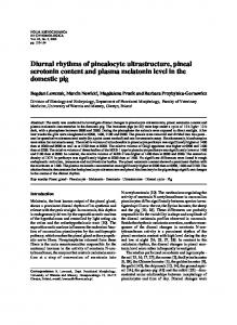
Brains were blocked at an angle corresponding to that of Paxinos and Watson (19) and 60 mm sections were cut with a vibratome (Technical Products, Inc., St. Louis, MO). All sections from the anterior border of the anterior commissural nucleus (ACN) to the posterior border of the arcuate nucleus were collected and were stained for the FOS protein as previously described (20). Sections were pretreated with 1% H2O2 in normal goat serum and incubated for 24 h at room temperature in antirat FOS antiserum (sc-52; Santa Cruz Biotechnology, Inc., Santa Cruz, CA) diluted 1:2000 in 0.4% Triton-X in PBS. This antibody recognizes an epitope specific to the FOS protein and does not bind to Fos-B, Fra-1, or Fra-2. Sections were then sequentially incubated in biotinylated goat antirat IgG, Vectastain Elite avidin-biotin complex (Vector Laboratories, Inc., Burlingame, CA) and DAB chromogen with nickel enhancement. For OT labeling, FOS-labeled sections were reexposed to 1% H2O2 and placed for 24 –36 h in antirabbit OT antiserum (#20068; 1:2000 in 0.4% Triton-X in PBS, INCSTAR Corp. Laboratories, Stillwater, MN). OT-IR was visualized with DAB peroxidase reaction as above in the absence of nickel enhancement. Using these procedures, the somata and processes of OTergic cells were stained brown and nuclei of FOS-labeled cells were black. FOS and OT double-labeled cells were defined by the presence of a black nucleus surrounded by brown cytoplasm. In the absence of FOS-IR, nuclei of OTergic cells were clear (Fig. 1). Cell counting. Cells were traced using a camera lucida by an experimenter blind to the treatment condition of the animal. To preclude the possibility of counting labeled cells in adjacent sections more than once, only cells with a distinct nucleus were counted. OT-IR, FOS-IR, and FOS/ OT-IR cells were counted bilaterally within either a single 60 mm section or 2 sections/rat in the anterior (antPVN, 21.40), middle (midPVN, 21.80) and posterior (postPVN, 22.12) PVN and in the ACN (approximate level 20.92 mm posterior to bregma; 21) and SON (21.40). As shown in Fig. 1, the PVN sections were also subdivided into magnocellular (PVNmag) and parvocellular (PVNparv) compartments based on cell size and anatomical location (19, 21). PVNparv and PVNmag cell counts from the ant-, mid-, and postPVN were summed for each animal; PVNmag cells in the antPVN were always 0. For all areas, the mean number of labeled cells observed unilaterally was used for analysis. For the SON and the ACN, the mean cell number was obtained from four single-side tracings from each animal. Mean unilateral cell numbers in the PVNmag and the PVNparv were obtained from 6 –12 (ant- plus midplus postPVN) sides counted. In this way PVNparv and PVNmag cell counts represented samples from the entire extent of their distribution within the nucleus. Because time of day effects have been observed previously in FOSrelated antigens (FRAs) immunoreactivity in the arcuate nucleus (ARC; 22), numbers of FOS-IR cells were measured in two subdivisions of the ARC (23.14; dorsomedial, ARCdm; ventrolateral, ARCvl) by superimposing a line 50o from the horizontal plane onto the camera lucida tracings as described by Ho¨kfelt et al. (23).
Plasma PRL and OT measurements within 1 h after mating To determine whether time of day effects on mating-induced OTergic activity were associated with alterations in pituitary PRL or OT secretion, repeated blood samples were obtained from animals bearing jugular catheters before mating and throughout the hour following mating at 0600 h, 1800 h, and 2400 h. Jugular catheterizations and blood collection procedures. On the day of ovx, 51 females were fitted with indwelling atrial catheters as previously described (1, 17). Catheterized females were treated postsurgically with atropine sulfate (0.1 ml sc; Webster, Inc., Sterling, MA) and received 10 daily antibiotic injections (Gentocin; 1.5 mg/rat sc; Henry Schein, Inc., Port Washington, NY). Catheters were cleared and flushed with heparinized saline (100 U/ml) daily for 10 days postoperatively. At the beginning of the mating session, a 70 cm extension was affixed to the atrial catheters to allow remote sample collection. Blood samples (0.3 ml) were obtained from freely behaving females as previously described 5 min before introduction of the male to the testing chamber and 5, 15, 30, and 60 min after the first mount or intromission. As previously reported, these procedures do not disrupt the expression of sexual behavior by the female nor notably influence the sexual behaviors
MATING-INDUCED OXYTOCIN AND PRL
4851
FIG. 1. Photomicrograph (1003) of the midPVN of a mated female. PVN sections were divided into magnocellular (PVNmag) and parvocellular (PVNparv) anatomical compartments for cell count analyses. Mean somal size in the 2 PVN compartments were 343.99 6 21.52 mm2 (10 cells/female, n 5 30) in the PVNmag and 260.24 6 15.85 mm2 (10 cells/female, n 5 30) in the PVNparv when measured at 2003 in 3 representative female brains using a CCD camera and the NIH Image program. Open arrowheads point to cells that are OTIR, and closed arrowheads indicate FOS-IR cells. Small arrows show OT-IR fibers, which run in a broad tract from the lateral PVN to the median eminence. v, Third ventricle.
displayed by the stimulus males (1). Following the mating session, catheter extensions were removed, and females were returned to their home cages. All blood samples were placed on ice immediately after collection and were centrifuged at 4 C at 2000 rpm for 20 min. Plasma was stored at 220 C until assay. PRL RIA. RIA for PRL was performed as previously described (1, 17) using antibody (anti-r-PRL-S-9) and standards (RP-3) provided by the National Hormone Pituitary Program and NIDDK and 125I-labeled PRL (18,000 cpm/100ul 1% BSA) from Covance Laboratories, Inc. (Vienna, VA). Precipitation of bound PRL was accomplished using antirabbit IgG (1:40 in 1% BSA; Antibodies Inc., Davis, CA). Measurements taken from plasma pools with known levels of PRL indicated a within-assay coefficient of variation of 8.9% and an interassay coefficient of variation of 12.4%. Assay sensitivity was 30 pg/tube. Oxytocin RIA. Methods for the OT RIA were adapted from those of Song et al. (24). Samples and standard amounts (0.1–100 pg/tube) of OT peptide (Peninsula Laboratories, Inc., Belmont, CA) were incubated with rabbit anti-OT primary antibody (no. 1733; Arnel Products Co., Inc., New York, NY) at a final tube concentration of 1:50,000 in PBS. Twelve hours later, 125I-labeled OT (3000 cpm, Covance Laboratories, Inc.) was added to each assay tube and incubation continued for 18 h. Separation of OT-bound antibody was accomplished by incubation with 70 ml Protein A (IgGSorb; The Enzyme Center, Malden, MA) for 20 min. All reagents and assay tubes were maintained at 4 C throughout the assay. Plasma from all samples were placed on a single assay. For validation of the assay, plasma from a recently hypophysectomized female rat was included. As previously shown in pituitary portal blood following pituitary stalk transection (25), plasma OT concentrations were markedly elevated in this female (54.8 pg/ml). Assay sensitivity was 1.5 pg/tube, and the intraassay coefficient of variation was 10.2%. Raw counts from all RIAs were analyzed using the Beckman Coulter, Inc. Immunofit EIA/RIA program.
Characterization of OTergic activity during daily PRL surges induced by mating To determine whether mating induced prolonged changes in OT activity that might influence PRL secretion during early pregnancy or PSP, FOS responses in OT cells were characterized as above in 15I and MO catheterized females from the previous experiment (n 5 16). Four days after the exposure to males, blood samples were obtained from these females at 0600 h, 1800 h, and 2400 h to confirm that PRL surges
were (15I) or were not (MO) present. On the following day, females were killed at either 0600 h or 2400 h and their brains collected for FOS- and OT-IR as above.
Statistics All data were analyzed using ANOVA (Superanova version 1.1; Abacus Concepts, Inc.). Cell count data from animals killed 1 h after mating were compared by two-way ANOVA (mating treatment 3 mating time). Three-way ANOVAs with repeated measures were applied to plasma OT and PRL values from samples taken acutely to mating and on day 4 (mating treatment 3 mating time 3 sample time). Immunocytochemical data from animals killed 5 days post mating were compared in a three-way analysis (mating treatment 3 mating time 3 time of kill). Effects of mating treatment and time of mating on behavioral parameters were compared separately in the catheterized and uncatheterized groups via two-way ANOVA. Student Newman-Keuls tests were used for post hoc comparisons between treatments, with a 5 0.05.
Results Mating-induced changes in brain OT-IR
OT-positive cells were segregated within the hypothalamus, with cell bodies located in the PVN, SON, and ACN as previously described (21, 26). Scattered OT-IR cells were also present in the POA, BNST, and along the third ventricle, particularly between the ACN and antPVN. Individual fibers with varicosities characteristic of OT neurons (26) were also observed throughout the hypothalamus, and a prominent OT-positive fiber tract was observed traveling ventrolaterally from the PVN (see Fig. 1). As shown in Fig. 2A, OT-IR in the PVNparv was influenced by both mating treatment [F(2, 13) 5 5.24, P # 0.05] and mating time [F(1, 13) 5 9.97, P # 0.01]. Post hoc tests revealed that females receiving 15I at 0600 h had significantly greater numbers of OT-IR cells than did HC animals (P # 0.05), with MO females showing intermediate levels that were not statistically different from the other two groups. In contrast, when females were tested at 2400 h, there were no differences
4852
MATING-INDUCED OXYTOCIN AND PRL
Endo • 1998 Vol 139 • No 12
FIG. 2. Mean (6 SEM) numbers of OT-IR cells seen in PVNparv (A), SON (B), PVNmag (C), and ACN (D) of females receiving HC, MO or 15I mating treatments at either 0600 h or 2400 h. Treatments assigned the same letter were not significantly different (P # 0.05).
seen in numbers of OT-positive cells across the three mating treatments. Numbers of OT-positive cells did not differ among treatment groups at either time within PVNmag (Fig. 2B) and were substantially lower than those seen in PVNparv. As in the PVNparv, both mating treatment [F(2, 13) 5 8.45. P # 0.005] and time of mating [F(1, 13) 5 8.80, P # 0.02] significantly influenced numbers of OT-IR cells in the SON (Fig. 2C). At 0600 h, receipt of 15I increased levels of OT-IR over both MO and HC levels (P # 0.05) and cell counts were higher overall at 0600 h than at 2400 h (P # 0.05). These statistical effects were due to a selective response among females receiving 15I at 0600 h, such that these animals had greater numbers of OT-positive cells in this area than those receiving any other treatment at either time (P # 0.05). Numbers of OT-IR cells in the ACN were influenced by neither mating treatment nor time of day (Fig. 2D). Mating-induced changes in FOS-labeled OT cells
Mating induced a significant increase in the absolute numbers of FOS/OT-IR cells observed and in the percent-
age of OT-IR cells colabeled for FOS. However, because treatment condition and time affected the numbers of OT-IR cells observed, the data presented are the mean (6 sem) numbers of FOS/OT-IR cells rather than the percentage double-labeled cells in each area. As shown in Fig. 3A, cell counts from the PVNparv revealed statistically significant effects of mating treatment [F(2, 13) 5 46.90, P # 0.001] and an interaction between mating treatment and time [F(2, 13) 5 4.42, P # 0.05] in the numbers of FOS/ OT-IR cells counted. At 0600 h, females receiving 15I had higher numbers of FOS/OT-colabeled cells than the other treatment groups (P # 0.05), and MO females showed significantly higher numbers of colabeled cells than did HC females (P # 0.05). This response was seen only at 0600 h; receipt of both 15I and MO induced equal elevations in numbers of FOS-positive OTergic cells over that produced by HC treatment at 2400 h. Though numbers of double-labeled cells in PVNmag also showed overall statistically significant increases after receipt of 15I and MO stimulation [F(2, 13) 5 4.54, P # 0.05; Fig. 3B], post hoc tests
MATING-INDUCED OXYTOCIN AND PRL
4853
FIG. 3. Mean (6 SEM) numbers of FOS/OT-colabeled cells seen in the PVNparv (A), SON (B), PVNmag (C), and ACN (D) of females receiving HC, MO, or 15I mating treatments at either 0600 h or 2400 h. Treatments assigned the same letter were not significantly different (P # 0.05).
revealed differences only between 15I and HC females (P # 0.05) combined over the 2 test times. Time of day did not influence FOS/OT-IR within PVNmag. As was observed in the PVNparv, both mating treatment [F(2, 13) 5 14.57, P # 0.001] and time [F(1, 13) 5 5.45, P # 0.05] significantly influenced numbers of FOS/OT-colabeled cells in the SON. As shown in Fig. 3C, expression of FOS in OTergic cells was significantly elevated following 15I at 0600 h compared with all other treatment groups (P # 0.05). In contrast, neither mating treatment nor mating time was found to influence numbers of FOS/OT-IR cells in the ACN, where numbers of FOS-positive OT-IR cells remained the same across all groups (Fig. 3D). Mating-induced changes in brain FOS-IR
Mean numbers of FOS-IR cells seen in the different treatment groups are presented in Fig. 4. Male exposure at 0600 h and 2400 h increased FOS expression in all brain areas ex-
amined. Effects of mating treatment were significant and similar within the PVNparv [F(2, 13) 5 15.27, P # 0.001] and the PVNmag [F(2, 13) 5 15.54, P # 0.001], and there was no significant overall effect of time of mating in either area. However, a significant interaction effect between mating treatment and time in the PVNmag (Fig. 4B; mating treatment 3 time: [F(2, 13) 5 5.69, P # 0.02] followed by post hoc analysis demonstrated that 15I induced significantly higher FOS-IR than did MO and HC treatments at 0600 h but not at 2400 h. Within the PVNparv, there was a nonsignificant trend for an interaction effect between mating treatment and time [F(2, 13) 5 3.13, P # 0.08]; 15I induced a significant elevation in FOS-IR cells over MO at 0600 h but not at 2400 h (Fig. 4A). Within the SON [F(2, 13) 5 20.02, P # 0.001] and the ACN, [F(2, 13) 5 5.45, P # 0.02], mating treatment also significantly influenced numbers of FOS-IR cells, and there were no time of day or treatment by time interaction effects. For these two areas, data from the two mating times were combined before
4854
MATING-INDUCED OXYTOCIN AND PRL
Endo • 1998 Vol 139 • No 12
FIG. 4. Mean (6 SEM) numbers of FOS-IR cells in the PVNparv (A), PVNmag (B), SON (C), ACN (D), ARCdm (E), and ARCvl (F) of females receiving HC, MO or 15I mating treatments at either 0600 h or 2400 h. For Fig. 4, A and B, treatments assigned the same letter were not significantly different (P # 0.05). For Fig. 4, C–F, post hoc analysis was carried out on data combined over the two treatment times. *, Significantly greater than HC groups over both times (P # 0.05); §, significantly greater than MO and HC groups over both times (P # 0.05).
post hoc analysis. In the SON (Fig. 4C), FOS-IR was significantly higher overall in 15I females than in MO females, and both groups showed significantly more FOS-IR than did the HC group (P # 0.05). In the ACN (Fig. 4D), 15I significantly raised FOS-IR over HC levels (P # 0.05), and levels seen
following MO treatment were intermediate to and not statistically different from the other two groups. Mating treatment significantly influenced the expression of FOS in both the ARCdm [Fig. 4E; F(2, 13) 5 10.99, P # 0.002] and the ARCvl [Fig. 4F; F(2, 13) 5 8.28, P # 0.005]. Because
MATING-INDUCED OXYTOCIN AND PRL
there were no significant effects of mating time or treatment by time interactions, data were again combined across time before post hoc analysis. In the ARCdm, 15I and MO animals showed significantly greater numbers of FOS-IR cells than did the HC group (P , 0.05). In the ARCvl, the 15I group had significantly greater numbers of FOS-IR cells than did either the MO or the HC groups (P , 0.05). Plasma OT and PRL responses to mating
Acute plasma OT and PRL responses to mating are presented in Fig. 5. Neither mating treatment, mating time, nor sample time had an effect on plasma OT levels. There was no significant effect of mating time on acute PRL secretion. However, there was a significant effect of mating treatment [F(1, 69) 5 11.73, P # 0.005]. Compared with MO treatment, plasma PRL was elevated at 5 and 15 min after receipt of 15I at 0600 h and 2400 h, and, in addition, at 30 min at 1800 h (P # 0.05).
4855
Brain OT- and FOS-IR associated with PRL secretion 4 –5 days post mating
Values from plasma samples taken 4 days after mating were used to verify the presence of mating-induced PRL surges in 15I females. Plasma PRL levels from these samples are presented in Fig. 6. Comparisons of 15I to MO females revealed strong effects of mating treatment [F(1, 40) 5 11.17, P # 0.005] and time of day [F(2, 40) 5 5.84, P # 0.01] on plasma PRL concentrations. Plasma PRL was significantly elevated at both 1800 h and 0600 h compared with 2400 h in 15I but not MO females (P # 0.05), confirming the induction of PRL surges by 15I treatment. There was no effect of the time of day at which mating had occurred on peak PRL concentrations observed 4 days later. Plasma OT levels were not affected by either mating treatment or sample time 4 days post mating (data not shown). As shown in Table 1, numbers of OT-IR and FOS/OT-IR cells were not influenced by mating treatment, mating time, or time of kill in any area examined in brains taken from 15I
FIG. 5. Plasma concentrations (mean 6 SEM) of OT (left panels) and PRL (right panels) seen within 1 h of mating at 1800 h (top), 2400 h (middle), and 0600 h (bottom). Arrows indicate time of mating. *, Hormone levels that were significantly higher than those seen in MO females (P # 0.05).
4856
Endo • 1998 Vol 139 • No 12
MATING-INDUCED OXYTOCIN AND PRL
and MO females 5 days post mating. Numbers of FOS-IR cells were substantially lower in all areas than observed an hour after mating, and FOS/OT-IR cells were rare. There was no correlation between plasma PRL levels 4 days after mating and OT and FOS labeling seen in brains taken after killing on the following day. Mating behavior
Results of the behavioral analyses are presented in Table 2. All females were highly receptive, displaying LQ’s of 90 – 100% (data not shown) and LRs ranging from 1.75 to 3.00. Within the uncatheterized animals, there was an effect of mating time on LR [F(1, 8) 5 19.05 P # 0.01], in that females mated at 0600 h had significantly higher LRs than those mated at 2400 h (2.94 6 0.03 vs. 2.42 6 0.15). This result was not seen in the catheterized females, where animals in all treatment groups displayed mean LRs of less than 2.50.
FIG. 6. Plasma PRL concentrations (mean 6 SEM) in blood samples taken 4 days after receipt of 15I or MO mating treatments. Values from samples taken at 0600 h and 1800 h show the presence of nocturnal and diurnal PRL surges in females receiving 15I treatment, verifying effective induction of PRL surges by mating in this group. Treatments assigned the same letter were not significantly different.
Discussion
Various mechanosensory stimuli have the ability to stimulate OT synthesis and release in mammals. The present results demonstrate facilitatory effects of mating on numbers of both OT-IR and FOS/OT-IR cells in parvocellular PVN and in SON neurons within 1 h following mating onset. These increases occurred specifically in response to intromissive stimulation from males because OT or FOS/OT labeling was greater in the 15I group than in the MO group in both areas. Insofar as VCS is also essential for initiation of the PRL surges of pregnancy and PSP (27), these results support the hypothesis that OTergic activity is important for surgetype PRL secretion. As proposed for the GnRH system (28), the increase in numbers of OT-IR cells may be the result of an increase in OT gene expression, a decreased movement of OT out of the soma, or a decrease in OT secretion; any of these alternatives singly or in combination might increase peptide content enough to result in measurable changes in OT-IR cell number. We do not know whether the increases in OT-IR cell number represent increases in OTergic activity in terminal areas of these neurons. However, increased numbers of FOSlabeled OT cells are likely to be associated with the release of OT, as shown by Arey and Freeman (6). The data also show that mating influences neural activity only in selected populations of OT neurons because OT cells of the ACN and magnocellular neurons within the PVN were not responsive to mating at any time of day. The mating-induced increases in OT-IR and FOS/OT-IR seen in these studies indicate that activation of both central and peripheral OTergic systems has occurred. Within the PVN, only PVNparv neurons that release OT onto central synapses and into the median eminence (26) were responsive to mating stimulation. There was no influence of mating on OT cells within the PVNmag neurons that are known to terminate in the neurohypophysis and release their product into the general circulation (26). Cells in the SON showed similar responses to those seen in PVNparv in that receipt of 15I induced a significant increase in both OT-IR and FOS/OT-IR above MO and HC levels. However, despite the high numbers of magnocellular OT cells in the SON, plasma OT concentrations did not diverge from baseline levels throughout 60 min following mating at any of the 3 sampling times. In the lactating rat, magnocellular OT responses to suckling are synchronized among cells through increased soma-soma apposition and formation of shared synapses (8), but these
TABLE 1. Numbers of OT-IR and FOS/OT-IR cells observed at the time of the nocturnal PRL surge (0600 h) and during a nonsurge period (2400 h) 5 days after 15I or MO mating stimulation Mating treatmentb
15I
MO
a b
Brain area
PVNparv PVNmag ACN SON PVNparv PVNmag ACN SON
Mean 6 SEM. n 5 4/time point in each mating treatment.
OT-IR Cellsa
FOS/OT-IR Cellsa
0600 h
2400 h
0600 h
2400 h
54.4 6 2.8 15.9 6 1.1 44.5 6 4.0 41.0 6 2.6 56.5 6 13.8 18.5 6 3.7 37.1 6 1.2 40.4 6 6.1
46.9 6 11.2 16.4 6 2.3 35.6 6 3.8 42.7 6 4.6 47.1 6 8.1 16.9 6 2.4 39.1 6 3.2 40.4 6 3.3
0.0 0.0 0.2 6 0.1 0.1 6 0.1 0.2 6 0.2 0.0 0.1 6 0.1 0.0
0.0 0.0 0.0 0.0 0.1 6 0.1 0.0 0.0 0.0
MATING-INDUCED OXYTOCIN AND PRL TABLE 2. Sexual receptivity (LR, mean 6 and uncatheterized females 1800 h
Uncatheterized 15I MO Catheterized 15I MO
— —
SEM)
in catheterized
Time of mating 2400 h
0600 h
2.17 6 0.19 (3) 2.94 6 0.06a (3) 2.67 6 0.13 (3) 2.95 6 0.02a (3)
2.26 6 0.04 (10) 2.20 6 0.02 (8) 2.25 6 0.07 (7) 2.28 6 0.09 (8) 2.21 6 0.05 (9) 2.12 6 0.14 (9)
LR was significantly elevated across both mating treatments in uncatheterized females mated at 0600 h compared with 2400 h (aP # 0.05). Number of animals is indicated in parentheses.
changes have not been shown in cycling females. In the absence of coordinated OT release from SON neurons at the time of mating, acute changes in plasma OT were not detected. Increases in peripheral OT levels following VCS have been demonstrated in other mammalian species including sheep (11) and rabbits (12). However, the current data verify a previous report that plasma OT levels are not altered following mating stimulation in the female rat (14). Notably, intromissive stimulation induced increases in both single- and double-labeled OT-positive cells within 1 h of mating at 0600 h but not at 2400 h. Our results demonstrate that the responsiveness of OT cells in the PVNparv and the SON is affected by the time of day at which afferent input is received, and suggest the existence of a diurnal fluctuation in sensitivity of central OTergic systems to activation by sensory input. Numbers of OT-IR or FOS/OT-IR cells were not influenced by time of day in either MO or HC control rats in this study, suggesting that the fluctuation is in responsiveness to intromissive stimulation rather than to an underlying rhythmicity in OTergic function. OT neurons in the PVNparv and SON appear to be particularly responsive to mating at 0600 h, because, at another time in another study, much larger amounts of mating stimulation at 2400 h induced increases in FOS expression in parvocellular PVN OT cells but did not increase total numbers of OT-IR cells at this time (13). The physiological significance of the elevated responsiveness of OT cells to mating at 0600 h is unknown. However, the sensitivity of the female specifically to intromissive stimulation suggests that the OT neurons may be involved in either neuroendocrine or behavioral sequelae to mating, such as induction of PSP or abbreviation of estrus (27), which are known to depend on such stimulation. Because females mated at 0600 h require fewer intromissions to become PSP than do those mated at 2400 h (15), it is possible that activation of OT neurons by mating at that time contributes to the initiation of PSP. Enhanced sexual responsiveness was seen in females mated at 0600 h compared with those mated at 2400 h. Previous studies have localized OT receptors in the ventromedial hypothalamus (VMH; 29), a brain site critical for the expression of lordosis (30), and infusion of OT either icv (31) or into the VMH (32) is facilitatory to female sexual behavior. Thus, central release of OT in animals receiving 15I at 0600 h may have contributed to their heightened receptivity at this time. Additionally, because progesterone is known to induce OT receptors within the VMH (32), it is possible that diurnal
4857
fluctuations in sensitivity to progesterone might have induced OT receptors in this area to a greater extent at 0600 h than at 2400 h. Because we administered progesterone 4 h before mating for all groups, fluctuations in OT receptor number would not be a consequence of the timing of progesterone administration. Influences of time of day on sexual behavior were not seen in catheterized females, whose LRs were slightly lower than those of uncatheterized females. Thus, low-level chronic stress from long-term catheterization may interfere with the enhancement of lordosis seen at 0600 h. Plasma PRL measurements taken acutely to mating confirmed our previous report that receipt of 15I but not MO induces rapid elevations in PRL secretion (1). Levels of PRL were significantly elevated within 5 min of mating and returned to baseline levels within 1 h. In contrast with the immunocytochemical results, time of mating was not found to influence the magnitude or timing of this response. Therefore, it is unlikely that centrally released OT acts as a PRF regulating PRL secretion in this circumstance. Time of day does influence PRL secretion in cycling (33), PSP (34) and lactating (16) females. The present data suggest that acute PRL responses to mating are subject to unique regulatory control, and that, at most, OT may be indirectly involved in controlling the acute PRL response through transduction or processing of genitosensory inputs. Short-term PRL release is seen in response to stress (35) or as a result of matinginduced release of endogenous opiate peptides (36), and it is possible that acute mating-induced PRL responses are reflective of one or more of these factors. Although we have observed modest increases in plasma corticosterone following similar amounts of mating stimulation (37), the striking difference between the acute PRL responses in 15I and MO animals suggests that stress may play a minor role in this response, because the treatment of animals in both groups included removal from the home cage, transport to the testing room, and exposure to and mounts from males. An unexpected finding in this study was that receipt of 15I, a treatment that induced PRL secretion within 5 min after mating, was not associated with an acute suppression of FOS-IR in the ARCdm. Dopamine released from tuberoinfundibular (TIDA) cells in the ARC is known to be an important PRL-inhibiting factor (PIF; 38), and it would be expected that FOS-IR in this area would be suppressed by mating. Decreases in constitutive expression of FRAs within dopaminergic neurons have been reported at the 2 times of day when PRL surges are expressed in ovx hormone-primed and PSP females (22, 39). There have been no studies in which dopamine release from the arcuate nucleus has been measured after mating, but decreased multiunit activity seen in the ARC following artificial VCS (40) suggests that, as is seen in the suckled female (41), mating may induce a transient inhibition of TIDA activity that could stimulate PRL secretion. It is therefore likely that the ARC cells that expressed increased FOS after mating in the present experiment are not DAergic neurons. Changes in numbers and distribution of FOS-IR cells following mating have been well characterized, and matinginduced increases in numbers of FOS-positive cells in the mPOA, MePD, BNSTpm and VMH were observed in the
4858
Endo • 1998 Vol 139 • No 12
MATING-INDUCED OXYTOCIN AND PRL
present study, which replicate those previously described (20, 42– 45; data not shown). We have now shown previously unreported increases in numbers of PVN FOS-positive cells that were selective only to females receiving intromissive stimulation. We had earlier shown differences between 15I and MO mating treatments in the expression of another immediate-early gene, egr-1 (20). As in our earlier data on the PVN (20), FOS responses to MO and 15I were equivalent among females mated at 2400 h; however, in the present study, FOS labeling in PVNparv was found to be significantly greater in 15I than in MO animals when mating occurred at 0600 h. The heightened responsiveness of PVN cells to 15I seen at 0600 h indicates that these cells exhibit differential sensitivity to intromissions across times of day, and mirror the results obtained for the OT and FOS/OT labeling. Increases in FOS-IR in the SON and ACN that have not been previously reported were seen following male exposure at both mating times, but the responses were not dependent upon intromissive stimulation. Because increases in numbers of FOS-positive cells in the PVN, SON, and ACN were far greater than those seen in FOS/OT-colabeled cells, activation by mating and diurnal changes in sensitivity to mating appear to occur within multiple cell types in these areas. The existence of endogenous circadian rhythms that regulate PRL secretion via control of hypothalamic (6, 22) and preoptic area (46) PRFs and PIFs has been hypothesized. Increases in constitutive FOS/OT-IR in the PVN (6) and decreases in FRAs/DA-IR in the ARCdm (22) have been observed in unmated ovx females at times of day coincident with the nocturnal and diurnal PRL surges. In the present experiment, there were no differences seen 5 days post mating in HC and MO control females in numbers of OT-IR or FOS/OT-IR cells across times of day that would suggest a rhythmic activity in OT cells. In addition, there was no difference in either single or double-labeled cells between animals showing (15I) or not showing (MO) PRL surges. Therefore, we did not observe rhythmic OT activity during early PSP associated with PRL surge secretion. There are several possible explanations for the discrepancy between the present results and those previously reported (6). One reason may be that the different antibodies used may have labeled different levels of constitutive FOS or may have been labeling different FOS/OT-IR cell populations due to differences in specificity. Another related possibility is that mating may induce immediate changes in one subpopulation of OT cells but suppress subsequent diurnal fluctuations in OTergic activity in the same or other populations of OT cells beginning 1 day after mating when PRL surges are normally initiated (17). In contrast to the present study, the earlier work was carried out in unmated animals (6). In light of these possibilities, the uniformly low levels of FOS/OT-IR seen in mated females 5 days after mating do not completely rule out the possibility that pulses of OTergic activity normally enhance PRL release at 0600 h and 1800 h. However, our data suggest that if parvocellular OT acts to stimulate PRL release during PSP, it does so around the time of mating by initiating the processes required for initiation and perseveration of the surges rather than by direct stimulation of PRL release.
Acknowledgments We would like to thank Dr. A. Parlow and the Hormone Pituitary Program, NIDDK, for generously supplying the antiserum (antirat-PRLS-9) and reference preparation (RP-3) used in the PRL RIAs. We would also like to thank John Wilkins of Covance Laboratories, Inc. and Dr. W. R. Crowley (University of Tennessee) for their helpful advice and Jane Willan for her technical assistance in completing these experiments.
References 1. Erskine MS, Kornberg E 1992 Acute luteinizing hormone and prolactin responses to paced mating stimulation in the estrous female rat. J Neuroendocrinol 4:173–179 2. Gunnet JW, Freeman ME 1983 The mating-induced release of prolactin: a unique neuroendocrine response. Endocr Rev 4:44 – 61 3. Lumpkin MD, Samson WK, McCann SM 1983 Hypothalamic and pituitary sites of action of oxytocin to alter prolactin secretion in the rat. Endocrinology 112:1711–1717 4. Samson WK, Lumpkin MD, McCann SM 1986 Evidence for a physiological role for oxytocin in the control of prolactin secretion. Endocrinology 119:554 –560 5. Morgan JI, Curran T 1991 Stimulus-transcription coupling in the nervous system: involvement of the inducible proto-oncogenes fos and jun. Annu Rev Neurosci 14:421– 451 6. Arey BJ, Freeman ME 1992 Activity of oxytocinergic neurons in the paraventricular nucleus mirrors the periodicity of the endogenous stimulatory rhythm regulating prolactin secretion. Endocrinology 130:126 –132 7. Arey BJ, Freeman ME 1990 Oxytocin, vasoactive-intestinal peptide, and serotonin regulate the mating-induced surges of prolactin secretion in the rat. Endocrinology 126:279 –284 8. Theodosis DT, Poulain DA 1992 Neuronal-glial and synaptic plasticity of the adult oxytocinergic system: factors and consequences. Ann NY Acad Sci 652:303–325 9. Negoro H, Visessuwan S, Holland RC 1973 Reflex activation of paraventricular nucleus units during the reproductive cycle and in ovariectomized rats treated with oestrogen or progesterone. J Endocrinol 59:559 –567 10. Higuchi T, Honda K, Fukuoka T, Negoro H, Wakabayashi K 1985 Release of oxytocin during suckling and parturition in the rat. J Endocrinol 105:339 –346 11. Kendrick KM, Keverne EB, Hinton MR, Goode JA 1991 Cerebrospinal fluid and plasma concentrations of oxytocin and vasopressin during parturition and vaginocervical stimulation in the sheep. Brain Res Bull 26:803– 807 12. Fuchs AR, Cubile L, Dawood MY 1981 Effects of mating on levels of oxytocin and prolactin in the plasma of male and female rabbits. J Endocrinol 90:245–253 13. Flanagan LM, Pfaus JG, Pfaff DW, McEwen BS 1993 Induction of FOS immunoreactivity in oxytocin neurons after sexual activity in female rats. Neuroendocrinology 58:352–358 14. Caldwell JD, Jirikowski GF, Greer ER, Stumpf WE, Pedersen CA 1988 Ovarian steroids and sexual interaction alter oxytocinergic content and distribution in the basal forebrain. Brain Res 446:236 –244 15. Frye CA, Erskine MS 1990 Influence of time of mating and paced copulation on induction of pseudopregnancy in cyclic female rats. J Reprod Fert 90:375–385 16. Arey BJ, Kanyicska B, Freeman ME 1991 The endogenous stimulatory rhythm regulating prolactin secretion is present in the lactating rat. Neuroendocrinology 53:35– 40 17. Kornberg E, Erskine MS 1994 Effects of differential mating stimulation on the onset of prolactin surges in pseudopregnant rats. Psychoneuroendocrinology 19:357–371 18. Hardy DF, DeBold JF 1971 The relationship between levels of exogenous hormones and the display of lordosis by the female rat. Horm Behav 2:287–297 19. Paxinos G, Watson C 1986 The Rat Brain in Stereotaxic Coordinates. Academic Press, San Diego 20. Polston EK, Erskine MS 1995 Patterns of induction of the immediate-early genes c-fos and egr-1 in the female rat brain following differential amounts of mating stimulation. Neuroendocrinology 62:370 –384 21. Swanson LW, Sawchenko PE 1983 Hypothalamic integration: organization of the paraventricular and supraoptic nuclei. Ann Rev Neurosci 6:269 –324 22. Lerant A, Freeman ME 1997 Dopaminergic neurons in periventricular and arcuate nuclei of proestrous and ovariectomized rats: endogenous diurnal rhythm of FOS-related antigens expression. Neuroendocrinology 65:436 – 445 23. Ho¨kfelt T, Meister B, Melander T, Everitt B 1987 Coexistence of classic transmitters and peptides with special reference to the arcuate nucleus-median emminence. Adv Biochem Psychopharmacol 43:21–34 24. Song SL, Crowley WR, Grosvenor CE 1988 Evidence for involvement of an adrenal catecholamine in the b-adrenergic inhibition of oxytocin release in lactating rats. Brain Res 457:303–309 25. Makara GB, Sutton S, Otto S, Plotsky PM 1995 Marked changes of arginine vasopressin, oxytocin, and corticotropin-releasing hormone in hypophysial portal plasma after pituitary stalk damage in the rat. Endocrinology 136:1864 –1868
MATING-INDUCED OXYTOCIN AND PRL 26. Armstrong WE 1995 Hypothalamic supraoptic and paraventricular nuclei. In: Paxinos G (ed) The Rat Nervous System: ed. 2. Academic Press, Inc., San Diego, pp 377–387 27. Erskine MS 1995 Prolactin release after mating and genitosensory stimulation in females. Endocr Rev 16:508 –528 28. Dellovade TL, Ottinger MA, Rissman EF 1995 Mating alters gonadotropinreleasing hormone cell number and content. Endocrinology 136:1648 –1657 29. Johnson AE, Coirini H, Ball GF, McEwen BS 1989 Anatomical localization of the effects of 17b-estradiol on oxytocin receptor binding in the ventromedial hypothalamic nucleus. Endocrinology 124:207–211 30. Pfaff DW, Schwartz-Giblin S 1988 Cellular mechanisms of female reproductive behaviors. In: Knobil E, Neill J (eds) The Physiology of Reproduction. New York, Raven Press, pp 1487–1568 31. Witt DM, Insel TR 1992 Central oxytocin antagonism decreases female reproductive behavior. In: Pedersen CA, Caldwell JD, Jirikowski GF, Insel TR (eds) Oxytocin in maternal, sexual, and social behaviors. Ann NY Acad Sci 652:445– 447 32. Schumacher M, Coirini H, Pfaff DW, McEwen B 1990 Behavioral effects of progesterone associated with rapid modulation of oxytocin receptors. Science 250:691– 694 33. Neill JD, Freeman ME, Tillson SA 1971 Control of the proestrus surge of prolactin and luteinizing hormone secretion by estrogens in the rat. Endocrinology 89:1448 –1452 34. Butcher RL, Fugo NW, Collins WE 1972 Semicircadian rhythm in plasma levels of prolactin during early gestation in the rat. Endocrinology 90:1125–1127 35. Neill JD 1970 Effect of stress on serum prolactin and luteinizing hormone levels during the estrous cycle of the rat. Endocrinology 87:1192–1197 36. Sirinathsinghji DJ, Audsley AR 1985 Endogenous opioid peptides participate in the modulation of prolactin release in response to cervicovaginal stimulation in the female rat. Endocrinology 117:549 –556
4859
37. Frye CA, McCormick CM, Coopersmith C, Erskine MS 1996 Effects of paced and non-paced mating stimulation on plasma progesterone, 3 a-diol and corticosterone. Psychoneuroendocrinology 21:431– 439 38. Ben-Jonathan N 1985 Dopamine: a prolactin-inhibiting hormone. Endocr Rev 6:564 –589 39. Lerant A, Herman ME, Freeman ME 1996 Dopaminergic neurons of periventricular and arcuate nuclei of pseudopregnant rats: semicircadian rhythm in fos-related antigens immunoreactivities and in dopamine concentration. Endocrinology 137:3621–3628 40. Kawakami J, Ibuke T 1972 Multiple unit activity in the brain correlated with induction and maintenance of pseudopregnancy in rats. Neuroendocrinology 9:2–19 41. de Greef WJ, Plotsky PM, Neill JD 1981 Dopamine levels in hypophyseal stalk plasma and prolactin levels in peripheral plasma of the lactating rat: effects of a simulated suckling stimulus. Neuroendocrinology 32:229 –233 42. Pfaus JG, Kleopoulos SP, Mobbs CV, Gibbs RB, Pfaff DW 1993 Sexual stimulation activated c-fos within estrogen-concentrating regions of the female rat forebrain. Brain Res 624:253–267 43. Rowe DW, Erskine MS 1993 c-fos proto-oncogene activity induced by mating in the preoptic area, hypothalamus and amygdala in the female rat: role of afferent input via the pelvic nerve. Brain Res 621:25–34 44. Tetel MJ, Getzinger MJ, Blaustein JD 1993 Fos expression in the rat brain following vaginal-cervical stimulation by mating and manual probing. J Neuroendocrinol 5:397– 404 45. Wersinger SR, Baum MJ, Erskine MS 1993 Mating-induced FOS-like immunoreactivity in the rat forebrain: a sex comparison and a dimorphic effect of pelvic nerve transection. J Neuroendocrinol 5:557–568 46. Lee Y, Arbogast LA, Voogt JL 1998 Semicircadian rhythms of c-Fos expression in several hypothalamic areas during pregnancy in the rat: relationship to prolactin secretion. Neuroendocrinol 67:83–93