RESEARCH COMMUNICATIONS molecular weight region (results not shown). The immunoblot results match well with the ELISA results. To conclude, an in vivo defect in T cell proliferation associated with enhanced level of T cell apoptosis was noted. T cell death by apoptosis may represent one of the important components of the ineffective immune response against M. tuberculosis-induced immunopathology in susceptible hosts14,22. Thus high concentration of M. tuberculosis antigens may incapacitate CMI in young susceptible mice by inducing thymic atropy.
1. Zhang, Y., Takahasi, K., Jiang, G., Kawai, M., Fukada, M. and Yokochi, T., Infect. Immunol., 1993, 61, 5044–5048. 2. Ozeki, Y., Kaneda, K., Fujiwara, N., Morimoto, M., Oka, S. and Yano, I., Infect. Immunol., 1997, 65, 1793–1799. 3. Hayashi, T., Catanzaro, A. and Rao, S. P., Infect. Immunol., 1997, 65, 5262–5271. 4. Jenkinson, E. J., Kingston, R., Smith, C. A., Williams, G. T. and Owen, J. T. T., Eur. J. Immunol., 1989, 19, 2175. 5. Hueber, A. and Evan, G. I., Trends Genet., 1998, 14, 364–367. 6. Anderson, P., Askgaard, D., Liungqvist, L., Bentson, M. W. and Heron, I., Infect. Immunol., 1991, 59, 1558–1563. 7. Esin, S., Butoni, G., Kallenius, G., Granes, H., Campa, M. and Svenson, S. B., Clin. Exp. Immunol., 1996, 140, 419. 8. Duke, R. C. and Cohen, J. J., Lymphokine Res., 1986, 5, 289. 9. Kaufmann, S. H. F., Annu. Rev. Immunol., 1993, 11, 129– 163. 10. Duhe, R. C. and Cohen, J. J., Annu. Rev. Immunol., 1992, 10, 267–293. 11. Wang, S. D., Huang, K. J., Lin, Y. S. and Lei, H. Y., Immunology., 1994, 152, 5014. 12. Placido, R. et al., J. Pathol., 1997, 181, 31–38. 13. Oddo, M., Reuno, T., Attinger, A., Bakker, T., MacDonald, H. and Meylan, P., J. Immunol., 1998, 160, 5448. 14. Das, G., Vohra, H., Saha, B., Agrewala, J. N. and Misra, G. C., Clin. Exp. Immunol., 1999, 115, 324. 15. Kornblum, H., Loughlin, S. E. and Leslie, F. M., Dev. Brain Res., 1987, 31, 45. 16. Egerton, M., Scollay, R. and Shortman, K., Proc. Natl. Acad. Sci. USA, 1990, 87, 2579–2582. 17. Noguchi, P. D., in Current Protocol in Immunology (eds Coligan, J. E. et al.), Library of Cataloging in Publication Data, USA, 1997, vol. 1. 18. Harlow, E. and David, L., in Antibodies – A Laboratory Manual, Cold Spring Harbor Lab. 19. MacDonald, H. R. and Kees, R. K., Nature, 1990, 343, 642– 644. 20. Morrison, D. C. and Ryan, J. L., Adv. Immunol., 1979, 28, 293– 450. 21. Reed, N. D., Manning, J. K. and Rudback, J. A., Infect. Dis., Suppl., 1973, 28 S70–S74. 22. Kremer, L., Estaquier, J., Wolowczuh, I., Biet, F., Ameisen, J. and Locht, C., Infect. Immunol., 2000, 68, 4264–4273. ACKNOWLEDGEMENTS. We thank Dr P. R. Narayanan, Director, Tuberculosis Research Centre, (TRC), Chennai, for his continuous enthusiasm and support. We also thank Dr V. Kumarasami, Head, Department of Immunology and Dr Alamelu Raja, Deputy Director, TRC, Chennai, for their support and encouragement. We thank members of the animal house department for their help. Received 18 October 2000; revised accepted 18 April 2001
304
Diversity and abundance of higher marine fungi on woody substrates along the west coast of India K. Prasannarai*,† and K. R. Sridhar*,‡ *Department of Biosciences, Mangalore University, Mangalagangotri, Mangalore 574 199, India † Present address: Department of Botany, St. Philomena College, Puttur 574 202, India
The diversity of marine fungi on intertidal wood collected from 13 locations in the west coast of India (beaches, islands and harbour locations) was assessed. Out of 3327 wood samples scanned, 72% possess sporulating marine fungi. Altogether 88 species belonging to 47 genera were encountered. Torpedospora radiata was the only fungus common to all locations. Five fungi were more frequent (> 10%): Antennospora quadricornuta, Clavatospora bulbosa, Crinigera maritima, Periconia prolifica and Torpedospora radiata. Twenty-four species were rare (0.04–0.5%); notable rare species were: Amylocarpus encephaloides, Arenariomyces majusculus, Calathella mangrovei, Carbosphaerella leptosphaerioides, Cirrenalia pseudomacrocephala, Crinigera maritima, Dryosphaera navigans, Dryosphaera tropicalis, Nimbospora bipolaris and Nimbospora effusa. Twenty-two species reported in the present study are new records for the Indian Peninsula. A. encephaloides was recorded from the tropical location, so also A. majusculus and C. pseudomacrocephala from the Indian Ocean. The species richness and diversity was highest in islands than in beaches and harbour locations. It has been predicted that islands adjacent to the west coast of India provide critical habitat for marine fungi. MARINE fungi are the important intermediaries of energy flow from detritus to higher trophic levels in the marine ecosystems1. They require sea water for the completion of their life cycle. More than 500 species of marine fungi have been described2. Higher marine fungi constitute Ascomycotina, Basidiomycotina and Deuteromycotina. Majority of them being ascomycetes, their spores show adaptation to the marine ecosystem in the production of appendages, which facilitate buoyancy in water, entrapment and adherence to substrates. Marine filamentous fungi have been reported on a variety of detritus: decaying wood, leaves, seaweeds, seagrasses, calcareous and chitinous substrates. Studies on marine fungi were initiated at the temperate parts of the world. Subsequently, tropical locations were the centres of interest to understand the abundance and diversity2,3. Tropical regions of Atlantic and Pacific Oceans were investigated more intensively than the Indian Ocean3. A few quantitative studies ‡
For correspondence. (e-mail:
[email protected]) CURRENT SCIENCE, VOL. 81, NO. 3, 10 AUGUST 2001
RESEARCH COMMUNICATIONS on marine fungi are available from the west coast of India4–6. Majority of the studies in the Indian Ocean were restricted to mangrove habitats7–10, so also from the Indian Peninsula4,11–13. Therefore the present study aims to understand the occurrence, species richness and diversity of filamentous marine fungi on woody substrates collected from the beaches, islands and harbour locations of the west coast of India. Intertidal wood samples were collected from 13 locations comprising seven beaches (Bambolim, Karwar, Kaup, Someshwara, Bekal Fort, Kovalam and Kanyakumari); two harbour sites (New Mangalore Port); and four islands (Kurmagadagudda, Anjadeep, Coconut and North Islands) adjacent to the Karnataka coast (Figure 1). Bambolim is largely a sandy bay with patches of laterite rocks. Similarly, Kaup, Someshwara and Kanyakumari are mainly sandy shores with hard rocky patches. Karwar and Kovalam too are sandy shores, while Bakel Fort is a rocky shore. Harbour St. 1 consists of granite wall, while Harbour St. 2 is a small sandy stretch. All the island shores are mainly rocky with small sandy stretches. From each station, based on the availability, 150–350 intertidal wood samples were randomly collected, brought
to the laboratory in sterile polythene bags and scanned for fungal structures within seven days of collection using stereomicroscope. After initial screening, each wood sample was separately incubated under fluorescent light (24– 32°C) in sterile airtight polythene bags containing sterile sand bed moistened with sea water. Each wood sample was re-examined for fungi once a month, up to 12 months. The fungi recovered were identified using the keys: Kohlmeyer and Kohlmeyer3, Kohlmeyer14, Kohlmeyer and Volkmann–Kohlmeyer15 and descriptions mainly by J. Kohlmeyer, B. Volkmann–Kohlmeyer, E. B. G. Jones and K. D. Hyde and A. Nakagiri. For each fungal species, the per cent frequency of occurrence and per cent relative abundance were determined (Table 1). One-way ANOVA was employed to analyse the differences in the frequency of occurrence in different locations for all species, as well as eight major species (frequency of occurrence: > 5%) (MICROSTAT, Ecosoft, Inc. 1984). The diversity of marine fungi in 13 locations was assessed based on the diversity indices16 and evenness17. To compare the species richness among samples of unequal size, rarefaction index18 was calculated for a random sample of 90 isolations. The expected number of species, E(s), in a random sample of n isolations taken from a total population of N isolations was calculated18. Variation in Shannon diversity (H′) in relation to species richness (S′) and Shannon evenness (J′) was determined by correlation analysis19 (1–2–3 Access System, Lotus Development Corporation, 1986). To compare the fungal composition between different habitats (beach, harbour and island) and species, the Jaccard index (JI) of association was calculated pair-wise among the habitats based on presence/absence of a species: JI = a/(a + b + c),
Figure 1. Map of the west coast of India indicating the sampling locations: 1, Bambolim; 2, Karwar; 3, Kaup; 4, Someshwara; 5, Bekal Fort; 6, Kovalam; 7, Kanyakumari; 8, Harbour St. 1; 9, Harbour St. 2; 10, Kurmagadagudda Island; 11, Anjadeep Island; 12, Coconut Island; 13, North Island. CURRENT SCIENCE, VOL. 81, NO. 3, 10 AUGUST 2001
where a is the number of species occurring in both habitats, b is the number of species unique to the first habitat and c is the number of species unique to the second habitat. Table 2 shows the pooled data of marine fungi in 13 locations. Out of a total of 3327 intertidal wood samples screened, 3577 isolations consisted of 72% sporulating marine fungi comprising 88 species belonging to 47 genera. Among them 24 species were recovered in any one of the locations, and were hence considered as an ‘exclusive species’. One-way ANOVA revealed no significant difference (P > 0.05) in the frequency of occurrence of all the species in 13 locations, so also for eight major species (frequency of occurrence: > 5%). Despite the heterogeneous composition of wood from 13 locations, the extent of collections and species consistently fell within an order of magnitude of each other, indicating that comparable number of fungi were obtained from each location (Table 3). Rarefaction index based on the number of species expected in a random sample of 90 collections 305
RESEARCH COMMUNICATIONS Table 1.
Occurrence and geographical distribution of marine fungi on driftwood samples of 13 locations in the west coast of India
Fungi
Locationa
FOb (%)
RAc (%)
13.9 12.3 12.2 7.7 7.6 7.3 4.9 4.9 3.3 2.9 2.3 1.6 1.5 1.4 1.0 0.8 0.8 0.8 0.8 0.8 0.7 0.7 0.7 0.6 0.6 0.6 0.6 0.5 0.5 0.5 0.5 0.4 0.4 0.4 0.3 0.3 0.3 0.3 0.3 0.3 0.3 0.2 0.2 0.2 0.2 0.2 0.2 0.2 0.1 0.1 0.1 0.1 0.1 0.1 0.1 0.1 0.1 0.04 0.04 0.04 0.04 0.04 0.04 0.04 0.04
9.3 8.2 8.1 5.1 5.1 4.9 3.3 3.2 2.2 1.9 1.5 1.1 1.0 0.9 0.7 0.5 0.5 0.5 0.1 0.6 0.5 0.5 0.5 0.4 0.4 0.4 0.4 0.3 0.3 0.4 0.3 0.3 0.2 0.3 0.2 0.2 0.2 0.2 0.2 0.2 0.2 0.1 0.1 0.1 0.1 0.1 0.1 0.1 0.1 0.1 0.1 0.1 0.1 0.1 0.1 0.1 0.1 0.03 0.03 0.03 0.03 0.03 0.03 0.03 0.03
GDd
Ascomycotina Antennospora quadricornuta (Cribb et Cribb) Johnson Torpedospora radiata Meyers Crinigera maritima Schmidt Corollospora maritima Werdermann Aniptodera chesapeakensis Shearer et Miller Corollospora filiformis Nakagiri Caryosporella rhizophorae Kohlm. Corollospora intermedia Schmidt *Didymosphaeria lignomaris Strongman et Miller Corollospora colossa Nakagiri et Tokura Corollospora angusta Nakagiri et Tokura Savoryella lignicola Jones et Eaton Verruculina enalia (Kohlm.) Kohlm. et Volkm.-Kohlm. Arenariomyces trifurctatus Höhnk Lulworthia sp. 1 (147–235 × 2.5 µm) Aniptodera sp. (22.5–25 × 7.5–10 µm) Corollospora sp. (27.5–32.5 × 6–7.5 µm) Halosarpheia marina (Cribb et Cribb) Kohlm. *Savoryella paucispora (Cribb et Cribb) Koch Swampomyces armeniacus Kohlm. et Volkmn.-Kohlm. *Corollospora quinqueseptata Nakagiri Dactylospora heliotrepha (Kohlm. et Kohlm.) Hafellner Halosarpheia viscosa (Schmidt) Shearer et Crane ex Kohlm. et Volkm.-Kohlm. *Corollospora indica Prasannarai Ananda et Sridhar Hypoxylon oceanicum Schatz Leptosphaeria australiensis (Cribb et Cribb) Hughes Lulworthia sp. 3 (255–295 × 2–2.5 µm) Lulworthia sp. 4 (274–333 × 2.5 µm) *Mycosphaerella salicorniae (Auerswald) Petrak *Nimbospora bipolaris Hyde et Jones Ascomycete sp. Arenariomyces parvulus Koch Corollospora pulchella Kohlm., Schmidt et Nair Pleospora avicenniae Borse *Aniptodera longispora Hyde Ceriosporopsis halima Linder *Coronopapilla mangrovei (Hyde) Kohlm. et Volkm.-Kohlm Halosphaeria salina (Meyers) Kohlm. Lulworthia grandispora Meyers Lulworthia sp. 2 (245–372 × 2.5 µm) *Nimbospora effusa Koch *Carbosphaerella leptosphaerioides Schmidt Didymosphaeria sp. (16.5–22.5 × 7.5–10 µm) Halosarpheia ratnagiriensis Patil et Borse Kallichroma tethys (Kohlm. et Kohlm.) Kohlm. et Volkm.-Kohlm. Lineolata rhizophorae (Kohlm. et Kohlm.) Kohlm. et Volkm.-Kohlm. Lophiostoma mangrovei Kohlm. et Vittal *Lulworthia kniepii Kohlm. *Amylocarpus encephaloides Currey *Arenariomyces majusculus Kohlm. et Volkm.-Kohlm. Corollospora lacera (Linder) Kohlm. Halosarpheia fibrosa Kohlm. et Kohlm. Halosarpheia retorquens Shearer et Crane Halosarphaeia sp. *Halotthia posidoniae (Durieu et Montagne) Kohlm. *Phaeosphaeria spartinaecola Leuchtmann Trematosphaeria striatispora Hyde Corollospora cinnamomea Koch *Dryosphaera novigans Koch et Jones *Dryosphaera tropicalis Kohlm. et Volkm.-Kohlm. *Leptosphaeria pelagica Jones Lulworthia sp. 5 (492–686 × 2.5 µm) Massarina velatospora Hyde et Borse *Mycosphaerella staticicola (Patouillard) Dias Phaeosphaeria sp. (12.5–17.5 × 6.5–7.5 µm)
306
1–9, 13 1–13 1–6, 9, 10, 13 1–12 1–5, 6–13 2–6, 8–11, 13 1, 3–6, 10, 11, 13 1–4, 6, 8–11 1–4, 8, 11 3–7, 9, 13 2–4, 6, 10–13 1, 2, 8–10, 12 1, 3, 12, 13 5, 8, 9 4, 5, 7 6, 12 4–6, 13 2, 5, 10, 11 1, 3, 4 3, 10, 11 2, 4, 6, 11 1, 6, 12, 13 2, 3, 7, 9 11, 13 1, 2, 7, 11, 13 10, 12 1, 7, 8, 13 1, 4 12 5, 6 4, 12 10–12 8, 12 1, 4 1, 2, 6 3, 6, 8 12 1, 6–8 9, 12 1 8, 9 7 2, 7 1, 12 1, 8, 10, 12 2, 5, 6, 12 3, 13 10 11 9 3, 10 1 2 2, 7 9 7 7, 13 4 11 11 1 1 4 12 7
S/T C C C C C S/T C C C C C S/T S/T
S/T S/T S/T C S/T C ? S/T S/T C S/T S/T C ? S/T C S/T S/T S/T S/T C S/T S/T S/T S/T S/T C ? C S/T C ? C S/T S/T C ? C S/T ?
CURRENT SCIENCE, VOL. 81, NO. 3, 10 AUGUST 2001
RESEARCH COMMUNICATIONS (Table 1.
Contd . . .) Locationa
Fungi
FOb (%)
RAc (%)
GDd
2.1 0.7 0.4
1.4 0.5 0.2
C S/T S/T
13.7 12.4 6.5 2.6 2.3 2.1 1.9 1.7 0.6 0.4 0.4 0.4 0.3 0.3 0.2 0.1 0.1 0.1 0.1 0.04
9.2 8.3 4.3 1.7 1.5 1.4 1.3 1.2 0.4 0.2 0.3 0.3 0.2 0.2 0.1 0.1 0.1 0.1 0.1 0.03
C S/T C C ? C
Basidiomycotina Nia vibrissa Moore et Meyers Halocyphina villosa Kohlm. et Kohlm. Calathella mangrovei Jones et Agerer
2, 4–6 1, 2, 4, 12, 13 1, 10, 13
Deuteromycotina Clavatospora bulbosa (Anast.) Nakagiri et Tubaki Periconia prolifica Anastasiou Zalerion varium Anastasiou Anastasiou Dictyosporium pelagicum (Linder) Hughes ex Johnson et Sparrow *Cladosporium algarum Cooke et Massee Trichocladium alopallonellum (Meyers et Moore) Kohlm. et Volkm.-Kohlm. Trichocladium sp. Zalerion maritimum (Linder) Anastasiou Trichocladium achrasporum (Meyers et Moore) Dixon Alternaria sp. Monodictys pelagica (Johnson) Jones Phoma sp. (5–10 × 2.5 µm) Cladosporium sp. Deuteromycete sp. 1 Periconia sp. (5–12.5 µm) *Cirrenalia pseudomacrocephala Kohlm. Cirrenalia sp. *Trichocladium constrictum Schmidt Deuteromycete sp. 2 Camarosporium sp.
2–7, 10–13 1–8, 10–13 1–12 1–3, 5, 6, 13 2–5, 7, 11 1–3, 7–9, 11–13 8, 9, 12 2, 6–10, 12 4, 6, 12, 13 12 3, 8, 9, 12 4, 6 13 2, 12 2, 6 12 8, 9 6 6, 12 8
C C C
S/T C
a
Beach: 1, Bambolim; 2, Karwar; 3, Kaup; 4, Someshwara; 5, Bekal Fort; 6, Kovalam; 7, Kanyakumari; Harbour location: 8, Harbour St. 1; 9, Harbour St. 2; Island: 10, Kurmagadagudda; 11, Anjadeep; 12, Coconut; 13, North. b Frequency of occurrence (FO): Number of isolations of a particular species divided by number of wood-supporting sporulating marine fungi X 100. c Relative abundance (RA): Number of isolations of a particular species divided by total number of isolations X 100. d Geographical distribution based on literature (see text for details) (GD): S/T, Subtropical and or tropical; C, Cosmopolitan; ?, insufficient data. *New records for Indian coast.
from each location indicated that Coconut Island is the richest (28 species) followed by Bambolim and Kovalam locations (25 species). The mean species per intertidal wood was least in Kaup (1.1) and highest in North Island (2.4). Exceptionally high species diversity was found in the samples of Coconut Island for Simpson’s index (D′), 0.955, and Shannon index (H′), 4.572, so also the evenness (J′), 0.914. Shannon diversity exhibits a significant correlation with evenness (r = 0.785; c.v.= 0.4779, 1 tail, 0.05) and species richness (r = 0.81; c.v. = 0.4779, 1 tail, 0.05). Pair-wise comparisons of similarities in species composition of 88 species in three habitats by Jaccard index revealed: beach vs island, JI = 0.49; harbour vs island, JI = 0.31; beach vs harbour, JI = 0.29. Distribution, per cent frequency of occurrence and per cent relative abundance of marine fungi in 13 locations in the west coast of India along with their global geographical distribution are given in Table 1. Altogether 88 species belonging to 47 genera were encountered (Ascomycotina, 65; Basidiomycotina, 3; Deuteromycotina, 20). Ascomycetes were maximum in Bambolim location followed by Someshwara, while the Dueteromycetes were highest at Coconut Island followed by Kovalam (Figure 2 a). The mean per cent frequency of occurrence was highest at Bekal Fort, while it was least at Kaup (Figure 2 b). Torpedospora radiata was the only fungus found in all locaCURRENT SCIENCE, VOL. 81, NO. 3, 10 AUGUST 2001
tions. Aniptodera chesapeakensis, Corollospora maritima, Periconia prolifica and Zalerion varium were found in all, except in one location. Five fungi showed more than 10% frequency of occurrence: Antennospora quadricornuta, Clavatospora bulbosa, Crinigera maritima, Periconia prolifica and Torpedospora radiata. Nineteen Ascomycotina (Amylocarpus encephaloides, Aniptodera longispora, Arenariomyces majusculus, Carbosporella leptosphaerioides, Corollospora indica, Corollospora quinqueseptata, Coronopapilla mangrovei, Didymosphaeria lignomaris, Dryosphaera navigans, Dryosphaera tropicalis, Halotthia posidoniae, Leptosphaeria pelagica, Lulworthia kniepii, Mycosphaerella salicorniae, Mycosphaerella staticicola, Nimbospora bipolaris, Nimbospora effusa, Phaeosphaeria spartinaecola and Savoryella paucispora) and three Deuteromycotina (Cirrenalia pseudomacrocephala, Cladosporium algarum and Trichocladium constrictum) were reported from the Indian Peninsula. The frequency of occurrence of these fungi ranged between 0.1 and 3.3%. Among the 24 ‘exclusive species’ (frequency of occurrence: 0.04– 0.5%), 13 species constitute new records from the Indian coast. The number of species of arenicolous (sand-inhabiting) fungi ranged from 5 to 10 in each location. Out of 65 species of Ascomycotina, 22 belonged to arenicolous fungi 307
RESEARCH COMMUNICATIONS (Corollospora spp, 11; Lulworthia spp, 7; Arenariomyces spp, 3; Carbosphaerella sp., 1). The location Someshwara possess the highest arenicolous fungi (10 species) followed by Kovalam, Harbour St. 1, Kurmagadagudda and Anjadeep Islands (7 species). Among the arenicolous fungi Corollospora maritima had the highest frequency of occurrence (7.7%) followed by Corollospora filiformis (7.3%). So far about 88 species belonging to 53 genera of marine fungi are known from the peninsular India (K. R. Sridhar, unpublished report). The present study added 22 species that were not earlier reported from the Indian coast. Our survey yielded maximum number of marine fungi compared to the surveys conducted in the Indian Ocean: Andaman and Nicobar Islands8, Maldives9, Seychelles20, east coast and west coast of India4,5,11,12,21.
Table 2.
Occurrence of marine fungi on driftwood collected from 13 locations along the west coast of India (n = 13; mean ± SE, range in parentheses) Total
Mean and range per location
Number of intertidal wood samples examined
3327
256 ± 54 (150–350)
Number of wood samples possessing sporulating marine fungi
2386
184 ± 40 (102–326)
Number of isolations
3577
275 ± 55 (106–783)
Number of species recovered
88
25 ± 1.1 (20–32)
Number of ‘exclusive species’
24
1.83 ± 0.4 (0–5)
Table 3.
Overall, the high fungal diversity observed in the present study is similar to other tropical locations: Maharashtra (west coast of India)4, Andaman and Nicobar islands8, Maldives9, Seychelles20, Brunei22, Malaysia and Singapore23. The pattern of dominance of A. quadricornuta, C. bulbosa, C. maritima, P. prolifica and T. radiata resembles the studies in the Indian Ocean5,23,24. It is evident that the intertidal wood of mangrove locations showed higher species richness and diversity compared to beach locations of the Indian Ocean5,7–9,13,23. In our study, Coconut Island showed high diversity, evenness and overall richness of marine fungi, indicating considerable heterogeneity. Surprisingly, Coconut Island is known for the diversity of intertidal flora and fauna (Sridhar, K. R. unpublished observations). Shannon diversity shows a higher correlation with species richness than evenness. The species richness is a very important component in the explanation of the ensuing values of diversity. Among the three major habitats (beach, harbour and island), Jaccard’s similarity was highest between beach vs island locations (0.49) and lowest between beach vs harbour locations (0.21). In addition to the Coconut Island, Kovalam and Bambolim locations also showed high fungal diversity. This might be due to some unique features: Coconut Island (Karnataka) possesses rocky and sandy shore with a few patches of mangrove vegetation; Kovalam (Kerala) consists of a vast sandy beach with shallow waters; Bambolim (Goa) is a bay with rich detritus on rocky (laterite) and sandy shore. Extent of salinity, kind of wood, position of intertidal region, nature of floor, pH and oceanic region affect the occurrence and diversity of marine fungi in mangrove ecosystem10. For the beach locations also, most of the
Species richness, diversity and evenness of marine fungi recorded from 13 locations in the west coast of India Species richness
Diversity index
Actual sp. recovered
E(s90)* (S′)
Simpson (D′)
Shannon (H′)
Evennes (J′)
Per cent wood colonized
Mean species per wood
Beach Bambolim Karwar Kaup Someshwara Bekal Fort Kovalam Kanyakumari
30 30 25 28 20 29 22
25 24 19 19 16 25 19
0.891 0.906 0.917 0.872 0.896 0.941 0.837
3.948 3.949 3.917 3.590 3.659 4.293 3.395
0.805 0.805 0.843 0.747 0.847 0.884 0.761
60 55 68 78 78 51 72
1.4 1.4 1.1 1.2 1.7 1.5 1.2
Harbour Station 1 Station 2
23 21
18 20
0.918 0.927
3.848 3.912
0.851 0.891
92 72
2.0 1.6
Island Kurmagada-Gudda Anjadeep Coconut North
21 22 32 24
19 19 28 20
0.907 0.852 0.955 0.870
3.744 3.497 4.572 3.688
0.852 0.784 0.914 0.804
87 94 68 71
1.6 1.7 1.9 2.4
Location
*Expected number of species out of a random sample of 90 isolations based on rarefaction index. 308
CURRENT SCIENCE, VOL. 81, NO. 3, 10 AUGUST 2001
RESEARCH COMMUNICATIONS above factors are applicable. In addition, exposure of beach wood to harsh dry conditions might influence the occurrence of fungi23,25. In beach locations unlike mangrove habitats direct sunlight, extremes of heat and wind lead to prolonged periods of desiccation of intertidal wood. Similar conditions prevail in rocky shores. The intertidal wood collected from such conditions requires long periods of incubation to get the wood imbibed with water and become sufficiently damp for fungal growth, sporulation/fruit body production. The rich and diverse marine fungi from the coastal locations devoid of mangrove vegetation in the present study may be attributed to the study of a variety of wood materials from the vast area and also due to the length of incubation period (12 months). For instance, intertidal wood samples were incubated and screened up to 12–30 months5,26. Overall comparison indicates higher isolation of marine fungi per wood in islands than in coastal locations (mean 1.9 vs 1.4), so also the mean number of species (25 vs 12–13 species). These observations suggest that islands provide better environment for the growth of marine fungi than coastal locations. Although a decline in fungal population
a
b
Figure 2. Number of species (a) and frequency of occurrence (b) of Ascomycotina, Deuteromycotina and Basidiomycotina at different locations: Bam, Bambolim; Kar, Karwar; Kau, Kaup; Som, Someshwara; Bek, Bekal Fort; Kov, Kovalam; Kan, Kanyakumari; Har1, Harbour St. 1; Har2, Harbour St. 2; Kur, Kurmagadagudda Island; Anj, Anjadeep Island; Coc, Coconut Island; Nor, North Island. CURRENT SCIENCE, VOL. 81, NO. 3, 10 AUGUST 2001
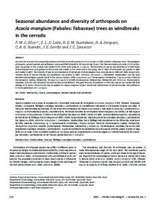
was seen at Mangalore Harbour Stations (21–23 species vs 21–32), A. quadricornuta, T. radiata and Z. varium were most frequent (> 20%). Five species: A. majusculus, H. posidoniae, Nia vibrissa, Camarosporium sp. and Cirrenalia sp., which were recorded exclusively in harbour locations, need special attention. Possibly these fungi withstand the hydrocarbon pollution stress in harbour locations. Previous studies in temperate locations: Denmark27 and Japan28; and tropical locations: Pacific Ocean26, Indian Ocean, Seychelles20 and South Africa29 yielded 4–19 species of arenicolous (sand-inhabiting) fungi. In our study, highest number of arenicolus fungi (22 species) was recorded. This might be due to the incubation of intertidal wood on sand bath. In addition to the formation of ascomata on sand grains, all arenicolous fungi produced ascomata on the wood materials. This corroborates the observations on arenicolous fungi on the beaches of Malaysia and Singapore23. The frequency of occurrence of arenicolous fungi in our study was also similar to that of the beaches of Malaysia and Singapore23. Corollospora spp. were common in almost all the locations, out of them either Corollospora maritima or Corollospora filiformis was most frequent. Corollospora spp. were more frequent (> 5%) in Kovalam, Harbour St. 1 and Anjadeep Island. Twenty-two species reported as new records to Indian Peninsula strengthened the data on biogeographic distribution of filamentous marine fungi. Kohlmeyer30 identified that four major factors control the global distribution of marine fungi: (i) availability of substrates/hosts; (ii) water temperatures; (iii) hydrostatic pressure; and (iv) the availability of oxygen. Kohlmeyer14, based on earlier studies and his own studies from tropical locations furnished the biogeographic distribution and distribution maps of higher marine fungi. Observations on mangrove ecosystem in tropical and subtropical locations helped to understand the biogeography of higher marine fungi more clearly in the tropics10. For instance, Hyde and Jones10 listed 90 higher marine fungi on 26 mangrove plant species. Jones2 classified higher marine fungi into: (i) cosmopolitan, and (ii) restricted to the subtropics and/or tropics. Based on the above facts as well as the studies from tropical locations: Pacific26, Malaysia and Singapore23; temperate locations: Japan28,31, Denmark32, Canada33 and England34, many species encountered in the present study were grouped into cosmopolitan, subtropical and or tropical in distribution (see Table 1). More biogeographic details are required to classify more authentically seven species found in the present study: A. majusculus, C. indica, D. tropicalis, H. posidoniae, M. staticicola, P. avicenniae and C. algarum. Figure 3 shows the spore morphology of some rare species of marine fungi encountered in our study. A. encephaloides was found as early as 1884 and 1888 at the off-Danish coast and along the New Zealand coast3. It was mainly recorded from temperate locations: Canada (New309
RESEARCH COMMUNICATIONS a c b
f e
d
g h
is known widely on woody debris from tropical locations of the Pacific Ocean (Hawaiian Islands), Atlantic Ocean (Trinidad), Indian Ocean (Sri Lanka) and South-East-Asia (Thailand)46. In addition, collections were also made from Queensland (Australia)46. N. bipolaris was reported by Hyde and Jones20 from the beaches of Seychelles. N. effusa was described from Sri Lanka45, subsequently reported from the Mare Anglaise (Seychelles)20. Diverse filamentous marine fungal flora was obtained on intertidal wood collected from 13 locations in the west coast of India. This study added 22 species of marine fungi to the Indian Peninsula as new records. The species richness and diversity were highest in Coconut Island followed by Kovalam and Bambolim. The lengthy period of incubation and intense scanning of the intertidal wood might have yielded higher species than mangrove locations of Indian Peninsula and islands of Indian Ocean. Arenicolous fungi constitute 25% of the total fungi recovered. Many rare fungi, e.g. A. encephaloides, A. majusculus, C. mangrovei, C. leptosphaerioides, C. pseudomacrocephala, C. maritima, D. navigans, D. tropicalis, Nimbospora bipolaris and N. effusa recorded in the present study strengthened the data on mycogeography of marine fungi.
a–c, g–i
i d–f
Figure 3. Ascospores of (a) Amylocarpus encephaloides, (b) Arenariomyces majusculus, (c) Carbosphaerella leptosphaerioides, (d) Crinigera maritima, (e) Dryosphaera navigans, (f) Dryosphaera tropicalis, (g) Nimbospora bipolaris and (h) Nimbospora effusa; (i) conidia of Cirrenalia pseudomacrocephala.
foundland and British Colombia), United States (Maine and Washington), Europe (Belgium, Denmark, Germany, Scotland and Sweden), South America (Chile)3. Recently, it is also known from the coast of Ireland35. The present record is from tropical region (Indian Ocean). A. majusculus was reported from the Pacific Ocean (Kauai, Hawaii)36. This is a report from the Indian Ocean. C. leptosphaerioides was known from United States, Hawaii, Denmark, Germany, Japan and Brunei25,28,36–40. It has been reported recently on the driftwood from South African coast (Indian Ocean)29, Malaysia and Singapore23. The present report constitutes a record from the Indian Peninsula. C. pseudomacrocephala has been reported from the Atlantic Ocean (Bermuda and Mexico)10. This is a report from the Indian Ocean. C. maritima was first reported on driftwood from Baltic Sea41 and later from Denmark42,43. It is also known from North Island of St. Mary’s Isles (west coast of India)5. D. navigans was described from Denmark on driftwood44. It is also known from Greenland, Sri Lanka and Thailand44,45. D. tropicalis 310
1. Hyde, K. D. and Lee, S. Y., Hydrobiologia, 1995, 295, 107–118. 2. Jones, E. B. G., in Aspects of Tropical Mycology (eds Isaac, S. et al.), Cambridge University Press, Cambridge, 1993, pp. 73–89. 3. Kohlmeyer, J. and Kohlmeyer, E., Marine Mycology: The Higher Fungi, Academic Press, New York, 1979. 4. Borse, B. D., Indian J. Mar. Sci., 1998, 17, 165–167. 5. Prasannarai, K. and Sridhar, K. R., Indian J. Mar. Sci., 1997, 26, 380–382. 6. Sridhar, K. R. and Kaveriappa, K. M., Mahasagar, 1991, 24, 66– 68. 7. Chinnaraj, S., Cryptogamie Mycol., 1992, 13, 312–319. 8. Chinnaraj, S., Indian J. Mar. Sci., 1993, 22, 141–142. 9. Chinnaraj, S., Sydowia, 1993, 45, 109–115. 10. Hyde, K. D. and Jones, E. B. G., PSZNI Mar. Ecolo., 1988, 9, 15–33. 11. Borse, B. D., Ch. Ramesh and Shrivastava, A. D., Indian Bot. Rep., 1988, 7, 18–25. 12. Ravikumar, D. R. and Vittal, B. P. R., Indian J. Mar. Sci., 1996, 25, 142–144. 13. Ravikumar, D. R. and Vittal, B. P. R., Kavaka, 1987, 15, 99–103. 14. Kohlmeyer, J., P.S.Z.N.I. Mari. Ecol., 1984, 5, 329–378. 15. Kohlmeyer, J. and Volkmann-Kohlmeyer, B., Bot. Mar., 1991, 34, 1–61. 16. Magurran, A. E., Ecological Diversity and its Measurement, Princeton University Press, New Jersey, 1988. 17. Pielou, F. D., Ecological Diversity, Wiley Interscience, New York, 1975. 18. Ludwig, J. A. and Reynolds, J. F., Statistical Ecology – A Primer on Methods and Computing, John Wiley and Sons, New York, 1988. 19. McGhee, J. W., Introductory Statistics, West Publishing Company, USA, 1985. 20. Hyde, K. D. and Jones, E. B. G., Bot. J. Linn. Soc., 1989, 100, 237–254. 21. Raghukumar, S., Kavaka, 1973, 1, 73–85. 22. Hyde, K. D., Bot. J. Linn. Soc., 1988, 98, 135–151. CURRENT SCIENCE, VOL. 81, NO. 3, 10 AUGUST 2001
RESEARCH COMMUNICATIONS 23. Sundari, R., Vikineswary, S., Yusoff, M. and Jones, E. B. G., Bot. Mar., 1996, 39, 327–333. 24. Hyde, K. D., in The Biology of Marine Fungi (ed. Moss, S. T.), Cambridge University Press, Cambridge, 1986, pp. 311–322. 25. Hyde, K. D., Hydrobiologia, 1989, 178, 199–208. 26. Volkmann-Kohlmeyer, B. and Kohlmeyer, J., Mycologia, 1993, 85, 337–346. 27. Farrent, C., Hyde, K. D. and Jones, E. B. G., Trans. Br. Mycol. Soc., 1985, 85, 164–167. 28. Nakagiri, A., Inst. Ferment. Osaka Res. Commun., 1989, 14, 52–79. 29. Steinke, T. D. and Jones, E. B. G., S. Afr. J. Bot., 1993, 59, 385– 390. 30. Kohlmeyer, J., Aust. J. Bot. Suppl. Ser., 1983, 10, 67–76. 31. Nakagiri, A. and Tokura, R., Trans. Mycol. Soc. Jpn., 1987, 28, 413–436. 32. Koch, J. and Petersen, K. R. L., Mycotaxon, 1996, 60, 397–414. 33. Strongman, D, Miller, J. D. and Whitney, N. J., Proc. N. S. Inst. Sci., 1985, 35, 99–105. 34. Haythorn, J. M., Jones, E. B. G. and Harrison, J. L., Trans. Br. Mycol. Soc., 1980, 74, 615–623. 35. Crumlish, B. and Curran, P., Mycologist, 1994, 8, 83–84. 36. Kohlmeyer, J. and Volkmann-Kohlmeyer, B., Mycol. Res., 1989, 92, 410–421.
Remobilizing P elements out of the stambh A locus of Drosophila melanogaster M. Paratpara Rao, Charu Jain and Shanti Chandrashekaran* Division of Genetics, Indian Agricultural Research Institute, New Delhi 110 012, India
P elements from two P-tagged alleles, stm AP1 and stm AP4, of the temperature-sensitive paralytic mutant stambh A (stm A) were remobilized to derive wild-type revertants and P excision lethals. stm AP1 remutated to lethality, while stm AP4 reverted to the wild type. The P excision lethals are not amorphic mutations, since they were weaker in paralytic phenotype (i) than the parental P-tagged allele and (ii) for deficiencies for stm A. The lethals collectively affected all stages of the life cycle, demonstrating that stm A+ is needed throughout fly development. P transposon elements serve as excellent tools for genetic analyses due to the ease with which mutations can be created at high frequencies, by way of insertions and excisions. Excision of P elements from an original insertion site can either be precise, resulting in reversion of the original mutation to wild type or imprecise (transposon leaves part of its sequence or takes away flanking nucleo-
*For correspondence. (e-mail:
[email protected]) CURRENT SCIENCE, VOL. 81, NO. 3, 10 AUGUST 2001
37. Koch, J., Friesia, 1974, 10, 209–250. 38. Kohlmeyer, J., Trans. Br. Mycol. Soc., 1971, 57, 473–492. 39. Schmidt, I., Hohere. Meerespilze der Ostsee. Biol. Rundsch., 1974, 12, 96–112. 40. Tokura, R., Bot. Mar., 1984, 27, 567–569. 41. Schmidt, I., Nat. Natursch. Meckl., 1969, 7, 5–14. 42. Rees, G., Johnson, R. G. and Jones, E. B. G., Trans. Br. Mycol. Soc., 1979, 72, 99–106. 43. Jones, E. B. G., Moss, S. T. and Koch, J., Trans. Br. Mycol. Soc., 1980, 74, 625–631. 44. Koch, J. and Jones, E. B. G., Can. J. Bot., 1989, 67, 1183–1197. 45. Koch, J., Nor. J. Bot., 1982, 2, 163–169. 46. Volkmann-Kohlmeyer, B. and Kohlmeyer, J., Can. J. Bot., 1993, 71, 992–995. ACKNOWLEDGEMENTS. We are thankful to Dr N. S. Raviraja for the statistical analysis, Dr S. K. Shyama for field collections at Goa and Mr A. B. Arun for technical assistance. We are also grateful to Mangalore University for granting permission to carry out this study and the referees for constructive suggestions to enrich the manuscript. Received 3 February 2001; revised accepted 31 March 2001
tides of the host gene), resulting in secondary mutations. Excision events involving more than one transposon often result in chromosomal rearrangements such as duplications, deletions, inversions and translocations1. Frequencies of precise excisions of P elements leading to wild-type revertants in Drosophila melanogaster are reported to vary widely from 4 × 10–13 at the white locus2,3 to 3.5 × 10⁻¹ at the singed locus 4. stambh A (stm A) was first identified through a recessive, reversible, temperature-sensitive (ts) paralytic mutation mapping to 56.8 cM on the second chromosome5. EMS-induced homozygous viable alleles, stm A1 and stm A2 paralyse at 38°C in 3–4 min and recover to normality at 23–24°C in 5–6 min. Later, isolated unconditional embryonic lethal alleles, stm A7 and stm A12 (also EMS-induced), showed hypotrophy of the anterior embryonic dorsal cuticle overlying the brain with a concomitant hypertrophy of the anterior dorsal neurogenic region, the brain6. The stm A7 and stm A12 alleles were weaker in their paralytic phenotype when heterozygous over stm A1 and stm A2. The time required for paralysis of 50% of flies (at 38°C) of stm A1 and stm A2 was 2.4 and 1.4 min respectively, while that for trans-heterozygote viable/lethal combinations of stm A1 (or stm A2)/stm A12 (or stm A7) ranged between 4 and 7 min6. stm A1/+ and stm A2/+ heterozygotes also show weak paralysis at 39°C, a temperature at which wild-type flies do not paralyse6. Based on these observations it was proposed that homozygous viable ts paralytic alleles stm A1 and stm A2, are not simple hypomorphs, but are semi-dominant gain of function neomorphs. Embryonic lethal alleles, stm A7 and stm A12 on the other hand, are extreme or complete loss 311