Articles in PresS. Am J Physiol Lung Cell Mol Physiol (September 18, 2015). doi:10.1152/ajplung.00293.2015
1
DNA damage response at telomeres contributes to lung ageing and chronic
2
obstructive pulmonary disease
3
Corresponding author: Dr. João F. Passos, Institute for Cell and Molecular
4
Biosciences, Newcastle University Institute for Ageing, Campus for Ageing and
5
Vitality, Newcastle University, Newcastle upon Tyne, NE4 5PL, UK. Phone +44 191
6
248 1222, Fax +44 191 248 1101, Email
[email protected]
7
Jodie Birch1,2, Rhys K Anderson1, Clara Correia-Melo1, Diana Jurk1, Graeme Hewitt1,
8
Francisco Madeira Marques1, Nicola J Green2, Elizabeth Moisey2, Mark A Birrell5,
9
Maria G Belvisi5, Fiona Black6, John J Taylor4, Andrew J Fisher2,3, Anthony De
10 11 12 13 14 15 16 17 18 19 20
Soyza2*, #João F. Passos1* 1
Newcastle University Institute for Ageing, Institute for Cell and Molecular
Biosciences, Newcastle upon Tyne, NE4 5PL, UK 2
Lung Immunobiology group, Institute of Cellular Medicine, Newcastle University,
Newcastle upon Tyne, NE2 4HH, UK 3
Institute of Transplantation, Freeman Hospital, Newcastle upon Tyne, NE7 7DN, UK
4
Institute of Cellular Medicine, Newcastle University, Newcastle upon Tyne, NE2
4HH, UK 5
Respiratory Pharmacology, Airway Disease Section, National Heart and Lung
Institute, Faculty of Medicine, Imperial College London, London, SW7 2A7, UK 6
Department of Pathology, Newcastle upon Tyne Hospitals Trust, Newcastle upon
21
Tyne, UK
22
Running title: Telomere dysfunction in lung ageing and COPD
23
Copyright © 2015 by the American Physiological Society.
1
24
Cellular senescence has been associated with the structural and functional decline
25
observed during physiological lung ageing and in chronic obstructive pulmonary
26
disease (COPD). Airway epithelial cells are the first line of defense in the lungs and
27
are important to COPD pathogenesis. However, the mechanisms underlying airway
28
epithelial cell senescence, and particularly the role of telomere dysfunction in this
29
process, are poorly understood. We aimed to investigate telomere dysfunction in
30
airway epithelial cells from patients with COPD, in the ageing murine lung and
31
following cigarette smoke exposure. We evaluated co-localization of γH2A.X and
32
telomeres and telomere length in small airway epithelial cells from patients with
33
COPD, during murine lung ageing and following cigarette smoke exposure in vivo
34
and in vitro. We found that telomere-associated DNA damage foci increase in small
35
airway epithelial cells from patients with COPD, without significant telomere
36
shortening detected. With age, telomere-associated foci increase in small airway
37
epithelial cells of the murine lung, which is accelerated by cigarette smoke exposure.
38
Moreover, telomere-associated foci predict age-dependent emphysema; and late-
39
generation Terc null mice, which harbour dysfunctional telomeres, show early-onset
40
emphysema. We found that cigarette smoke accelerates telomere dysfunction via
41
reactive oxygen species in vitro and may be associated with ATM-dependent
42
secretion of inflammatory cytokines IL-6 and IL-8. We propose that telomeres are
43
highly sensitive to cigarette smoke-induced damage and telomere dysfunction may
44
underlie decline of lung function observed during ageing and in COPD.
45
Word count: 233
46
Key words: senescence, airway epithelial cells, cigarette smoke
47
2
48
Introduction
49
Chronic obstructive pulmonary disease (COPD) is a major global health problem that
50
is becoming increasingly prevalent (26). COPD is characterized by chronic
51
inflammation of the peripheral airways and lung parenchyma and involves airway
52
fibrosis, mucous hypersecretion (chronic bronchitis) and destruction of alveolar
53
airspaces (emphysema). The key risk factor for COPD is cigarette smoking (19).
54
Accelerated lung ageing and cellular senescence have been associated with COPD
55
(38, 48). Senescence, defined as the irreversible loss of division potential in somatic
56
cells, plays important roles in vivo: on the one hand, it protects against cancer
57
progression yet on the other hand contributes to age-dependent tissue dysfunction
58
(10). Evidence is mounting that cells bearing senescent markers accumulate in
59
tissues with age (23) and in age-related diseases (13).
60
Telomeres are specialized structures at the ends of chromosomes consisting of
61
tandem TTAGGG repeats stabilized by a complex of proteins, known as shelterin
62
(15). Shelterin is thought to arrange telomeric DNA into a loop structure known as
63
the T-loop. It is believed that during replicative senescence the progressive loss of
64
telomere repeats destabilizes T-loops, increasing the probability of telomere
65
uncapping, i.e. loss of shelterin (21). Telomere uncapping, whether by inhibition of
66
shelterin or telomere shortening due to extensive replication, has been shown to
67
activate the DNA damage response (DDR) in a manner similar to double strand
68
breaks (DSBs) (14). Uncapped telomeres become associated with DDR factors such
69
as phosphorylated forms of the histone protein 2A.X (H2A.X) and ataxia
70
telangiectasia mutated (ATM), which can activate a signaling cascade leading to
71
culmination of senescence (14). More recently, it has been shown that a DDR can
3
72
induce senescence irrespectively of telomere length, which has been attributed to
73
telomeres being particularly susceptible to oxidation-induced damage and to the
74
inability of telomeres to repair DSBs (20, 25, 30). Moreover, it has been shown in
75
vivo that with age, telomeres co-localizing with DDR proteins increase in the skin of
76
baboons (23) and in the liver, brain and gut of mice, which can occur irrespectively of
77
length (20, 25).
78
Telomere shortening has been associated with COPD in circulating leukocytes (45),
79
alveolar epithelial cells (27, 36) and pulmonary vascular endothelial cells (5).
80
However, it is unclear whether activation of a DDR at telomeres contributes to
81
senescence and tissue dysfunction in the ageing lung and to COPD-associated
82
accelerated lung ageing. In our study, we investigate the role of telomere dysfunction
83
in the ageing mouse lung and its potential role in cigarette smoke-induced COPD.
84
Methods
85
Study subjects
86
Patients undergoing lung resection for localised lung tumours were recruited as
87
controls from the Freeman Hospital, Newcastle upon Tyne, UK (Table 1). Samples
88
from patients with advanced COPD were obtained from an archive of explant lung
89
tissue taken at time of lung transplantation at Freeman Hospital. A smaller number of
90
cases were used for immuno-FISH analysis due to limited availability of tissue at
91
time of staining. All samples were parenchymal and only airways with a diameter of
92
less than 2 mm and without cartilage were included in the analysis. The clinical
93
characteristics of these subjects are the same as those listed in Table 1. All subjects
94
gave written informed consent prior to inclusion in the study. This work was
4
95
approved by the County Durham and Tees Valley 2 Research Ethics Committee
96
(Res-11/NE/0291)
97
Animals
98
Wild-type C57BL/6 male mice were used (n=3-5 per age group (6.5, 15 and 24
99
months)). TERC-/- C57BL/6 male mice were bred to produce successive generations
100
of mice with decreasing telomere length. Lungs from fourth-generation (G4) mice
101
were collected. All work was compiled with the guiding principles for the care and
102
use of laboratory animals. The project was approved by the Faculty of Medical
103
Sciences Ethical Review Committee, Newcastle University. Project license number:
104
60/3864.
105
Lung tissues from mice exposed to either room air or cigarette smoke were a kind
106
gift from Dr Mark Birrell, Imperial College London, UK. Male C57BL/6 mice
107
(n=5/group) at 10 weeks of age were subjected to a whole body cigarette smoke
108
exposure system or room air, as previously described (18). Briefly, cigarette smoke
109
was generated using 3R4F cigarettes (Cigarette filter removed, Tobacco Health
110
Research Institute, University of Kentucky, Lexington, KY) and pumped into a
111
Teague chamber (136L) for 1 hour, twice daily (500ml/min) for 14 days. Mice were
112
sacrificed 24 hours after the final exposure.
113
Cell culture and treatments
114
Human embryonic lung MRC5 fibroblasts were obtained from ECACC (Salisbury,
115
UK) and cultured in Dulbecco’s Modified Eagle’s Medium (DMEM) (Sigma, Dorset,
116
UK) supplemented with foetal bovine serum (FBS) (10% v/v), L-glutamine (2mM),
117
penicillin/streptomycin and maintained at 37°C, 5% CO2.
5
118
Primary human bronchial epithelial cells (PBECs) were isolated from bronchial
119
brushings carried out during research bronchoscopy (normal controls) or from
120
explant lung tissue specimens (COPD) (Table 2). The work was performed under
121
approval of the Newcastle 1 Research Ethics Committee. PBECs were cultured on
122
0.5% Purecol (Invitrogen, Carlsbad, CA)-coated dishes in small airway epithelial cell
123
growth medium (L/SABM) supplemented with 2% FBS, 100 U ml-1 penicillin and 100
124
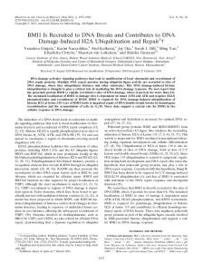
mg ml-1 streptomycin (Lonza, Basel, Switzerland).
125
MRC5 fibroblasts (PD 20-25) were grown until replicative senescence and cultured
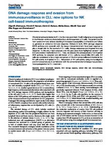
126
with DMEM plus 5% cigarette smoke extract (CSE) or DMEM alone. CSE was
127
generated by bubbling smoke from one research grade cigarette (University of
128
Kentucky, 4A1) into 25 ml DMEM. The solution was filtered (0.2 μm) and the
129
resulting CSE designated 100%. The CSE solution was diluted to 5% in sterile
130
DMEM and used immediately. CSE or DMEM alone was replenished every 48 hours.
131
Identical experiments under hypoxic conditions (3% O2) were run in parallel. Human
132
primary small airway epithelial cells (P1-3) were treated with two exposures of 5%
133
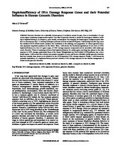
CSE or media alone (control), 48 hours apart.
134
Chemical inhibitors used: KU55933 (ATM chemical inhibitor) (10μM, diluted in
135
DMSO) (R&D, 3544). Inhibitors were replaced every 48 hours along with 5% CSE or
136
fresh DMEM.
137
Immunofluorescence
138
Cells grown on coverslips were fixed with 2% paraformaldehyde (PFA),
139
permeabilized with PBG-Triton and incubated with the primary antibody at 4°C
140
overnight. The following day, cells were incubated with fluorescein-conjugated
141
secondary antibody (Alexa Fluor® 488 or 594) (Invitrogen) for 45-minutes at room
6
142 143
temperature. Primary antibodies used: rabbit polyclonal anti-Ki67 (ab15580; 4 μg ml– 1
Abcam), mouse monoclonal anti-γH2AX (#05-636; 0.25 μg ml–1 Millipore), mouse
144
monoclonal anti-p16 (SC-81156; 1:500 Santa Cruz).
145
Immuno-FISH
146
Immuno-FISH was performed as described (25). Briefly, cells grown on sterile
147
coverslips were fixed with 2% paraformaldehyde and incubated with anti-γH2AX
148
antibody (mouse monoclonal, #05-636, Millipore) overnight at 4°C. After application
149
of the secondary antibody, cells were washed with PBS and FISH was performed.
150
10μl of Cy-3-labelled telomere specific (C3TA2)3 peptide nucleic acid (PNA) probe
151
(4 ng μl-1) (Panagene) was applied to the cells followed by denaturation at 80 °C and
152
hybridization for 2 hours at room temperature in the dark. Cells were washed three
153
times for 10 min with wash buffer (70 ml formamide (70%), 30 ml SSC 2%) and three
154
times for 5 min with TBS-Tween 0.05%. Cells were incubated with 4,6-diamidino-2-
155
phenylindole (DAPI), mounted and imaged using a Leica DM5500B fluorescence
156
microscope. In depth Z-stacking was used (images were captured as stacks
157
separated by 0.247 μm with ×100 objective) followed by Huygens (SVI)
158
deconvolution.
159
For immuno-FISH in formalin-fixed paraffin-embedded murine and human lung
160
tissues, sections were de-waxed in 100% Histoclear and hydrated in 100%, 90% and
161
70% ethanol (2 x 5 minute incubations) and in distilled water (2 x 5 minutes). For
162
antigen retrieval, the slides were placed in 0.01 M citrate buffer and heated until
163
boiling for 10 min. After cooling down to room temperature, the slides were washed
164
twice with distilled water for 5 min. After blocking in normal goat serum (NGS) (1:60)
165
in BSA/PBS, primary antibody (rabbit polyclonal anti- γH2AX 1:400)(Cell Signaling,
7
166
9718) was applied and incubated at 4 °C overnight. The next day, slides were
167
washed three times in PBS, incubated with secondary antibody for 30 min, washed
168
three times in PBS and incubated with Avidin DCS (1:500) for 20 min. Following
169
incubation, slides were washed three times in PBS and dehydrated with 70, 90 and
170
100% ethanol for 3 min each. Sections were denatured for 5 min at 80 °C in
171
hybridization buffer (70% formamide (Sigma), 25 mM MgCl2, 1 M Tris pH 7.2, 5%
172
blocking reagent (Roche)) containing 2.5 μg ml − 1 Cy-3-labelled telomere specific
173
(CCCTAA) peptide nuclei acid probe (Panagene), followed by hybridization for 2
174
hours at room temperature in the dark. The slides were washed twice, for 15 min
175
each, with wash buffer, followed by 2×SSC and PBS washes for 10 min. Sections
176
were incubated with DAPI, mounted and imaged. In depth Z-stacking was used (a
177
minimum of 40 optical slices with ×100 objective) followed by Huygens (SVI)
178
deconvolution.
179
Immunohistochemistry
180
Sections were dewaxed in xylene (5 min), rehydrated through graded ethanol
181
solutions (100%, 90% and 70%) and washed in distilled H2O. Endogenous
182
peroxidase activity was blocked by immersing sections in 0.3% H2O2 (Sigma, H1009)
183
diluted in methanol for 30 minutes. To retrieve antigens, sections were boiled in
184
0.01M citrate (pH 6.0) or 0.01M EDTA buffer (pH 8.0). Sections were blocked in 5%
185
non-fat milk protein (NFMP) diluted in TBS (human) or NGS diluted 1:60 in 0.1%
186
BSA in PBS (mouse). Sections were incubated with the primary antibody overnight
187
at 4°C or for 45 min – 1 hour at room temperature. For human tissue, anti-mouse
188
(Dako, K4006) or anti-rabbit (Dako, K4010) secondary antibody conjugated with a
189
horseradish-peroxidase (HRP)-labelled polymer was added (Envision+ System-HRP,
8
190
Dako) and slides were treated with 3,3’-diaminobenzidine (DAB) for 5-10 min. For
191
mouse tissue, biotinylated secondary antibody was added and detected using the
192
rabbit peroxidase ABC kit (Vector lab, PK-4001) according to the manufacturer’s
193
instructions. Substrate was developed using the NovaRed kit (Vector lab, SK-4800).
194
Nuclei were counterstained with Carrazzi haematoxylin and sections were
195
dehydrated through graded ethanol solutions, cleared in xylene and mounted in Di-
196
N-Butyle Phthalate in Xylene (DPX) (Thermo Scientific, LAMB-DPX). Staining was
197
analysed with a NIKON ECLIPSE-E800 microscope and images were captured with
198
a Leica DFC420 camera using the LAS software (Leica). Primary antibodies used:
199
mouse monoclonal anti-p16 (1:500, SC-81156; Santa Cruz), rabbit polyclonal anti-
200
SIRT1 (1:100, ab13749; Abcam), rabbit polyclonal anti-p21 (1:200, ab7960; Abcam).
201
Senescence-associated β-galactosidase staining
202
Senescence-associated β-Galactosidase staining was carried out as previously
203
described (17). Briefly, cells were fixed with 2% formaldehyde in PBS for 5 min.
204
Following fixation, cells were washed once with PBS and incubated at 37°C for 16
205
hours in freshly prepared senescence-associated β-Galactosidase staining solution
206
containing 2 mM magnesium chloride, 150 mM sodium chloride, 40 mM citric acid,
207
12 mM sodium phosphate dibasic, 5 mM potassium ferrocyanide, 5 mM potassium
208
ferricyanide and 1 mg/L, 5-bromo-4-chloro-3-inolyl-β-D-galactoside (X-Gal) at pH
209
6.0. Following staining, nuclei were stained with DAPI and cells were imaged.
210
Analysis of telomere length by Real-time PCR
211
Telomere length of isolated epithelial cells was measured by quantitative real-time
212
PCR, as described (34). Telomere length was measured as abundance of telomeric
213
template versus a single gene by quantitative real-time PCR. Measurements were
9
214
performed in quadruplicate. Three DNA samples with known telomere lengths (3.0,
215
5.5 and 9.5 kbp) were run as internal standards, allowing us to estimate telomere
216
length in base pairs.
217
Detection of ROS levels
218
Superoxide anion levels were determined by flow cytometric analysis of MitoSOX
219
fluorescence as described (40).
220
Detection of SASP factors
221
A Quantibody Human Cytokine Array for 20 cytokines (RayBiotech; QAH-CYT-1)
222
was performed. Concentrations of IL-6 and IL-8 in cell culture media were
223
determined using sandwich ELISA (R&D Systems; DY206/DY208) according to the
224
manufacturer’s instructions. Limits of detection for these assays were 10 pg/ml-1.
225
Western blotting
226
Western blotting was conducted using routine protocol.
227
Statistical analysis
228
Data are represented as mean +/- SEM or median +/- range. Where data were
229
normally distributed, statistically significant differences between groups were
230
assessed using ANOVA and significant differences between two groups were
231
evaluated using an Independent samples t-test. Where data were not normally
232
distributed, statistical significant differences between groups were assessed using
233
the Kruskal Wallis test and significant differences between two groups were
234
evaluated using the Mann-Whitney U test. P values less than 0.05 were considered
10
235
significant. Data were analyzed with GraphPad Prism version 6.0, GraphPad
236
Software, San Diego California USA, www.graphpad.com
237
Results
238
Patients with COPD show increased telomere-associated foci in small airway
239
epithelial cells
240
To assess telomere dysfunction, we obtained explant lung tissue from patients
241
undergoing transplantation for COPD (n=10) and from controls (n=9) undergoing
242
pulmonary resection for localized lung cancer (Table 1). We performed telomere
243
specific Q-FISH together with immunofluorescence staining against DNA damage
244
protein γH2A.X (immuno-FISH). Analysis revealed a significant increase in % of
245
small airway epithelial cells containing telomere-associated DNA damage foci (TAF)
246
in patients with COPD (Figure 1a & 1b). No significant differences in telomere FISH
247
intensity were detected (Figure 1c). Similarly, analysis of individual telomeres in
248
small airway epithelial cells in the COPD lung revealed no differences in FISH
249
intensity between co-localizing (with γH2A.X) and non-co-localizing telomeres
250
(Figure 2), suggesting that TAF can occur independently of telomere length. To
251
determine whether other senescence markers were increased in patients with
252
COPD, we conducted immunohistochemistry against p16, p21 and SIRT1. p16 and
253
p21 are cyclin-dependent kinase inhibitors and tumour suppressors, expressed in
254
most senescent cells (31). Decreased expression of SIRT1 (a NAD-dependent
255
deacetylase), has been associated with cellular senescence (50). Consistent with a
256
senescent-associated phenotype, using a semi-quantitative scoring method, we
257
observed increased p16 and decreased SIRT1 expression in small airway epithelial
258
cells from patients with COPD (Figure 1d). No significant differences in p21
259
expression were observed (data not shown). Using Immuno-FISH (p16 and γH2A.X),
11
260
we found that p16 positive cells have more TAF than p16 negative cells (Figure 1e),
261
suggesting that TAF may be involved in senescence induction.
262
Following ex vivo analysis, we investigated whether TAF were increased in small
263
airway epithelial cells isolated from the COPD lung (Table 2). By Immuno-FISH, we
264
found a significant increase in % of cells positive for TAF from patients with COPD
265
(Figure 3a), without significant differences in telomere FISH intensity (Figure 3b).
266
Because we found no significant differences in telomere length using Q-FISH, we
267
compared telomere length in small airway epithelial cells isolated from COPD
268
patients and age-matched controls (Table 3), using quantitative real-time PCR.
269
Similarly, we detected no statistically significant differences (Figure 3b). Small
270
airway epithelial cells isolated from the COPD lung had increased positivity of Sen-β-
271
Gal, however this failed to reach statistical significance, with extensive inter-patient
272
variability observed (Figure 3c and d).
273
Telomere-associated foci increase in small airway epithelial cells in mice with
274
age and following cigarette smoke exposure
275
Following our observation that TAF were increased in small airway epithelial cells of
276
patients with COPD, we investigated whether TAF increased in small airway
277
epithelial cells during physiological ageing. Mice have long telomeres and express
278
ubiquitously the enzyme telomerase; hence it was believed that telomere dysfunction
279
did not play a role in cellular senescence in murine tissues (39). However, our group
280
demonstrated that TAF accumulate in liver and intestine with age (25) and TAF have
281
been shown to quantitatively predict mean and maximum lifespan in both short- and
282
long-lived mice cohorts (29).
12
283
We found a significant increase in % of cells positive for TAF from 6.5 until 24
284
months of age (as well as mean number of TAF per cell, not shown) (Figure 4a &
285
4b). No significant changes in telomere FISH intensity were found, however, a
286
tendency for decreased FISH intensity in older animals was observed (Figure 4c).
287
Telomere dysfunction has been associated with increased expression of p21 (12).
288
Consistently, we found with increasing age that a greater % of small airway epithelial
289
cells stained positive for p21 (Figure 4d). The ageing lung is associated with
290
structural changes similar to those that occur in emphysema, including distal
291
airspace enlargement (28). Consistent with this, we found increased airspace size in
292
mice with age, indicated by a decreasing number of airspaces per visual field
293
(Figure 4e). Interestingly, telomere FISH intensity did not correlate with airspace
294
number, however there was an inverse correlation between % of cells positive for
295
TAF and number of airspaces (P = 0.02) (Figure 4e). These results suggest that
296
telomere dysfunction may play a role in age-related lung tissue decline.
297
Cigarette smoke has been associated with early onset-senescence and induction of
298
H2A.X phosphorylation in human pulmonary endothelial cells in vitro (3) and
299
telomere length is reduced in small airway epithelial cells isolated from healthy
300
smokers (49). However, the role of cigarette smoke in activation of a DDR
301
specifically at telomeres has not been fully elucidated. We found that 3-month-old
302
mice exposed to cigarette smoke, twice daily for 2 weeks, had an increased % of
303
small airway epithelial cells positive for TAF, similar to levels observed in mice at 15
304
months of age. While this increase was not significant (P = 0.06), we found that
305
mean number of TAF increased significantly (P = 0.03) (Figure 4f). No significant
306
differences in telomere FISH intensity were observed (Figure 4f). Altogether, these
13
307
results suggest that small airway epithelial cells accumulate TAF with age, which can
308
be accelerated by cigarette smoke exposure.
309
Late-generation TERC-/- mice show increased telomere-associated foci in
310
small airway epithelial cells and early-onset emphysema
311
At late generations, mice deficient in the RNA component of telomerase (mTERC-/-),
312
exhibit a number of phenotypes indicative of premature ageing, thought to be due to
313
early onset of senescence (8). Late generation mTERC-/- mice show critically short
314
telomeres in most tissues and premature incidence of TAF. We found at 6 months of
315
age that 4th generation mTERC-/- mice have an increase in % of small airway
316
epithelial cells containing TAF and decreased telomere FISH intensity (Figure 5a, 5b
317
& 5c). Consistent with the hypothesis that telomere dysfunction contributes to loss of
318
alveolar integrity, we found a significant reduction in number of airspaces in G4
319
mTERC-/- mice (Figure 5d and 5e). The correlation between TAF and number of
320
airspaces we report is strengthened when G4 mTERC-/- are added (Figure 5f),
321
however telomere FISH intensity still does not correlate (not shown).
322
Cigarette smoke extract induces telomere-associated foci and senescence
323
markers in primary human airway epithelial cells and MRC5 fibroblasts
324
In vitro exposure to cigarette smoke has been shown by several groups to result in
325
expression of senescence-associated markers (3, 38). Nevertheless, the role of
326
telomere dysfunction in cigarette smoke-induced senescence is less clear. Recent
327
data from our group and others have revealed that stress-induced activation of a
328
DDR at telomeres is persistent when compared to non-telomeric damage, mostly
329
because of inhibition of DNA repair mechanisms at telomere regions (20, 25). This
14
330
suggests that TAF, given their persistence, may be excellent markers for age-related
331
accumulated damage.
332
In order to determine whether cigarette smoke extract (CSE) induced TAF in isolated
333
small airway epithelial cells, we cultured cells isolated from healthy non-smoking
334
controls (n=5) and exposed them to 5% CSE for 48 hours. Small airway epithelial
335
cells had increased TAF following CSE exposure (Figure 6a & 6b), however
336
analysis of Sen-β-Gal expression revealed no significant increases (not shown). Our
337
data shows that TAF are induced as a consequence of CSE and may precede the
338
induction of other senescence markers.
339
Epithelial cells cannot be cultured for prolonged periods of time without induction of
340
epithelial to mesenchymal transition (EMT), a process whereby epithelial cells lose
341
their epithelial features and acquire mesenchymal characteristics. This limits our
342
ability to determine the effects of chronic cigarette smoke exposure on telomere
343
dysfunction and other senescence-associated phenotypes. Therefore, we used
344
normal human foetal lung fibroblasts (MRC5), which can be cultured for longer
345
periods of time and are not overly-sensitive to the effects of cigarette smoke
346
exposure. MRC5 cells were cultured for 60 days in the presence or absence of 5%
347
CSE to determine the effects of long-term cigarette smoke exposure. Consistent with
348
previous observations, we show that long-term exposure to CSE induces
349
accelerated senescence in MRC5 cells, evidenced by reduced population doublings
350
(PD) (Figure 6c), increased Sen-β-Gal activity (Figure 6d) and reduction in
351
proliferation marker Ki67 (Figure 6d). Senescence is characterised by increased
352
secretion of bioactive, pro-inflammatory peptides; the so-called senescence-
353
associated secretory phenotype (SASP). We first conducted a cytokine array
354
analysis of 20 pro-inflammatory mediators and found that CSE exposure for 39 days
15
355
leads to increased secretion of interleukin-6 (IL-6), interleukin-8 (IL-8), GRO,
356
monocyte chemotactic protein 1 (MCP-1) and vascular endothelial growth factor
357
(VEGF) (Figure 6e). Most other cytokines or growth factors analysed were secreted
358
below detection level. This is consistent with previous reports suggesting that pro-
359
inflammatory cytokines IL-6 and IL-8 are major components of the SASP and have
360
been shown to contribute to the induction and maintenance of senescence in
361
autocrine and paracrine fashions (1, 2). ELISAs for IL-6 and IL-8 detection
362
independently confirmed the findings of the cytokine array (Figure 6e). This rise in
363
cytokine secretion following CSE exposure was first observed at day 13, but became
364
enriched after 39 days in culture when cells reached premature senescence.
365
Consistent with a potential role for telomere dysfunction in the process, immuno-
366
FISH revealed a significant increase in the % of cells containing TAF and in the
367
mean number of TAF following CSE exposure, preceding other markers of
368
senescence (Figure 6f & 6g). At day 67 in culture, all cells were positive for TAF, but
369
mean number of TAF was increased in CSE-exposed cells (Figure 6f).
370
Cigarette smoke-induced pro-inflammatory phenotype is accelerated by ROS-
371
dependent telomere dysfunction
372
Mechanistically, it is unclear how CSE induces telomere dysfunction. Telomeres are
373
highly sensitive to oxidative stress compared to the bulk of the genome, and less
374
efficiently repaired when subjected to single- and DSBs (25, 42). Moreover, it has
375
been shown that exposure to CSE increases ROS and oxidative stress markers in
376
human fibroblasts and airway epithelial cells (9, 16). To determine whether TAF were
377
induced by oxidative stress, we cultured MRC5 fibroblasts under low oxygen
378
pressure (3% O2) and exposed them for 25 days to 5% CSE. Culturing cells at low
16
379
oxygen pressure is an accepted method of reducing intracellular ROS and under
380
these conditions we and others have previously reported reduced mitochondrial and
381
cellular oxidative stress (39, 40, 43), which resulted in reduced DNA damage and
382
delayed onset of senescence. We found that CSE significantly increased
383
mitochondrial-derived ROS (measured by MitoSOX fluorescence), which was
384
suppressed when culturing cells under 3% O2 (Figure 7a). Consistent with a causal
385
role for oxidative stress in TAF induction, we found that low oxygen was able to
386
supress smoke-induced increases in TAF (Figure 7b). Similarly, treatment of
387
primary small airway epithelial cells with antioxidant N-Acetyl-Cysteine (NAC)
388
significantly reduced short-term CSE-induced increases in TAF (Figure 7c). As
389
shown previously, CSE exposure increased % of cells positive for Sen-β-Gal,
390
however, cultivation of cells at 3% O2 drastically reduced frequencies of Sen-β-Gal-
391
positive cells (Figure 7d). Furthermore, we found that CSE driven growth arrest was
392
suppressed upon cultivation of MRC5 fibroblasts at low oxygen (not shown).
393
Telomere-dysfunction and resulting DDR activation result in increased expression of
394
IL-6 and IL-8 (44). Consistent with a role for ROS-dependent telomere dysfunction
395
contributing to the SASP, we found that low oxygen significantly reduced IL-6 and IL-
396
8 secretion in MRC5 fibroblasts, irrespective of smoke exposure (Figure 7e).
397
Mechanistically, it has been shown that persistent ATM activation is necessary for
398
induction of the SASP (44). In order to test the hypothesis that CSE-dependent
399
activation of a DDR results in increased IL-6 and IL-8, we treated smoke exposed
400
MRC5 fibroblasts with an ATM inhibitor (KU55933). We first demonstrated that
401
KU55933 supresses phosphorylation of H2AX (a target of ATM kinase), confirming
402
its role in DDR inhibition (Figure 7f). At 14 days we found that ATM inhibition
403
represses smoke-induced TAF increase (not shown) as well as IL-6 and IL-8
17
404
secretion (Figure 7f) supporting the hypothesis that smoked-induced DDR activation
405
results in increased SASP.
406
Altogether, the results from MRC5 cells suggest that cigarette smoke exposure
407
causes telomere dysfunction, possibly through increased oxidative stress, which
408
leads to senescence induction and SASP activation.
409
DISCUSSION
410
Increased cellular senescence is a major feature of ageing and has been implicated
411
in COPD pathogenesis. Short telomeres, known activators of cellular senescence,
412
have been implicated in COPD, mostly in circulating leukocytes (45).
413
Recent data has questioned the utility of telomere length in circulating leukocytes as
414
a biomarker of ageing. While some studies suggest that leukocyte telomere length
415
may act as a proxy for telomere length in other somatic cell types, there is evidence
416
suggesting that this is not true for some tissues (47). Furthermore, recent studies
417
have suggested telomere dysfunction can be induced independently of length. In
418
fact, data suggests that senescence can be induced by activation of a DDR at
419
relatively long telomeres in human fibroblasts during stress- (20, 25), replicative (30),
420
and oncogene-induced senescence in vitro (46) and in mice in vivo (25). While the
421
mechanisms driving telomere dysfunction are still unclear, these data suggest that a
422
“critical” telomere length may not be the sole determinant in the activation of a
423
persistent DDR.
424
Using Q-FISH and real-time PCR in small airway epithelial cells, we failed to detect
425
robust differences in telomere length between controls and patients with COPD. This
426
contrasts with previous reports where telomere shortening is described in other lung
427
cells from patients with COPD, including alveolar type II cells and endothelial cells
18
428
(5, 48). It is possible that our study failed to detect differences in telomere length due
429
to a relatively small sample size. Decreased telomere length in smokers (49) and
430
patients with COPD (5) has been described, using larger sample sizes than in our
431
study. However, only small differences in telomere length have been reported (less
432
than 15% in most cases). We observe significant increases in the frequency of cells
433
positive for TAF in patients with COPD, even with smaller sample sizes. Moreover,
434
we have found clear evidence for increased TAF in small airway epithelial cells in
435
lung tissue from COPD patients, which are younger than controls, demonstrating that
436
TAF are robust indicators of COPD-associated damage, despite the age
437
discrepancy. Consistently, another study also failed to find differences in telomere
438
length between lung fibroblasts isolated from patients with emphysema and aged-
439
matched controls, despite increased expression of senescence-associated markers
440
(37). In addition to increased TAF, we observed increased p16, which is considered
441
a hallmark of senescence. Moreover, TAF content was greater in p16-positive cells,
442
suggesting that TAF correlate with expression of senescence markers. We did not
443
detect differences in p21 positivity between patients with COPD and controls.
444
However, the p16-pRB pathway may be activated following activation of, or
445
independent to, the p21 pathway (24).
446
Our study cannot eliminate telomere shortening as a mechanism driving COPD-
447
associated telomere dysfunction because i) in age-matched isolated small airway
448
epithelial cells our sample number is relatively small and ii) in lung tissue sections,
449
where our numbers are greater, the controls were older. Nonetheless, comparison of
450
individual telomere lengths co-localising and not with γH2A.X in small airway
451
epithelial cells present in COPD lung tissue revealed no significant differences,
452
suggesting that telomeres can signal a DDR irrespectively of length. This data is in
19
453
accordance with several recent publications reporting telomere dysfunction,
454
irrespective of length, in a variety of cells (20, 25). We have used γH2A.X
455
immunoreactivity alone to determine damage both in genomic DNA and at telomere
456
regions. It has previously been shown that γH2A.X immunoreactivity can be detected
457
independent of DNA damage. For example, one study described two distinct γH2A.X
458
populations during cell division: one that formed large foci that co-localize with DSB
459
repair proteins and another forming abundant small foci dissociated from repair
460
proteins, which may have a role in the mitotic process (35). It is therefore possible
461
that the presence of γH2A.X foci at telomeres that we observe occur independently
462
of oxidative DNA damage and DSBs to the sequence. However, based on our data,
463
we have reasons to believe that the TAF we observe are not those described small
464
foci associated with mitosis. Firstly, γH2A.X foci which generally co-localize with
465
telomeres are the largest in size both in small airway epithelial cells and in human
466
fibroblasts (25); secondly, the presence of TAF inversely correlates with decreased
467
proliferation and downstream pathways of senescence in both fibroblasts and small
468
airway epithelial cells. However, since we have not analyzed co-localization between
469
γH2A.X, telomeres and DSB repair proteins, it is possible that the TAF we observe
470
may not be the outcome of DSBs, but due to activation of another signaling event.
471
Our study suggests that cigarette smoke enhances oxidative stress and contributes
472
to telomere dysfunction in vitro and in vivo. Data indicates that telomeres are
473
particularly susceptible to oxidative stress when compared to the rest of the genome
474
(25, 42); however the mechanisms are not completely understood. Telomere repeats
475
contain guanine triplets, which are susceptible to oxidative modifications, and are
476
less efficiently repaired when subjected to different types of DNA damaging agents
477
(20, 25). Whilst cigarette smoke has been shown to induce γH2A.X (3), this is, to our
20
478
knowledge, the first time TAF have been observed. The importance of this finding
479
lies on the fact that when a DDR is induced at telomeric regions, it is persistent and
480
unresolved, which is characteristic of senescence (25). We did not determine
481
whether cigarette smoke exposure increased levels of oxidative stress in vivo.
482
However, it has been shown by other groups that both short- and long-term
483
exposures to cigarette smoke increase markers of oxidative stress in the lungs of
484
mice, including, 8-OHdG and 4HNE (6, 51, 52). The importance of oxidative DNA
485
damage to the pathogenesis of COPD has been underscored by a number of studies
486
showing that patients with COPD have different types of oxidative DNA damage in
487
both the nuclear and mitochondrial genomes (7, 11, 33, 41). However, this is the first
488
report, to our knowledge, describing possible oxidative damage to telomere regions
489
(independently of telomere shortening) in the context of physiological lung ageing
490
and cigarette smoke-induced accelerated lung ageing. While we do not disregard the
491
role of other forms of oxidative damage in the pathogenesis of COPD or following
492
cigarette smoke exposure, we hypothesize that telomere-associated damage is
493
highly important in the context of senescence, since telomeres are particularly
494
susceptible to oxidative damage and are irreparable.
495
Telomere length in COPD patients has been shown to inversely correlate with mRNA
496
expression of inflammatory cytokines (5), however it is still unclear whether there is a
497
causal link between telomere dysfunction and the SASP as a result of cigarette
498
smoke exposure. We demonstrate that: i) inhibition of ROS suppresses smoke-
499
induced telomere dysfunction, along with decreased secretion of IL-6 and IL-8 and ii)
500
inhibition of ATM, one of the main initiating factors of a DDR, diminishes CSE-
501
induced IL-6 and IL-8 release. Altogether, these data suggest a causal link between
502
ROS, activation of a DDR at telomeres and the pro-inflammatory phenotype
21
503
characteristic of senescence. However, it is not possible to delineate from these
504
experiments whether telomeric damage specifically is responsible for ATM-
505
dependent SASP induction, since ATM inhibition with KU55933 will have global
506
effects. Technically, it would be very difficult to inhibit ATM activity only at telomere
507
regions but this would allow any causal link between telomere dysfunction and SASP
508
activation to be identified. Moreover, it is not possible to extrapolate the findings from
509
MRC5 cells to primary airway epithelial cells, as we were unable to culture these
510
cells for longer than 5 days without development of EMT-related phenotypic
511
changes, which is a limitation of our study.
512
Our data proposes that TAF correlate with development of lung emphysema more
513
strongly than telomere length in ageing mice and could play a causal role in age-
514
related lung degeneration, given that late generation mTERC-/- mice show early
515
onset of emphysematous-like changes. There is still uncertainty regarding the role of
516
telomere length in emphysema: A previous study using G4 mTERC-/- mice failed to
517
observe lung emphysema (4). However, the authors reported very small differences
518
in telomere length of less than 15% between wild-type and G4 mTERC-/- mice, in
519
contrast to an almost 4-fold difference we observed. This may explain the
520
discrepancies in the data as another study using G4 mTERC-/- reported loss of
521
alveolar integrity coupled with similar telomere signal loss as observed in our mice
522
(32).
523
In summary, whilst our data do not invalidate the role of telomere shortening in
524
COPD-associated senescence, it suggests that TAF are a more robust marker of
525
senescence in COPD, as compared to telomere length. We observe increases in
526
percentage of cells positive for TAF in COPD, despite relatively small samples sizes.
527
Moreover, although we observed good associations between number of γH2A.X foci
22
528
alone and mean number of TAF with age and in the context of cigarette smoke
529
exposure, we consistently observe more significant increases in TAF. Telomeres
530
occupy just 0.02% of the genome and thus the probability of damage occurring at
531
telomeres is extremely low. Despite this, we observed robust increases in TAF with
532
age and even following short-term cigarette smoke exposure, suggesting that
533
telomeres may have particular properties that render them susceptible to damage. In
534
fact, it has been shown that telomeres accumulate more single stranded breaks than
535
the bulk of the genome in response to oxidative stress (42). It has been argued that
536
this may be due to the fact that telomeric repeats contain guanine triplets, which are
537
remarkably sensitive to oxidative modifications (22). These factors, coupled with the
538
reported protection of telomeres from repair activities may contribute to their specific
539
targeting and persistent damage as a consequence of cigarette smoke exposure and
540
during the ageing process. Further work needs to be performed in order to establish
541
whether TAF are associated with COPD susceptibility and severity or have any
542
prognostic value. We propose that TAF may be causal to structural decline and
543
increased inflammatory processes that occur during physiological lung ageing and in
544
COPD.
545 546
References
547 548 549 550 551 552 553 554 555
1. Acosta JC, Banito A, Wuestefeld T, Georgilis A, Janich P, Morton JP, Athineos D, Kang TW, Lasitschka F, Andrulis M, Pascual G, Morris KJ, Khan S, Jin H, Dharmalingam G, Snijders AP, Carroll T, Capper D, Pritchard C, Inman GJ, Longerich T, Sansom OJ, Benitah SA, Zender L, and Gil J. A complex secretory program orchestrated by the inflammasome controls paracrine senescence. Nat Cell Biol 15: 978-990, 2013. 2. Acosta JC, O'Loghlen A, Banito A, Guijarro MV, Augert A, Raguz S, Fumagalli M, Da Costa M, Brown C, Popov N, Takatsu Y, Melamed J, d'Adda di Fagagna F, Bernard D, Hernando E, and Gil J. Chemokine signaling via the CXCR2 receptor reinforces senescence. Cell 133: 1006-1018, 2008.
23
556 557 558 559 560 561 562 563 564 565 566 567 568 569 570 571 572 573 574 575 576 577 578 579 580 581 582 583 584 585 586 587 588 589 590 591 592 593 594 595 596 597 598 599 600 601 602 603 604 605 606
3. Albino AP, Huang X, Jorgensen E, Yang J, Gietl D, Traganos F, and Darzynkiewicz Z. Induction of H2AX phosphorylation in pulmonary cells by tobacco smoke: a new assay for carcinogens. Cell Cycle 3: 1062-1068, 2004. 4. Alder JK, Guo N, Kembou F, Parry EM, Anderson CJ, Gorgy AI, Walsh MF, Sussan T, Biswal S, Mitzner W, Tuder RM, and Armanios M. Telomere length is a determinant of emphysema susceptibility. Am J Respir Crit Care Med 184: 904-912, 2011. 5. Amsellem V, Gary-Bobo G, Marcos E, Maitre B, Chaar V, Validire P, Stern JB, Noureddine H, Sapin E, Rideau D, Hue S, Le Corvoisier P, Le Gouvello S, Dubois-Rande JL, Boczkowski J, and Adnot S. Telomere dysfunction causes sustained inflammation in chronic obstructive pulmonary disease. Am J Respir Crit Care Med 184: 1358-1366, 2011. 6. Aoshiba K, Koinuma M, Yokohori N, and Nagai A. Immunohistochemical evaluation of oxidative stress in murine lungs after cigarette smoke exposure. Inhal Toxicol 15: 1029-1038, 2003. 7. Aoshiba K, Zhou F, Tsuji T, and Nagai A. DNA damage as a molecular link in the pathogenesis of COPD in smokers. Eur Respir J 39: 1368-1376, 2012. 8. Blasco MA, Lee HW, Hande MP, Samper E, Lansdorp PM, DePinho RA, and Greider CW. Telomere shortening and tumor formation by mouse cells lacking telomerase RNA. Cell 91: 25-34, 1997. 9. Caito S, Rajendrasozhan S, Cook S, Chung S, Yao H, Friedman AE, Brookes PS, and Rahman I. SIRT1 is a redox-sensitive deacetylase that is post-translationally modified by oxidants and carbonyl stress. FASEB J 24: 3145-3159, 2010. 10. Campisi J, and d'Adda di Fagagna F. Cellular senescence: when bad things happen to good cells. Nature reviews Molecular cell biology 8: 729-740, 2007. 11. Ceylan E, Kocyigit A, Gencer M, Aksoy N, and Selek S. Increased DNA damage in patients with chronic obstructive pulmonary disease who had once smoked or been exposed to biomass. Respir Med 100: 1270-1276, 2006. 12. Choudhury AR, Ju Z, Djojosubroto MW, Schienke A, Lechel A, Schaetzlein S, Jiang H, Stepczynska A, Wang C, Buer J, Lee HW, von Zglinicki T, Ganser A, Schirmacher P, Nakauchi H, and Rudolph KL. Cdkn1a deletion improves stem cell function and lifespan of mice with dysfunctional telomeres without accelerating cancer formation. Nat Genet 39: 99-105, 2007. 13. Correia-Melo C, Hewitt G, and Passos JF. Telomeres, oxidative stress and inflammatory factors: partners in cellular senescence? Longevity & healthspan 3: 1, 2014. 14. d'Adda di Fagagna F, Reaper PM, Clay-Farrace L, Fiegler H, Carr P, Von Zglinicki T, Saretzki G, Carter NP, and Jackson SP. A DNA damage checkpoint response in telomere-initiated senescence. Nature 426: 194-198, 2003. 15. de Lange T. Shelterin: the protein complex that shapes and safeguards human telomeres. Genes & development 19: 2100-2110, 2005. 16. Deslee G, Adair-Kirk TL, Betsuyaku T, Woods JC, Moore CH, Gierada DS, Conradi SH, Atkinson JJ, Toennies HM, Battaile JT, Kobayashi DK, Patterson GA, Holtzman MJ, and Pierce RA. Cigarette smoke induces nucleic-acid oxidation in lung fibroblasts. Am J Respir Cell Mol Biol 43: 576584, 2010. 17. Dimri GP, Lee X, Basile G, Acosta M, Scott G, Roskelley C, Medrano EE, Linskens M, Rubelj I, Pereira-Smith O, and et al. A biomarker that identifies senescent human cells in culture and in aging skin in vivo. Proc Natl Acad Sci U S A 92: 9363-9367, 1995. 18. Eltom S, Stevenson CS, Rastrick J, Dale N, Raemdonck K, Wong S, Catley MC, Belvisi MG, and Birrell MA. P2X7 receptor and caspase 1 activation are central to airway inflammation observed after exposure to tobacco smoke. PLoS One 6: e24097, 2011. 19. Forey BA, Thornton AJ, and Lee PN. Systematic review with meta-analysis of the epidemiological evidence relating smoking to COPD, chronic bronchitis and emphysema. BMC pulmonary medicine 11: 36, 2011. 20. Fumagalli M, Rossiello F, Clerici M, Barozzi S, Cittaro D, Kaplunov JM, Bucci G, Dobreva M, Matti V, Beausejour CM, Herbig U, Longhese MP, and d'Adda di Fagagna F. Telomeric DNA damage
24
607 608 609 610 611 612 613 614 615 616 617 618 619 620 621 622 623 624 625 626 627 628 629 630 631 632 633 634 635 636 637 638 639 640 641 642 643 644 645 646 647 648 649 650 651 652 653 654 655 656
is irreparable and causes persistent DNA-damage-response activation. Nat Cell Biol 14: 355-365, 2012. 21. Griffith JD, Comeau L, Rosenfield S, Stansel RM, Bianchi A, Moss H, and de Lange T. Mammalian telomeres end in a large duplex loop. Cell 97: 503-514, 1999. 22. Henle ES, Han Z, Tang N, Rai P, Luo Y, and Linn S. Sequence-specific DNA cleavage by Fe2+mediated fenton reactions has possible biological implications. J Biol Chem 274: 962-971, 1999. 23. Herbig U, Ferreira M, Condel L, Carey D, and Sedivy JM. Cellular senescence in aging primates. Science 311: 1257, 2006. 24. Herbig U, Jobling WA, Chen BP, Chen DJ, and Sedivy JM. Telomere shortening triggers senescence of human cells through a pathway involving ATM, p53, and p21(CIP1), but not p16(INK4a). Mol Cell 14: 501-513, 2004. 25. Hewitt G, Jurk D, Marques FD, Correia-Melo C, Hardy T, Gackowska A, Anderson R, Taschuk M, Mann J, and Passos JF. Telomeres are favoured targets of a persistent DNA damage response in ageing and stress-induced senescence. Nat Commun 3: 708, 2012. 26. Hogg JC, and Timens W. The pathology of chronic obstructive pulmonary disease. Annu Rev Pathol 4: 435-459, 2009. 27. Houben JM, Mercken EM, Ketelslegers HB, Bast A, Wouters EF, Hageman GJ, and Schols AM. Telomere shortening in chronic obstructive pulmonary disease. Respir Med 103: 230-236, 2009. 28. Janssens JP, Pache JC, and Nicod LP. Physiological changes in respiratory function associated with ageing. Eur Respir J 13: 197-205, 1999. 29. Jurk D, Wilson C, Passos JF, Oakley F, Correia-Melo C, Greaves L, Saretzki G, Fox C, Lawless C, Anderson R, Hewitt G, Pender SL, Fullard N, Nelson G, Mann J, van de Sluis B, Mann DA, and von Zglinicki T. Chronic inflammation induces telomere dysfunction and accelerates ageing in mice. Nat Commun 2: 4172, 2014. 30. Kaul Z, Cesare AJ, Huschtscha LI, Neumann AA, and Reddel RR. Five dysfunctional telomeres predict onset of senescence in human cells. EMBO Rep 13: 52-59, 2012. 31. Krishnamurthy J, Torrice C, Ramsey MR, Kovalev GI, Al-Regaiey K, Su L, and Sharpless NE. Ink4a/Arf expression is a biomarker of aging. J Clin Invest 114: 1299-1307, 2004. 32. Lee J, Reddy R, Barsky L, Scholes J, Chen H, Shi W, and Driscoll B. Lung alveolar integrity is compromised by telomere shortening in telomerase-null mice. American journal of physiology Lung cellular and molecular physiology 296: L57-70, 2009. 33. Maluf SW, Mergener M, Dalcanale L, Costa CC, Pollo T, Kayser M, da Silva LB, Pra D, and Teixeira PJ. DNA damage in peripheral blood of patients with chronic obstructive pulmonary disease (COPD). Mutation research 626: 180-184, 2007. 34. Martin-Ruiz C, Saretzki G, Petrie J, Ladhoff J, Jeyapalan J, Wei W, Sedivy J, and von Zglinicki T. Stochastic variation in telomere shortening rate causes heterogeneity of human fibroblast replicative life span. J Biol Chem 279: 17826-17833, 2004. 35. McManus KJ, and Hendzel MJ. ATM-dependent DNA damage-independent mitotic phosphorylation of H2AX in normally growing mammalian cells. Molecular biology of the cell 16: 5013-5025, 2005. 36. Mui TS, Man JM, McElhaney JE, Sandford AJ, Coxson HO, Birmingham CL, Li Y, Man SF, and Sin DD. Telomere length and chronic obstructive pulmonary disease: evidence of accelerated aging. J Am Geriatr Soc 57: 2372-2374, 2009. 37. Muller KC, Welker L, Paasch K, Feindt B, Erpenbeck VJ, Hohlfeld JM, Krug N, Nakashima M, Branscheid D, Magnussen H, Jorres RA, and Holz O. Lung fibroblasts from patients with emphysema show markers of senescence in vitro. Respir Res 7: 32, 2006. 38. Nyunoya T, Monick MM, Klingelhutz A, Yarovinsky TO, Cagley JR, and Hunninghake GW. Cigarette smoke induces cellular senescence. Am J Respir Cell Mol Biol 35: 681-688, 2006. 39. Parrinello S, Samper E, Krtolica A, Goldstein J, Melov S, and Campisi J. Oxygen sensitivity severely limits the replicative lifespan of murine fibroblasts. Nat Cell Biol 5: 741-747, 2003.
25
657 658 659 660 661 662 663 664 665 666 667 668 669 670 671 672 673 674 675 676 677 678 679 680 681 682 683 684 685 686 687 688 689 690 691 692 693 694 695
40. Passos JF, Saretzki G, Ahmed S, Nelson G, Richter T, Peters H, Wappler I, Birket MJ, Harold G, Schaeuble K, Birch-Machin MA, Kirkwood TB, and von Zglinicki T. Mitochondrial dysfunction accounts for the stochastic heterogeneity in telomere-dependent senescence. PLoS Biol 5: e110, 2007. 41. Pastukh VM, Zhang L, Ruchko MV, Gorodnya O, Bardwell GC, Tuder RM, and Gillespie MN. Oxidative DNA damage in lung tissue from patients with COPD is clustered in functionally significant sequences. International journal of chronic obstructive pulmonary disease 6: 209-217, 2011. 42. Petersen S, Saretzki G, and von Zglinicki T. Preferential accumulation of single-stranded regions in telomeres of human fibroblasts. Exp Cell Res 239: 152-160, 1998. 43. Richter T, and von Zglinicki T. A continuous correlation between oxidative stress and telomere shortening in fibroblasts. Exp Gerontol 42: 1039-1042, 2007. 44. Rodier F, Coppe JP, Patil CK, Hoeijmakers WA, Munoz DP, Raza SR, Freund A, Campeau E, Davalos AR, and Campisi J. Persistent DNA damage signalling triggers senescence-associated inflammatory cytokine secretion. Nat Cell Biol 11: 973-979, 2009. 45. Savale L, Chaouat A, Bastuji-Garin S, Marcos E, Boyer L, Maitre B, Sarni M, Housset B, Weitzenblum E, Matrat M, Le Corvoisier P, Rideau D, Boczkowski J, Dubois-Rande JL, Chouaid C, and Adnot S. Shortened telomeres in circulating leukocytes of patients with chronic obstructive pulmonary disease. Am J Respir Crit Care Med 179: 566-571, 2009. 46. Suram A, Kaplunov J, Patel PL, Ruan H, Cerutti A, Boccardi V, Fumagalli M, Di Micco R, Mirani N, Gurung RL, Hande MP, d'Adda di Fagagna F, and Herbig U. Oncogene-induced telomere dysfunction enforces cellular senescence in human cancer precursor lesions. EMBO J 31: 2839-2851, 2012. 47. Thomas P, NJ OC, and Fenech M. Telomere length in white blood cells, buccal cells and brain tissue and its variation with ageing and Alzheimer's disease. Mech Ageing Dev 129: 183-190, 2008. 48. Tsuji T, Aoshiba K, and Nagai A. Alveolar cell senescence in patients with pulmonary emphysema. Am J Respir Crit Care Med 174: 886-893, 2006. 49. Walters MS, De BP, Salit J, Buro-Auriemma LJ, Wilson T, Rogalski AM, Lief L, Hackett NR, Staudt MR, Tilley AE, Harvey BG, Kaner RJ, Mezey JG, Ashbridge B, Moore MA, and Crystal RG. Smoking accelerates aging of the small airway epithelium. Respir Res 15: 94, 2014. 50. Yao H, Chung S, Hwang JW, Rajendrasozhan S, Sundar IK, Dean DA, McBurney MW, Guarente L, Gu W, Ronty M, Kinnula VL, and Rahman I. SIRT1 protects against emphysema via FOXO3-mediated reduction of premature senescence in mice. J Clin Invest 122: 2032-2045, 2012. 51. Yao H, Sundar IK, Ahmad T, Lerner C, Gerloff J, Friedman AE, Phipps RP, Sime PJ, McBurney MW, Guarente L, and Rahman I. SIRT1 protects against cigarette smoke-induced lung oxidative stress via a FOXO3-dependent mechanism. American journal of physiology Lung cellular and molecular physiology 306: L816-828, 2014. 52. Yao H, Yang SR, Edirisinghe I, Rajendrasozhan S, Caito S, Adenuga D, O'Reilly MA, and Rahman I. Disruption of p21 attenuates lung inflammation induced by cigarette smoke, LPS, and fMLP in mice. Am J Respir Cell Mol Biol 39: 7-18, 2008.
696 697 698
Acknowledgements: We would like to acknowledge Thomas von Zglinicki for
699
providing ageing mouse tissues, Samuel Sharp (BSc student) for providing technical
700
assistance, the Biomarkers’ Lab (particularly Claire Kolenda) for conducting telomere
701
Real-time PCR and Gail Johnson and Kasim Jiwa from the Sir William Leech Centre
26
702
for Lung research, Freeman Hospital, UK, for providing technical assistance and
703
advice with immunohistochemical staining. We would also like to acknowledge Chris
704
Ward for technical advice and support.
705
Author’s contributions: JB conducted majority of the experiments and contributed
706
to the design and writing of the manuscript; RKA performed Immuno-FISH in tissues
707
and following hydrogen peroxide treatment; CM performed Sen-β-Gal assay; GH
708
conducted western blotting; DJ contributed with image deconvolution and analysis,
709
FMM conducted Ki-67 immunofluorescence and quantification; NJG and EM were
710
responsible for the recruitment of patients and acquiring, isolation and culturing of
711
primary airway epithelial cells; MAB and MGB contributed to study design, provided
712
tissue sections and revision of manuscript; JJT contributed to study design and
713
manuscript; AJF contributed to the design of the study, recruitment of patients and
714
revision of the manuscript; ADS; designed and supervised the study, contributed to
715
the recruitment of patients and revision of manuscript; JFP designed and supervised
716
the study and wrote the manuscript. All authors read and commented on the
717
manuscript.
718
Acknowledgements of support: The research was funded/supported by the
719
National Institute for Health Research (NIHR) Biomedical Research Centre for
720
Ageing and Chronic Disease based at Newcastle Upon Tyne Hospitals NHS
721
Foundation Trust. The views expressed are those of the author(s) and not
722
necessarily those of the NHS, the NIHR or the Department of Health. JB is funded
723
partly by a UK medical research council (MRC) studentship. NJG and EM are funded
724
by the Welcome Trust. AF is part supported by the MRC/ABPI COPDMAP
725
consortium. Work in JFP lab is supported by a BBSRC David Phillips Fellowship and
27
726
a BBSRC grant BB/K017314/1. ADS acknowledges a HEFCE senior lectureship and
727
the support of Northumbria Tyne and Wear Comprehensive local research network
728
(NIHR-CLRN).
729
Figure legends:
730
Figure 1 COPD patients show increased TAF in small airway epithelial cells without
731
significant telomere shortening. Explant lung tissue sections from patients with COPD and
732
lung resection specimens from control subjects containing small airway material were
733
analyzed for expression of senescence-associated markers by immuno-FISH and
734
immunohistochemistry. (a) Representative images of immuno-FISH staining for γH2A.X
735
(green) and telomeres (red) in small airway epithelial cells from patients with COPD and
736
controls captured using X100 oil objective and following Huygens (SVI) deconvolution.
737
Arrows point to γH2A.X foci co-localizing with telomeres (TAF), depicted by associated
738
histograms and shown at higher magnification on the right (images are from one single Z
739
plane). (b) Immuno-FISH images of small airway epithelium in patients with COPD and
740
controls color-coded according to number of TAF (blue: low number; red: high number). (c)
741
Q-FISH images of small airway epithelium in patients with COPD and controls color-coded
742
according to telomere length (blue: short; red: long). Dot plots represent percentage of cells
743
positive for TAF and mean telomere length for each individual generated by quantifying Z-
744
stacks of at least 50 cells per subject. The horizontal line represents group median. (d)
745
Representative images of immuno-staining for p16 and SIRT1 (brown) in small airway
746
epithelial cells captured using X40 objective. Arrows point to positive cells. Bar graphs
747
represent the level of positive staining in the airway epithelium quantified using a semi-
748
quantitative scoring method; each column and error bar represents median + range. (e)
749
Representative image of double immunofluorescence staining for γH2A.X (green) and p16
750
(yellow) combined with Q-FISH (red), carried out on lung tissue samples from patients with
751
COPD (n = 8) to determine whether TAF and p16 expression co-localise. Dot plot represents
752
mean number of TAF in both p16 positive and p16 negative cells per individual with the
28
753
horizontal line representing group median. Statistics: Mann-Whitney U test *P < 0.05, **P