Mar 1, 1994 - pBR322 DNA (4361 bp; New England Biolabs) with Hae III. (Boehringer Mannheim), which recognizes 22 restriction sites within this DNA.
Proc. Nati. Acad. Sci. USA Vol. 91, pp. 6904-6908, July 1994 Biochemistry
DNA-dependent protein kinase (Ku protein-p350 complex) assembles on double-stranded DNA (tnscription factor/hum autoant-gen Ku)
AKIRA SUWA*t, MICHITO HIRAKATA*, YOSHIHIKo TAKEDA*, STEPHEN A. JESCH§, TSUNEYO MIMORIu, AND JOHN A. HARDIN*¶ *Institute for Molecular Medicine and Genetics, Medical College of Georgia, Augusta, GA 30912-3100; tDepartment of Internal Medicine, Keio University School of Medicine, 35 Shinanomachi, Shinjuku-ku, Tokyo 160, Japan; and IDepartment of Chemistry and Biochemistry, Campus Box 205, University of Colorado, Boulder, CO 80309
Communicated by Roger D. Kornberg, March 1, 1994 (received for review July 20, 1993)
purified components are recombined in the presence of DNA. We conclude that the DNA-PK holoenzyme is a stable complex of Ku protein and p350 that forms on DNA.
The Ku protein Is an autoantigen that c si AMBSTRACT of 70- and 80-kDa polypeptides. It a ates with doublestranded DNA at free ends. In the present study, we examined the ability of anti-Ku antibodies to hnmupr tate various structures from extracts ofHeLa cells prepared at different salt concentrations. Under physiological conditions, these antibodies identified a complex coinin the Ku protein and the 350-kDa component (p350) of DNA-dependent protein kinase (DNA-PK), which appeared to be dosely asscited on the DNA strand. In reconstitution experiments with cell extracts and biochemically purified components, the Ku protein-Sp30 complex formed only in the presence of double-stranded DNA. The reconstituted complex was catalytically active. Together with previous studies, these results indicate that the Ku protein interacts with DNA to create a binding site for p350 as the DNA-PK holoenzyme assembles.
MATERIALS AND METHODS AntiSera. Patient anti-sera containing anti-Ku antibodies were obtained from Japanese patients with various rheumatic diseases (1, 5). Mouse monoclonal antibodies specific for p80 (mAb 111) were a gift from Westley H. Reeves (University of North Carolina) (3>. Anti-p350 rabbit polyclonal antibodies (serum 9543-2) were a gift from Stephen P. Jackson (Wellcome/CRC Institute). Anti-RNAP II antibodies (Promega) were mouse monoclonal antibodies that recognize the CTD of RNAP II. Anti-DNA anti-serum was from a patient with systemic lupus erythematosus. This serum gave a strong positive result in the Farr assay but immunoprecipitated no proteins (including histones) from [35S]methionine-labeled cell extracts. A mouse serum to p350 was prepared by immunizing BALB/c mice with biochemically purified p350. Preparation of DNA. Restriction fragments of doublestranded DNA were prepared by digestion of plasmid pBR322 DNA (4361 bp; New England Biolabs) with Hae III (Boehringer Mannheim), which recognizes 22 restriction sites within this DNA. Single-stranded DNA was prepared from bacteriophage M13mpl8. Radiolabeled Cell Extracts. HeLa cells were labeled with [35S]methionine (Amersham), and cell extracts were prepared as described (5). Immunoprecipitation (IPP) buffer was used for cell extractions and consisted of 10 mM Tris HC1, pH 7.5/0.1% Nonidet P-40/0.5 mM phenylmethylsulfonyl fluoride/i pg of leupeptin per ml/i ug of aprotinin per ml/ variable amounts ofNaCl (ranging in 0.05-M increments from 0.05 to 0.5 M NaCl). These buffer conditions were used for all of the cell extractions described below. In some experiments, cell extracts were incubated with EtdBr (50 pg/ml) at 40C for 30 min. Reassembly Experiments. Cell extracts prepared with 0.5 M IPP buffer were dialyzed into 0.15 M IPP buffer at 40C for 8 hr prior to immunoprecipitation. In some experiments, cell extracts were digested with DNase I (150 units per 2 x 10⁶ cells; Pharmacia) at 37C for 30 min. To remove residual DNA fragments, cell extracts were incubated with the silica matrix of Geneclean II and Mermaid kits (BIO 101) at 40C for 10 min. In add-back experiments, DNA was restored with addition of 300 ng of Hae III-digested pBR322 doublestranded DNA or M13mpi8 single-stranded DNA. After
It has recently become apparent that the Ku protein is functionally related to a 350-kDa polypeptide (p350) associated with an enzymatic activity known as DNA-dependent protein kinase (DNA-PK) (1-15) or template-associated protein kinase (16). This enzyme is an abundant nuclear component that phosphorylates a number of transcription factors as well as the C-terminal domain (CTD) of the largest subunit of RNA polymerase II (RNAP II) (17-20). In this capacity, it is thought to play a key role in initiation of transcription (21-24). Active preparations of DNA-PK that have been purified biochemically contain p350 along with the p70 and p80 Ku subunits (25, 26). Moreover, recent studies by Dvir et al. (16) and Gottlieb and Jackson (15) have demonstrated that biochemically purified p350 is catalytically active only in the presence of the Ku protein and that anti-Ku antibodies inhibit active DNA-PK preparations. In UV light crosslinking experiments, Ku protein mediated direct interaction of p350 with DNA (15). In gel mobility-shift assays, p350 produced a supershift of DNA-Ku protein complexes but when tested alone did not alter mobility of free DNA (16). These studies led to the conclusion that Ku protein regulates binding of p350 to DNA. It remained to be determined whether this process involved a stable interaction of Ku protein with p350, occurred via an intervening DNA segment, or involved a transient interaction of Ku protein with either DNA or p350 that triggered the latter to bind DNA. In the present studies, we demonstrate an immunoprecipitable Ku protein-p350 complex. This complex assembles in the presence of double-stranded DNA, is stable in physiologic buffers, is resistant to DNase digestion and the presence of EtdBr, and can be reconstituted when its separately
Abbreviations: DNA-PK, DNA-dependent protein kinase; CTD, C-terminal domain; RNAP II, RNA polymerase II. tPresent address: Tokyo Metropolitan Ohtsuka Hospital, 2-8-1 Minamiohtsuka, Toshima-ku, Tokyo 170, Japan. ITo whom reprint requests should be addressed.
The publication costs of this article were defrayed in part by page charge payment. This article must therefore be hereby marked "advertisement" in accordance with 18 U.S.C. §1734 solely to indicate this fact. 6904
Biochemistry: Suwa et aL
Proc. NatL. Acad. Sci. USA 91 (1994)
addition ofthese DNAs, cell extracts were maintained at 4TC for 30 min, and immunoprecipitation was carried out immediately thereafter to minimize any further DNase I activity. Ku protein and p350 were isolated and used in kinase assays as described (20, 27). Immunoblot and immunoprecipitation assays were performed according to standard protocols (28-30) with bound antibodies detected enzymatically. In some immunoprecipitation experiments, IgG was linked covalently to Sepharose beads with dimethyl pimelimidate.
6905
by the corresponding antiserum in immunoblots. Also, we observed that V8 protease digestion products (31) were identical for the 350-kDa polypeptides that immunoprecipitate with either anti-p350 or anti-Ku antibodies (data not shown). The rabbit anti-p350 antiserum did not coimmunoprecipitate the Ku polypeptides, possibly because it binds epitopes that are accessible only after denaturation or that are blocked in the Ku protein-p350 complex. As shown in Fig. 2, six different anti-Ku anti-sera immunoprecipitated a polypeptide of 350 kDa along with the Ku polypeptides when tested under conditions of 0.15 M NaCl. In contrast, antibodies to a variety of other nuclear autoantigens and a normal human serum did not immunoprecipitate the Ku protein-p350 complex. The exception is anti-DNA antibodies used in lane 8. Here it can be seen that p70, p80, and p350 are included along with an array of other polypeptides, presumably because they coimmunoprecipitate with the DNA fragments that are solubilized as cell extracts are prepared. The Ku Protein-p350 Complex Assembles in Vitro in the Presence of DNA. To explore the salt sensitivity of Ku protein-p350 complexes, we prepared cell extracts under conditions of 0.5 M NaCl and subsequently dialyzed them into 0.15 M IPP buffer. As shown in Fig. 3, immunoprecipitations were carried out periodically with anti-Ku and antiDNA antibodies. It can be seen that p70 and p80 were readily immunoprecipitated at each time point (Fig. 3A). However, after 8 hr of dialysis the immunoprecipitates incorporated p350, indicating that Ku protein-p350 complexes reassemble as physiologic salt conditions are approached. Immunoprecipitations were also carried out with anti-DNA antibodies to confirm that these proteins were associating with DNA. As shown in Fig. 3A Right, anti-DNA antibodies immunopre-
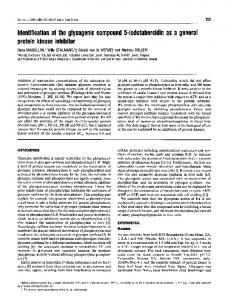
RESULTS Inununoprecipitation of Ku Protehn-p350 Complexes. Initial studies were carried out with IPP buffer. A broad array of nonspecifically incorporated proteins appeared in anti-Ku antibody-mediated immunoprecipitates prepared at 0.05 M NaCl. In contrast, immunoprecipitates prepared at a salt concentration > 0.3 M NaCl contained little other than the p70 and p80 Ku heterodimer. Thus, in subsequent experiments, cell extractions and immunoprecipitations were performed with buffers containing 0.15 M NaCl (physiologic salt) or 0.5 M NaCl (high 'salt), which is a standard immunoprecipitation condition (5, 28, 29). As shown in Fig. 1, anti-p350 antibodies immunoprecipitated a single polypeptide of %350 kDa under conditions of 0.15 M and 0.5 M NaCl (Fig. 1A). At physiologic salt conditions, various anti-Ku antibodies immunoprecipitated p70 and p80 as well as the 350-kDa polypeptide (lanes 3 and 5). At 0.5 M NaCi, immunoprecipitates contained only the Ku components (lanes 4 and 6). The absence of the 350-kDa band in the latter experiments argues strongly that none of the sera contains antibodies that bind this polypeptide directly. The 350-kDa polypeptide is identified as the catalytic component (p350) of DNA-PK in Fig. 1B because it is recognized cm~ Lo
I-
CYI
Co IL
0 A
C:L CX5
C/)
IC
C6
ClD
I;Ct
e75
B 0) CM)
CC)
C2;x
-
ZC2 X
a.
)
CD
C7
CtX
ZC_}
_ z
Cal Cal lr M
z .
2 i
n
C-)
CZ
Cd)
CD
CD C)~
CC
CD
CD
xX
-
L
E)
CD
n
-
L) T-
C)}X Xr U)
CO
LO
C f° CCD
&
C
kDa
-p350 200-
200
-
46
p)35
--
97.4--
97.4-
69 -
kDa
--
p80 p70
69-
p80 *
;i- n
46 30-
30 FIG. 1. Anti-Ku antibodies immunoprecipitate a dissociable complex of Ku protein and p350. (A) Autoradiogram demonstrating polypeptides immunoprecipitated under different salt conditions. mAb 111 is a mouse monoclonal antibody that recognizes p80. Anti-Ku antiserum OM also contains anti-Ro antibodies. (B) Identification of the 350-kDa polypeptide as the p350 component of DNA-PK. Immunoprecipitates were prepared with serum OM at 0.15 M NaCl and used as substrate in immunoblots. Individual strips were probed with antibodies as indicated. NHS, normal human serum; NRS, normal rabbit serum.
_s.E,:.-soSfAri
Biochemistry: Suwa et aL
6906 CN)
Ln C)
.__
E~]
CO
C)L
M IU
1--
lz
.-
r
t
=
*
1m
kDa
=
a
_
INm
CZ
C
_
_
c
To assess the extent of DNA cleavage in these experiments, immunoprecipitation was also carried out using antiDNA antibodies. Again as in Fig. 3A, immune complexes from undigested or lightly digested cell extracts contained a number of polypeptides including the Ku subunits and p350 (Fig. 3B, lanes 5 and 6). More intense DNase I digestion or treatment with EtdBr prevented incorporation of Ku protein and p350 into anti-DNA immunoprecipitates (lanes 7 and 8). From these experiments, we conclude that (i) DNase I removed DNA that could simultaneously associate with proteins and could be recognized by antibodies; (ii) EtdBr destabilizes the Ku protein-p350 complex, possibly because Ku protein does not bind to DNA that has intercalated EtdBr, and (iii) an apparently fragile complex of Ku protein-p350 may persist after it exits from the DNA strand. In interpreting these experiments, it should be recalled that DNA fragments as short as 12 bp activate DNA-PK activity (19). DNA fragments of this size may be produced by DNase I and could account for the apparent increase in Ku protein-p350 complexes observed (Fig. 3B, lane 2). Such short DNA fragments are not likely to be recognized well by anti-DNA antibodies. These experiments indicated that DNA serves to stabilize the Ku protein-p350 complex and suggested that DNA might be required for its initial assembly. To test this possibility, we prepared cell extracts under 0.5 M NaCl salt conditions, digested them with DNase I, and adjusted salt conditions to 0.15 M NaCl with dialysis. In some cases, extracts were treated further with silica gels to remove any residual DNA fragments prior to dialysis into 0.15 M IPP buffer. As shown in Fig. 4, a Ku protein-p350 complex formed as the salt content of buffer was lowered (lanes 1 and 2). Formation of this complex appears to depend on the remaining presence of small fragments of DNA because it was not observed when extracts were treated with silica matrix (lane 3), which removes DNA fragments of >10-bp. Subsequent addition of double-stranded DNA (lane 4) promoted formation of the complex, but single-stranded DNA (ane 5) had no effect. The concern remained that the complexes detected in HeLa cell extracts might not be analogous to active forms of DNA-PK. To address this issue, Ku protein and p350 purified biochemically from HeLa cells were combined with GC147 (GAL4-CTD fusion protein) and the AdUAS DNA template (which contains GAL4 binding sites) in the presence of [y32PJATP. Immunoprecipitations were performed with
0
0 a
c:"
-
ICm
__
200 -
=
Proc. NatL. Acad. Sci. USA 91 (1994)
IC3
(