Published on Web 02/22/2007
DNA Hybridization Detection with Blue Luminescent Quantum Dots and Dye-Labeled Single-Stranded DNA Hui Peng, Lijuan Zhang, Tanja H. M. Kja¨llman, Christian Soeller, and Jadranka Travas-Sejdic* Polymer Electronic Research Centre, The UniVersity of Auckland, PriVate Bag 92019, Auckland, New Zealand Received November 28, 2006; E-mail:
[email protected]
Quantum dots (QDs) have become an important photonic tool in the past two decades due to their unique properties, such as high chemical stability, resistance to photodegradation, and readily tunable optical properties.1-3 QD colloids are often prepared using organometallic routes at high temperature.4-6 The most widely used surface-capping ligands are trioctylphosphine/trioctylphosphine oxide (TOP/TOPO) and long-chain alkylamine. The resulting QDs are hydrophobic, and further chemical modifications are generally required for compatibility with biological applications. Since the first reports on the designs of hydrophilic QDs and QD-protein conjugates,1,2 a number of surface functionalization schemes have been developed to make QDs water soluble and thus suitable for biological applications.3,7,8 An alternative approach is to synthesize QDs directly in an aqueous medium. Since the original report on the aqueous synthesis of mercaptoethanol- and thioglycerol-capped CdTe QDs,9 significant progress has been made in the preparation of thiol-capped CdTe QDs that exhibit very stable luminescence.10 Due to their tunable narrow-band emission and broad excitation spectra QDs are excellent donors for fluorescence resonance energy transfer (FRET)-based biosensors. Several sensor designs based on FRET between QDs and dye-labeled biomolecules have been developed.11-13 For example, Mauro et al. designed a maltosebinding assay based on FRET between CdSe/ZnS QDs and dye acceptors.11 A FRET TNT sensor was developed based on a hybrid QDs antibody fragment,12 and Gill et al. used FRET in CdSe/ZnSDNA conjugates to probe hybridization and DNA cleavage.13 More recently, QD and FRET-based protease sensors to measure extracellular matrix metalloproteinase activity have been reported.14 Homogeneous DNA fluorescence essays are a particularly versatile way to detect hybridization, but generally, labeling of two nucleic acids or dual modification of the same strand is necessary to achieve sequence specificity. Here we report a simple sensing platform to evaluate specific hybridization based on the FRET between luminescent CdTe QDs and dye-labeled single-stranded DNA (ssDNA) probes through a cationic polymer acting as an electrostatic linker (Scheme 1). Unlike the FRET-based sensors mentioned above, in this design covalent immobilization of the probe molecules is not required, and DNA sequence specificity was achieved with minimal probe modification. Water-soluble CdTe QDs were prepared in aqueous solution by using thioglycolic acid as the capping ligand.9 After refluxing for 10 min, the resulting CdTe QD colloid was irradiated for 12 h with a 150-W xenon lamp at room temperature (see Supporting Information for details). During irradiation, the emission of CdTe QDs colloid progressively blue shifted, and the PL intensity increased due to the oxidation of Te atoms and the formation of a CdS shell. The negatively charged CdTe QDs were dissolved in a cationic polymer solution, poly(diallyldimethylammonium chloride) (PDADMAC), for 10 min to acquire positive surface charge. These positively charged CdTe QDs (CdTe+) had an emission peak at 497 nm with a fwhm of 45 nm (Figure 1). 10.1021/ja0685452 CCC: $37.00 © xxxx American Chemical Society
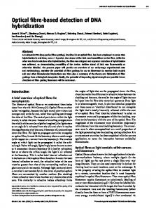
Scheme 1. Principle of DNA Hybridization-Detection System Based on the QD/Cy3-labeled DNA FRET
Cy3-labeled ssDNA (Cy3-DNA) was chosen as the acceptor. Although Cy3 is widely used for DNA labeling, it is rarely employed in FRET measurements with QDs because its emission peak overlaps with those of most commercially available QDs, and effective separation cannot be obtained. The absorption and emission spectra of Cy3-DNA in 100 mM NaCl and 50 mM sodium citrate (SSC buffer, pH 7.8) are shown in Figure 1. The emission of CdTe+ partly overlaps with the absorption spectrum of Cy3, suggesting that efficient FRET between them can take place. There is a little overlap between CdTe+ and Cy3 emission, allowing effective separation. Indeed, in a solution of CdTe+ and Cy3-labeled ssDNA, excitation of CdTe+ at 360 nm results in efficient FRET to Cy3 (Figure 1). (Note that there is negligible direct emission from Cy3 upon excitation at 360 nm.) There is a 10 nm red-shift in the Cy3 emission peak that can be explained by an increase in polarity in the vicinity of Cy3 due to the interaction with the cationic polymer.15,16 It is interesting to note that this is accompanied by a 10-nm blue-shift of the CdTe+ emission. In a control experiment, no FRET between negatively charged CdTe QDs and Cy3 was detected (see Figure 2 of Supporting information), suggesting the electrostatic interactions play a key role to ensure proximity between donor and acceptor. The calculated FRET efficiency is 92% according to E ) 1 - FDA/FD, where FDA and FD are integrated fluorescence intensities of CdTe+ in the presence or absence of the acceptor Cy3, respectively.17 In order to obtain high FRET efficiencies the excess of unbound cationic polymer PDADMAC in the CdTe+ solution had to be minimized. An experiment was carried out by adding PDADMAC
Figure 1. Normalized spectra of (a) emission of CdTe+ excited at 360 nm, (b) absorption of Cy3-DNA, (c) emissions of Cy3-DNA excited at 488 nm, and (d) emission of CdTe+/Cy3-DNA excited at 360 nm; All spectra were recorded in SSC buffer. J. AM. CHEM. SOC. XXXX, XXX, PAGE EST: 1.8
9
A
COMMUNICATIONS
Figure 2. (A) Emission spectra of CdTe+/Cy3-DNA hybrid after hybridization with different concentrations of target. (a) 0 nM; (b) 6.7 nM; (c) 13.3 nM; (d) 20.2 nM; (e) 26.8 nM; (f) 33.6 nM. (B) Normalized fluorescence changes of Cy3-DNA after incubation with complementary (a) or noncomplementary DNA (b) samples. A0 and A are integrated areas of Cy3 before and after the incubation, respectively.
to a solution containing unmodified CdTe QDs and Cy3-DNA (see Figure 3 of Supporting information). Increasing the concentration of PDADMAC from 5.8 × 10⁻⁶ wt % to 1.7 × 10⁻⁵ wt % caused a small increase in the fluorescence intensity of Cy3, but when the concentration of PDADMAC reached 4.1 × 10⁻⁵ wt %, the fluorescence intensity of Cy3 began to decrease with a maximal FRET efficiency of 77%. This result implies that Cy3-DNA prefers to interact with free PDADMAC molecules in the solution. In solution, the polymer chains have a conformation of a flexible random coil, which facilitates their interaction with ssDNA through electrostatic and hydrophobic interactions. To study how the conjugation of CdTe+ and Cy3-DNA was affected by the presence of sample DNA, the hybrid of CdTe+ and Cy3-DNA was incubated with different concentrations of complementary DNA at room temperature, and spectra were obtained after 30 min (Figure 2A). While the fluorescence intensity of Cy3 progressively decreased with increased concentration of complementary DNA (Figure 2A), the normalized integrated area showed a sample concentration-dependent decrease as shown in Figure 2B, indicating an increased distance between QDs and dsDNA. This may be explained by the more rigid DNA duplex structure as compared to ssDNA, which may increase the distance between polymer and dye, thus decreasing the FRET efficiency. Additionally, the increase in negative charge density due to the formation of a DNA duplex will increase the repulsive electrostatic forces between negatively charged CdTe QDs and DNA-Cy3, also resulting in larger distances and lower FRET efficiency. After incubation with noncomplementary DNA, the fluorescence intensity of Cy3-DNA also decreases slightly (Figure 2B). This is probably due to the competition of added negatively charged noncomplementary DNA with Cy3-DNA in the CdTe+/Cy3-DNA duplex. It is clear, though, that noncomplementary DNA causes a much smaller change in the fluorescence intensity of Cy3 than corresponding concentrations of complementary DNA. As noted above, the luminescent intensity of the CdTe+/Cy3DNA complex generally decreased with increasing concentrations of DNA, largely independent of the general FRET behavior. In order to understand this phenomenon, the interaction between
B J. AM. CHEM. SOC.
PAGE EST: 1.8
CdTe+ and ssDNA was investigated. It was found that the addition of complementary DNA caused the decrease in luminescent intensity of CdTe+ in the presence of high concentration of DNA probe (see Figure 4 of Supporting information) probably due to aggregation of QDs caused by hybridization. Longer sequences of DNAs were used to evaluate the general usefulness of this sensing platform. Generally, similar results were obtained (see Figures 5 and 6 of Supporting information). In summary, a simple DNA-sensing platform was developed on the basis of the FRET between blue-luminescent CdTe QDs and dye-labeled ssDNA. A cationic polymer acts as an “electrostatic linker” to achieve efficient energy transfer from the QD donor to the dye acceptor. The differential interaction of single-stranded and double-stranded DNA with CdTe+ results in differential changes of FRET efficiency, which is used here to recognize the hybridization event. This platform provides a homogeneous DNA assay that has all the advantages of a solution-based fluorescence detection method, but requires only minimal DNA modification. Acknowledgment. We greatly thank the Royal Society of New Zealand Marsden Fund for financial support of this research. Supporting Information Available: Synthesis and modification of CdTe QDs, general experimental procedure, interaction between CdTe+ and ssDNA, measurement of the amount of bound Cy3-DNA, and detection of longer DNA. This material is available free of charge via the Internet at http://pubs.acs.org. References (1) Bruchez, M., Jr.; Moronne, M.; Gin, P.; Weiss, S.; Alivisatos, A. P. Science 1998, 281, 2013-2016. (2) Chan, W. C.; Nie, S. Science 1998, 281, 2016-2018. (3) Mattoussi, H.; Mauro, J. M.; Goldman, E. R.; Anderson, G. P.; Sundar, V. C.; Mikulec, F. V.; Bawendi, M. G. J. Am. Chem. Soc. 2000, 122, 12142-12150. (4) Murray, C. B.; Norris, D. J.; Bawendi, M. G. J. Am. Chem. Soc. 1993, 115, 8706-8715. (5) Peng, X.; Schlamp, M. C.; Kadavanich, A. V.; Alivisatos, A. P. J. Am. Chem. Soc. 1997, 119, 7019-7029. (6) Reiss, P.; Bleuse, J.; Pron, A. Nano Lett. 2002, 2, 781-784. (7) Dubertret, B.; Skourides, P.; Norris, David, J.; Noireaux, V.; Brivanlou, Ali, H.; Libchaber, A. Science 2002, 298, 1759-1762. (8) Uyeda, H. T.; Medintz, I. L.; Jaiswal, J. K.; Simon, S. M.; Mattoussi, H. J. Am. Chem. Soc. 2005, 127, 3870-3878. (9) Rogach, A. L.; Katsikas, L.; Kornowski, A.; Su, D.; Eychmueller, A.; Weller, H. Ber. Bunsen-Ges. 1996, 100, 1772-1778. (10) Gaponik, N.; Talapin, D. V.; Rogach, A. L.; Hoppe, K.; Shevchenko, E. V.; Kornowski, A.; Eychmueller, A.; Weller, H. J. Phys. Chem. B 2002, 106, 7177-7185. (11) Medintz, I. L.; Clapp, A. R.; Mattoussi, H.; Goldman, E. R.; Fisher, B.; Mauro, J. M. Nat. Mater. 2003, 2, 630-638. (12) Goldman, E. R.; Medintz, I. L.; Whitley, J. L.; Hayhurst, A.; Clapp, A. R.; Uyeda, H. T.; Deschamps, J. R.; Lassman, M. E.; Mattoussi, H. J. Am. Chem. Soc. 2005, 127, 6744-6751. (13) Gill, R.; Willner, I.; Shweky, I.; Banin, U. J. Phys. Chem. B 2005, 109, 23715-23719. (14) Shi, L.; De Paoli, V.; Rosenzweig, N.; Rosenzweig, Z. J. Am. Chem. Soc. 2006, 128, 10378-10379. (15) Gaylord, B. S.; Heeger, A. J.; Bazan, G. C. J. Am. Chem. Soc. 2003, 125, 896-900. (16) Peng, H.; Soeller, C.; Travas-Sejdic, J. Chem. Commun. 2006, 37353737. (17) Clapp, A. R.; Medintz, I. L.; Mauro, J. M.; Fisher, B. R.; Bawendi, M. G.; Mattoussi, H. J. Am. Chem. Soc. 2004, 126, 301-310.
JA0685452
Supporting Information for DNA Hybridization Detection with Blue Luminescent Quantum Dots and Dye-Labeled Single-Stranded DNA Hui Peng, Lijuan Zhang, Tanja H. M. Kjällman, Christian Soeller and Jadranka Travas-Sejdic* Polymer Electronic Research Centre, The University of Auckland, Private Bag 92019, Auckland, New Zealand
Materials Thioglycolic acid (≥ 98%), CdCl2 (99.99%), tellurium powder (99.997%), NaBH4 (95%), poly(diallyldimethylammonium chloride) (PDADMAC) (very low molecular weight