DNA Methylation and Cancer Phillippa C. Taberlay and Peter A. Jones
Abstract DNA methylation acts in concert with other epigenetic mechanisms to regulate normal gene expression and facilitate chromatin organization within cells. Aberrant DNA methylation patterns are acquired during carcinogenic transformation; such events are often accompanied by alterations in chromatin structure at gene regulatory regions. The expression pattern of any given gene is achieved by interacting epigenetic mechanisms. First, the insertion of nucleosomes at transcriptional start sites prevents the binding of the transcriptional machinery and additional cofactors that initiate gene expression. Second, nucleosomes anchor all of the DNMT3A and DNMT3B methyltransferase proteins in the cell, which suggests a role for histone octamers in the establishment of DNA methylation patterns. During carcinogenesis, epigenetic switching and 5-methylcytosine reprogramming result in the aberrant hypermethylation of CpG islands, reducing epigenetic plasticity of critical developmental and tumor suppressor genes, rendering them unresponsive to normal stimuli. Here, we will discuss the importance of both established and novel molecular concepts that may underlie the role of DNA methylation in cancer.
1 Overview The eukaryotic genome is complex and has evolved to enable large amounts of DNA to be contained within the boundary of the nucleus. The structural organization of DNA into chromatin involves several orders of compaction and creates an environment that is generally repressive for gene transcription. However, chromatin is a highly dynamic structure that must be modified to accommodate the transcriptional P.C. Taberlay and P.A. Jones Department of Urology, Biochemistry and Molecular Biology, USC/Norris Comprehensive Cancer Center, Keck School of Medicine, University of Southern California, Los Angeles, California 90033, USA e-mail:
[email protected]
S.M. Gasser and E. Li (eds.), Epigenetics and Disease, Progress in Drug Research 67, DOI 10.1007/978-3-7643-8989-5_1, # Springer Basel AG 2011
1
2
P.C. Taberlay and P.A. Jones
machinery when gene expression is required, to facilitate DNA repair mechanisms, or to allow DNA replication [1]. Epigenetic regulation of these processes is typically driven in a cell type-dependent manner during and following differentiation from totipotency. It has also been established that epigenetic mechanisms, such as DNA methylation, govern many aspects of embryonic growth from conception and are necessary for the survival of mammals. Since several enzymatic systems coordinate epigenetic modifications, a high level of combinatorial control must be maintained to ensure the correct chromatin conformation and identity of each cell. To this end, it is now apparent that alterations to normal epigenetic processes deregulate biological signaling pathways, contributing to carcinogenesis and disease. Here we will discuss mechanisms that may be involved in establishing aberrant DNA methylation patterns in carcinogenesis.
2 Mechanisms of Silencing by DNA Methylation The biology of DNA methylation events in cancer is currently the best characterized epigenetic aberration in disease [2]. DNA methylation is a relatively stable modification that occurs in the context of CpG dinucleotides in mammalian cells. The presence of CpG sites within the genome is irregular, with some regions containing a high frequency of CpG dinucleotides (CpG islands) in contrast to areas where this dinucleotide is underrepresented. The distribution of CpG sites throughout the genome has implications for cellular gene expression profiles. First, CpG rich regions are often situated in promoters that are proximal to the transcription start sites of many genes while the remainder of the genome is relatively CpG poor, including sites of viral integration as well as intergenic and intronic regions [3]. Second, not all CpG sites in the genome are methylated. CpG islands are resistant to de novo methylation in normal cells [4, 5], while CpG poor regions are predisposed to this process [6]. Distinct methylation patterns are established during embryonic development and are mitotically heritable through many cellular divisions. The faithful maintenance of normal DNA methylation patterns is disrupted in cancer, where CpG islands become susceptible to methyltransferase activity and CpG poor regions undergo hypomethylation during transformation. Consistent with this, the overall level of genomic 5-methylcytosine is decreased in cancer cells [7, 8]. Hypomethylation of bulk cellular DNA might result in genomic and chromosomal instability [9, 10] and is perhaps suggestive of a global switch mechanism that directs changes in chromatin structure concomitant with aberrations in DNA methylation patterns. The change in DNA methylation patterns is considered to be common in most cancers [11], with significant effects on gene expression patterns, cellular growth, and selective advantage. These changes can be the result of silencing of tumor suppressor genes and alterations to associated downstream pathways [2, 12], such as repression of the p53 tumor suppressor pathway [13]. It is important to emphasize
DNA Methylation and Cancer
3
that epigenetic mechanisms act in concert to coordinate normal gene regulatory processes and that cellular deregulation in disease involves many systems. DNA methylation is a mediator of long-term silencing [6] and contributes to the regulatory mechanisms of tissue-specific gene expression in normal cells. The covalent addition of a methyl group to DNA can influence gene transcription [14] by varying the binding of transcription factors [15, 16] or through the recruitment of methyl-binding proteins [17, 18] and chromatin modifiers such as histone deacetylases [14, 19]. These studies provided some of the first evidence that there was interplay between different epigenetic modifications. Despite this evidence, the exact mechanism for gene silencing by DNA methylation is still debated. Foremost, while covalent histone modifications are sufficient to repress gene expression, they are considered to be a less stable and reversible process [3, 20]. Therefore, it is likely that there are additional determinants that specify the establishment of permanently silenced and hypermethylated CpG islands in cancer. CpG islands remain unmethylated in normal cells and are frequently sites of DNase hypersensitivity on a global scale [21–23]. DNase hypersensitivity has been used as a marker for genomic regions that are free of nucleosomes, suggesting that the extent of nucleosome occupancy may be correlated with gene silencing and expression. Given these data, it has been hypothesized for many years that inactive genes exhibit a closed, compact chromatin structure in contrast to active gene promoters that are less condensed to allow for the binding of transcriptional machinery. Technological advances have confirmed and extended these data, and it is now clear that the role of nucleosome positioning in gene silencing through DNA methylation is critical for gene control. DNA accessibility is a requirement for transcription [1, 24] and can be considered to be either a constitutive state [25, 26] or one that is generated following extensive chromatin remodeling, as demonstrated for the PHO5 promoter in yeast [27]. Genome-wide screens in several organisms have shown that the regions upstream of many transcriptional start sites are devoid of nucleosomes [28–32], indicative of the specific gene expression patterns in these eukaryotes. In support of a model whereby nucleosomes are central to gene control, it has been shown that a nucleosome depleted yeast PHO5 promoter is maintained through DNA replication [33]. The inheritance of a nucleosome depleted state was shown to be independent of coactivator complexes, and transcription of PHO5 was not required to maintain the nucleosome depleted region [33]. Despite such striking associations, the significance of these findings was not directly correlated with events of gene silencing during carcinogenesis until recently [24]. Extensive analyses of the MLH1 promoter, which is frequently hypermethylated in cancers, reveal that the formation of a nucleosome depleted region is required for gene expression [24]. The precise positioning of nucleosomes can be determined at individual promoters by using a high-resolution single-molecule assay called methyltransferase-based single-promoter analysis (M-SPA) [34]. The M-SPA assay has also been utilized to confirm the requirement for a nucleosome depleted region at the GRP78 [35] and BRCA1 [24] promoters. By extension, a nucleosome depleted region is likely to be characteristic of expressing genes containing a CpG
4
P.C. Taberlay and P.A. Jones
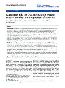
island promoter. A nucleosome is inserted immediately upstream of the transcriptional start site of an inactive MLH1 promoter, which becomes permanently silenced by DNA methylation in cancer cell lines [24]. These data suggest that changes in nucleosome occupancy contribute to the epigenetic silencing of CpG islands during transformation (Fig. 1). The mechanisms that then ensure that CpG islands remain permanently silenced are unclear, but it is feasible that this process involves protein complexes that facilitate the addition and removal of other epigenetic marks.
3 DNA Methylation, Covalent Histone Modifications, and Histone Variants 3.1
Histone Variants
Beyond the physical positioning of nucleosomes, the composition and posttranslational modification of these core particles must also be considered. Histone variants, including H2A.Z and H3.3 (Fig. 2a), have altered amino acid sequences compared with the canonical histone proteins [36] and have been shown to have profound effects on gene expression [32, 37, 38] as well as being associated with distinct chromosomal regions [39, 40]. Importantly, H2A.Z is enriched at transcriptional start sites of both active and inactive genes [41], suggesting that H2A.Z also has roles that are independent of transcription. One such function may be to maintain genes in a poised state [42] and to prevent the permanent silencing of these loci by DNA methylation in cancer [40, 43]. Alternatively, H2A.Z may contribute to the over-expression of oncogenes or cell cycle regulators during transformation. It has recently been demonstrated that the over-expression of H2A.Z is linked to the progression of estrogen-responsive breast cancers [44]. In this study, c-MYC was shown to bind to the H2A.Z promoter in response to estrogen, increasing H2A.Z protein expression [44]. This observation correlated with altered proliferation properties of MCF7 cells [44]. Despite this, a conclusive mechanistic link between H2A.Z and cancer progression remains to be established. DNA methylation and H2A.Z are mutually exclusive epigenetic marks in plants [40]. Altered DNA methylation patterns are mirrored by changes in H2A.Z localization and vice versa [40], suggesting a high level of interaction between the mechanisms underlying these two epigenetic modifications. Specifically, genomic regions that exhibit a loss of DNA methylation become enriched for H2A.Z [40], which is proposed to be a direct effect of DNA hypomethylation events rather than changes in the levels of transcription [40]. The insertion of H2A.Z into nucleosomes is reliant on the Snf-2-related CREB-binding protein activator (SRCAP) chromatin remodeling complex in humans [45, 46]. A mutation in plants of the equivalent complex, PIE1, results in genome-wide DNA hypermethylation in Arabidopsis thaliana [40]. While the distribution of DNA methylation patterns
DNA Methylation and Cancer
5
Fig. 1 Nucleosomes contribute to the epigenetic silencing of genes in concert with DNA methylation in cancer cells. In normal cells (above), active promoters are depleted of nucleosomes immediately upstream of the transcriptional start site. Nucleosome-depleted regions are flanked by nucleosomes that are enriched for active marks and are permissive for transcription, such as H3K4me3. In addition, these nucleosomes contain histone variants shown to correlate with transcription, such as H2A.Z. During the silencing process (below), a nucleosome is inserted into the nucleosome depleted region, physically interfering with the process of gene expression. DNA methylation and the acquisition of repressive histone marks, such as H3K9me3, permanently silence genes in cancer cells. DNMT3A and DNMT3B are anchored to nucleosomes associated with methylated DNA. Removal of DNA methylation leads to the eviction nucleosomes from reactivated loci after treatment of cancer cells with DNMT inhibitors such as 5-Aza-CdR (not shown; [24]) agent. Small white circle unmethylated CpG site; small black circle methylated CpG site; large circle nucleosome; X silenced transcriptional start site; 4 trimethylation of histone H3 at lysine 4 (H3K4me3); 9 trimethylation of histone H3 at lysine 9 (H3K9me3); DNMT DNA methyltransferase; 5-Aza-CdR 5-Aza-20 -deoxycytidine demethylating agent