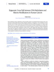
[DNA
methylation in embryo development: Epigenetic impact of ART --Assisted Reproductive Technologies] 2017
Canovas S1,2, Ross PJ3, Kelsey G4, Coy P,1,2 1
Physiology of Reproduction Group, University of Murcia; 2IMIB-Arrixaca, Spain. 3 Department of Animal Science, UC Davis, Davis,CA, USA 4 Epigenetics Programme, The Babraham Institute, Cambridge, UK.
[email protected]
Impact on DNA methylation dynamics in preimplantation embryos DNA methylation errors
CAUSES
ART
Lifestyle insults
Me
Me
Me
Me
Genomic imprinting errors
Phenotypic abnormalities
Imprinting syndromes
CONSEQUENCES
Infertility
Gene expression alterations
[DNA
methylation in embryo development: Epigenetic impact of ART --Assisted Reproductive Technologies] Canovas et al.
2017
Summary DNA methylation can be considered a component of epigenetic memory with a critical role during embryo development, and which undergoes dramatic reprogramming after fertilization. Though it has been a focus of research for many years, the reprogramming mechanism is still not fully understood. Recent results suggest that absence of maintenance at DNA replication is a major factor, and that there is an unexpected role for TET3-mediated oxidation of 5mC to 5hmC in guarding against de novo methylation. Base-resolution and genome-wide profiling methods are enabling more comprehensive assessments of the extent to which ART might impair DNA methylation reprogramming, and which sequence elements are most vulnerable. Indeed, as we also review here, studies showing the effect of culture media, ovarian stimulation or embryo transfer on the methylation pattern of embryos emphasize the need to face ART-associated defects and search for strategies to mitigate adverse effects on the health of ART-derived children. Keywords: epigenetic, embryo, in vitro fertilization, ICSI, embryo culture, DNA methylation, infertility
2
[DNA
methylation in embryo development: Epigenetic impact of ART --Assisted Reproductive Technologies] Canovas et al.
2017
1. Introduction In the era of “omic” technologies, we still have limited knowledge of the mechanisms surrounding fertilization, a process that entails the union of two morphologically and functionally dissimilar cells (oocyte and sperm) to create an entire organism, and moreover, about the biology of the germinal cells that will be responsible for transmitting genetic and epigenetic information to the next generation. Epigenetic marks (BOX 1), which differ between cell lineages and are responsible for the maintenance of different cell types within the same organism, reinforce cell-fate decisions and establish barriers against reversion to preceding cellular states.[1] These marks show asymmetrical distribution between male and female gametes and some of the differences persist in the early embryo. After fertilization in the pre-implantation embryo and during primordial germ cell (PGC) specification epigenetic information, particularly in the form of DNA methylation, is erased to a basal state. Later, it is reacquired during epigenetic reprogramming at blastocyst stage (Figure 1), but not all regions of the genome are affected equally (Figure 2).
Figure 1. a) DNA methylation landscape during preimplantational embryo development. A 5mC asymmetric demethylation occurs in male and female pronucleus and subsequently an increase of 5hmC and other oxidized derivates appears, with a lower intensity in the female pronucleus. b) Comparative CpG global methylation levels along preimplantational embryo development. In mouse, global DNA methylation shows a continuous decrease from zygote to blastocyst stage. In contrast, human embryos show a stabilization, and monkey even an increase, of the global DNA methylation around 8-cell stage.
Protective mechanisms that prevent DNA demethylation of specific regions exist, at the same time that DNA methyltransferase DNMT1 and de novo DNA methyltransferases (DNMT3A,B) are expressed in mouse early embryonic development.[2, 3] The process of DNA methylation remodeling is linked directly to the viability of the embryo, as revealed by embryonic lethality after DNMT1 or DNMT3B mouse knockouts, and to the capacity of germinal cells to transmit information to perpetuate the species, as evidenced by the knockout of DNMT3A or the recently
3
[DNA
methylation in embryo development: Epigenetic impact of ART --Assisted Reproductive Technologies] Canovas et al.
2017
discovered DNMT3C in the male germline in mouse.[5-7] Defects during the first wave of epigenetic remodeling affect embryo viability and could entail defects in the organism, but errors during PGC reprogramming can lead to reproductive failure. In mammals, DNA methylation and covalent modifications of histone tails are epigenetic marks with key roles during embryonic development.[4],[8-13] Classical studies by immunofluorescence analyses showed that in the mouse zygote the parental genomes undergo asymmetric loss of DNA methylation with the active involvement of oxidation reactions and subsequent lineage-specific reacquisition of methylation.[14, 15] Nonetheless, the wave of demethylation does not affect some specific regions, such as parental imprints and active retrotransposons, that are protected from demethylation to ensure embryonic viability (Figure 2).[16] Modifications on histone tails, some of which correlate with DNA methylation in some genomic contexts, also show asymmetric distribution in male and female pronuclei, in part because of the conservation of some of the histone marks inherited from the oocyte in the female pronucleus (PN), versus the replacement of sperm protamines by acetylated histones in the male PN.[17, 18] Recent technological progress has allowed single-base genome-wide analysis of DNA methylation in small numbers of cells, providing a more precise view of the epigenetic landscape and shedding some light on the mechanisms of epigenetic regulation during embryonic development.
Figure 2. Degree of demethylation during early fertilization/primordial germ cells specification in human a) and mouse b). Representative examples of specific regions (ERV1:HERV9-INT and SVA_A in human; IAP in mouse) showing demethylation resistance during demethylation wave at the early embryo. Dashed line represents male gametes.
In this context, there is increasing evidence in several species that epigenetic alterations in ART-derived offspring may be responsible for some phenotypic and gene expression abnormalities. These alterations might occur during reprogramming in the
4
[DNA
methylation in embryo development: Epigenetic impact of ART --Assisted Reproductive Technologies] Canovas et al.
2017
early embryo, just when the epigenetic transitions are most dramatic and when, as they are manipulated in vitro, the embryo may be most vulnerable.[19] In humans, according tothe World Health Organization (WHO), infertility affects around 20% of couples and over 6 million babies have been born using ART. Although the majority of them seem healthy, a greater incidence of adverse long-term effects has been reported among ART-conceived infants, suggesting that conditions inherent to the treatments have an epigenetic cost and aberrant DNA methylation during preimplantation reprogramming has been considered a key factor. Here, we review the latest knowledge about DNA methylation reprogramming after fertilization, the possible impact of ART on this epigenetic mark and, also, propose some alternatives to reduce the potential epigenetic cost for some of these procedures. 2. Maternal and paternal pronuclei are only partly demethylated through 5mC oxidation by TET enzymes. After fertilization, there is a loss of 5mC signal and accumulation of the oxidized form, (5-hydroxymethylcytosine, 5hmC) in the paternal pronucleus, two observations that were initially interpreted to indicate a role of oxidation of 5mC in active DNA demethylation (Figure 1).[20] The Ten-eleven-translocation enzymes (TET1, TET2 and TET3) oxidize 5mC to 5hmC and further to 5-formylcytosine (5fC) and 5carboxylcytosine (5caC). TET3 is expressed in oocytes and preimplatation embryos and was first considered responsible for the paternal genome demethylation after fertilization.[21-24] Recent studies in mice using more precise techniques, such as RRBS PBAT or TAB-seq to profile 5mC and 5hmC, and in other species, such as cows, rabbits and pigs ], have shown that 5hmC and 5fC are detected in both parental genomes.[24-29] It suggests that maternal and paternal genomes could both undergo TET3 dependent demethylation.[2], [27] In rhesus monkeys, it has also been reported that both paternal and maternal genomes undergo active DNA demethylation.[30] In mouse, pronuclear DNA demethylation is now considered a complex process with major contributions from DNA replicationand DNA repair pathways and only partially dependent on TET3 activity, with a greater role in the paternal than the maternal pronucleus.[2], [25-27], [31] Initially, a key role of TET3 in DNA-demethylation in pronucleus was proposed, based mainly on the first TET3 knockout report showing embryonic sublethality.[22] But this interpretation is inconsistent with the latest evidence. Attenuation of 5mC oxidation in zygotes by genetic ablation of maternal TET3 has been reported as compatible with embryonic development, although it entails neonatal sublethality. The phenotype is attributable to compensation by replication-dependent passive demethylation of 5mC, as well as by oxidation of 5mC mediated by TET1 or TET2.[32] Curiously, the genetic ablation of Tet3 does not lead to complete absence of 5hmC in the maternal pronucleus, because a small portion of this mark is produced early during oocyte development before the germinal vesicle stage.[32] Moreover, recent results revealed that TET3-mediated paternal 5mC oxidation is dispensable for
5
[DNA
methylation in embryo development: Epigenetic impact of ART --Assisted Reproductive Technologies] Canovas et al.
2017
mouse development and suggests the existence of a compensatory mechanism for defective 5mC oxidation in preimplantation embryos.[33] Quantitative assessment of 5mC by ultrasensitive liquid chromatography-mass spectrometry (LC-MS) and chemical inhibition of TET activity (DMOG) showed that loss of paternal 5mC and appearance of 5hmC are temporally disconnected and that 5hmC is not required for removal of most 5mC, confirming that ablation of maternal Tet3 did not block paternal 5mC erasure in early mouse zygotes.[20] Previously reported developmental defects in maternal Tet3 knockouts could be attributed to Tet3haplo-insufficiency, but not to defective paternal 5mC oxidation as initially proposed.[22],[33] In this context, the relevance of de novo DNA methylation has been studied in mouse by analyzing the DNA methylation patterns on both strands at single nucleotide resolution (CpG dyads), using aphidicolin to block cell division in embryos or SAMase to disrupt de novo and maintenance DNA methylation. With these approaches, it was confirmed that DNA demethylation requires DNA replication and it is mainly the result of impaired maintenance of DNA methylation, as illustrated by the increase in hemimethylated CpG dyads.[34] During early embryo development, the demethylation process is discontinuous, with two drops in methylation: at the zygotic stage (mainly in the paternal genome) and around blastocyst formation. Control of methylation maintenance during the first zygotic DNA replication is selective, with high methylation at active retrotransposons such as Intracisternal A-particles (IAP) and a proportion of specific long interspersed nucleotide elements-1 (LINE1), although methylated CpG dyads lose 5mC and more than 50% are hemimethylated CpG dyads. In the zygote, there is de novo DNA methylation by DNMT3A, which generates new 5mC that is hydroxylated to 5hmC by TET3, confirming the necessity of DNA replication for DNA demethylation.[20] Therefore, TET3 seems to be more important for preventing aberrant de novo methylation from the abundant DNMT3A inherited from the oocyte. Recently, a new role for TET3 has been reported. TET enzymes also catalyze the formation of hydroxymethylcytosine in RNA (hmrC). 5-hmrC is present at levels of one 5-hmrC per 5000 5-methylcytidine in RNA of mammalian cells and tissues, which suggests that RNA oxidation at this level could modulate the epigenetic regulation of gene expression.[35] In fact, a recent study in Drosophila showed that RNA hydroxymethylation can favour mRNA translation, and genetic ablation of dTet (the unique Tet ortholog that is conserved in Drosophila) resulted in defects in brain development accompanied by decreased RNA hydroxymethylation.[36] 3. Maternal and paternal pronuclei suffer asymmetrical impairment of DNA methylation Classically, passive replication-dependent demethylation has been considered the mechanism responsible for maternal genome methylation erasure, by exclusion of DNMT1, the enzyme responsible for methylation maintenance at replication, from the nucleus during preimplantation development, while the paternal genome undergoes
6
[DNA
methylation in embryo development: Epigenetic impact of ART --Assisted Reproductive Technologies] Canovas et al.
2017
active demethylation prior to formation of the 2-cell embryo.[37-38] Although the mechanism that controls differential susceptibility to demethylation in male and female pronuclei is not fully understood, some factors involved in this process have been identified. DPPA3 (STELLA/PGC7) protects 5mC in the maternal pronucleus from TET3-mediated conversion to 5hmC through its binding to H3K9me2-containing chromatin, although this histone modification also participates in the protection of imprinted loci in mature sperm that avoid demethylation.[39-41] In humans, TET2 and TET3 activity is inhibited by DPPA3, which localizes to specific loci and is controlled by H3K9me2 marks and DNA sequence.[42] In mice, Dppa3-null embryos show impaired DNA replication, ectopic micronuclei and abnormal segregation of maternal chromosomes, and aberrant H2AX phosphorylation, which inhibits DNA replication.[43] These results demonstrate that DPPA3 protects maternal DNA from aberrant epigenetic modifications to ensure early embryogenesis, but the question remains: why not paternal DNA? Recent studies in mice and monkeys, using genome-wide methylation profiling and single-nucleotide polymorphisms(SNPs) to track paternal and maternal genomes, revealed active demethylation also in the maternal genome, with the presence of 5hmC in the female pronucleus. Nonetheless, a large portion of CpG sites maintain their methylation levels confirming that only a portion of both parental genomes undergo active demethylation right after fertilization.[2],[27],[30] These latest findings suggest a complex interplay between the mechanisms involved in DNA methylation reprogramming, that is, demethylation, de novo methylation and methylation maintenance. Methylation patterns change differently in maternal and paternal pronuclei, as revealed by deep hairpin bisulfite sequencing (DHBS) and conventional single-stranded bisulfite sequencing (BS). While there is a reduction in DNA methylation in paternal LINE1 elements, which is linked to an increase of hemimethylated CpG dyads, the global level of methylation of this retroelement remains stable in the maternal PN.[34] Although minor, active demethylation also contributes to the drop in DNA methylation during the zygote stage, as demonstrated by changes in DNA methylation after treatment with aphidicolin to block DNA synthesis. While different groups have reported that excision of 5fC and 5caC by thymine DNA glycosylase (TDG) is a downstream event in the TET-mediated oxidation of 5mC, this mechanism seems dispensable for DNA demethylation in early embryos and germ cell reprogramming. [44] 4. Mechanisms and degree of demethylation during early fertilization vary depending on genomic context Despite the considerable efforts made during the last years, a key question that remains unresolved is the mechanism(s) responsible for specifying the methylation state of a given sequence at a given time in the genome. Recent findings are providing evidence that the DNA sequence itself is one of the determinants of the DNA
7
[DNA
methylation in embryo development: Epigenetic impact of ART --Assisted Reproductive Technologies] Canovas et al.
2017
methylation profile, but crosstalk between DNA methylation, histone modifications and ncRNAs suggest that they also play prominent roles.[45, 46] Methylation erasure during the zygotic stage does not affect all regions of the genome equally (Figure 2). Methylation persists at certain sequences, such as active retrotransposons (IAPs) and imprinted genes, which are protected from global demethylation, a requirement for embryonic viability.[16] Also, exceptionally rare intragenic regions, including some exons, promoters, splice sites and CpG islands (CpGi)that were methylated in gametes, maintain DNA methylation. Around 7% of CpGs in the mouse, which correspond mainly with unmethylated (80%) cytosines, maintain a consistent state ofmethylation from gametes to E7.5 embryos.[27] A large number of genome-scale DNA methylation profiling studies have employed RRBS, which is limited to CG-rich elements like CpGi, while the majority of 5mC in sperm corresponds with non-CpGi.[47] More recently unbiased approaches, such as PBAT and MethylC-Seq , complemented by highly sensitive methods to quantify total CpG byLC-MS, have been used to obtain a more detailed picture of methylation dynamics during early embryo development.[20],[26],[27] These results revealed that the paternal DNA undergoes significant demethylation at fertilization, with more than 90% of intergenic and gene body sequences with demethylation of over 10%, and 70% of these regions losing over 25% methylation, and with non-CpGi promoters showing a similar tendency. In contrast, little demethylation was found at CpGi-associated sequences and 5´untranslated regions (UTRs), which are already hypomethylated in sperm, but when such sequences were demethylated, it was mainly by an active process.[26],[27] Regarding repetitive elements, IAPs show similar methylation levels in oocyte and sperm (78%), with only a slight decrease to around 60% in inner cell mass (ICM).[27] In the zygote, following replication, LINE1 and major satellites (mSat) copies are hemimethylated in more than 50% of all methylated CpG dyads. However, at IAPs the majority of methylated CpG dyads remains fully methylated, which confirms selective maintenance of DNA methylation, but also that the early embryo has the capacity for methylation maintenance.[34] Mouse endogenous retroviral (MERV-L) elements, which are considered as controlling elements of gene expression during pre-implantation development, and which are not well assayed by RRBS, also undergo substantial demethylation in the zygote.[26] 4.1 Non-CG Methylation in Early Embryos Besides CpG methylation, methyl groups are also added to non-CpG positions: CHG and CHH (where H = A, G, T), most frequently in CAG or CAC contexts. The biological function of non-CG methylation is not fully understood, particularly because methylation at CHG/CHH sites cannot be reinstated at DNA replication, as the maintenance methylation transferase DNMT1 specifically recognizes hemimethylated CpG sites. However, non-CpG methylation is prominent in mammalian oocytes, where
8
[DNA
methylation in embryo development: Epigenetic impact of ART --Assisted Reproductive Technologies] Canovas et al.
2017
it accounts for more than half of the genomic 5mC content, although the proportion of CHG or CHH sites methylated is much lower than CpG sites (3.6%, 3.1% and ~38%, respectively). Non-CpG methylation has also been detected, at substantially lower levels, in mammalian embryos.[48] In oocytes, non-CpG methylation correlates very strongly with CpG methylation and, like CpG methylation, occurs predominately at transcribed gene bodies.[47,49] Non-CpG methylation generally occurs later than CpG methylation during oocyte development.[50] This, and the fact that non-CpG sites are incompletely methylated, suggests that it is the product of sustained DNMT3A activity in a non-dividing cell. After fertilization, non-CpG methylation decreases progressively to undetectable levels in the ICM. At promoters, non-CpG methylation negatively correlates with transcription in oocytes, but whether it has an effect independent of CpG methylation, which is more complete, is unclear.[27] The current state of knowledge about the dynamics of DNA methylation in the early embryo illustrates the complexity in the regulation of this epigenetic mark, and suggests that sophisticated studies are required to evaluate the possible impact of external factors on DNA methylation, and the significance of such effects, during embryo development. 5. Assisted Reproductive Technologies Impact Offspring Assisted reproductive technologies (ART) encompass fertility treatments that handle the eggs, sperm or embryos outside the body, and provide a helpful approach to treat infertility, which is estimated to affect around 186 million people worldwide.[51] ART include a wide variety of techniques, from intrauterine insemination to intracytoplasmic sperm injection (ICSI) and embryo freezing, which substitute for different stages of the reproductive cycle, and could be considered more or less invasive. Globally, ART use has doubled during the last decade, with a mean annual increase around 10% worldwide, and a progressive rise in banking cycles in which all eggs and/or embryos are frozen for future ART cycles. Nowadays children conceived using ART account for 2% of all births, which has brought to the society a growing interest in their long-term health. Although most babies conceived by ART seem healthy, they are at increased risk of fetal growth restriction, premature birth, low birthweight, congenital anomalies (including genetic and non-genetic) and perinatal complications. Also, ART has been linked to imprinting disorders, such as Beckwith-Wiedemann syndrome (BWS), Angelman syndrome (AS) and Silver-Russell syndrome (SRS) (Table 1).[52-57] Even the use of less invasive fertility treatments, such as ovarian stimulation and intra-uterine insemination, could increase the risk of multiple pregnancy and certain types of major congenital malformations.[58] In addition, the risk of spontaneous miscarriage is higher in ART cohorts than in spontaneous pregnancies.[59] Although the mechanisms underlying these alterations are not fully understood, deregulation of DNA methylation needs to be considered as a candidate mediator, because embryos
9
[DNA
methylation in embryo development: Epigenetic impact of ART --Assisted Reproductive Technologies] Canovas et al.
2017
experience whole sale reprogramming of DNA methylation and this epigenetic mark can be affected by nutritional, environmental and lifestyle cues.[60-63] The risk of birth defects in infants born after ART was first reported in The Lancet (Lancaster,1987) just 10 years after the first birth after in vitro fertilization (IVF, Louise Brown), by showing an increased incidence of spina bifida and transposition of the great vessels in neonates born after IVF. The risk of ART-derived birth defects has been estimated at 30% higher than the incidence of congenital anomalies in children born after spontaneous conception. Nonetheless, it still remains moderately low (4-6%) and some studies have shown no significant effect of ART.[64, 65] This discrepancy could be attributed to the wide spectrum of techniques and protocols employed in ART, parental characteristics, the small size of some studies and over-representation of multiple births in ART pregnancies, which are selectively included or not in the studies. To overcome some of these limitations, large population- and national registry-based cohorts with accurate information on ART procedures and meta-analysis have been proposed. The Division of Reproductive Health of the Center for Disease Control and Prevention-CDC (USA) analyzed data from more than 4.6 million infants (including 64,861 conceived using ART), and concluded that singleton infants conceived using ART from 2000 to 2010 were 40% more likely to have a non-chromosomal birth defect compared with naturally-conceived singleton births [66]. Another study, using the Swedish national health registers, analyzed data from more than 2.5 million infants born (including 30,959 conceived by IVF)which were followed up for a mean 10 years. In this study, ART treatment (IVF and ICSI)overall was associated with a small increased risk of mental retardation and ICSI for paternal infertility was associated with a small increase in the relative risk for autistic disorder and mental retardation.[67] Both studies appear to confirm adverse health consequences in ART-conceived infants. Nonetheless, careful evaluation of the long-term outcomes of children conceived using these technologies has been neglected, and is necessary. But, is this increased risk a consequence of ART per se, or is it derived from higher frequency of twin pregnancies, or from parental characteristics (infertility, aging, other)?. The answer is not straightforward for several reasons. First, despite the large number of individuals who have been conceived by ART (around than 6million), it is still very challenging to identify risks of different ART protocols for conditions that have a very low incidence in the general population (e.g., mental retardation 60.8 per 100,000 person-years; autism 29.3 per 100,000 person-years). Second, a control group of fertile couples willing to undergo ART is not practical in human. With the aim to distinguish if an increased risk is a consequence of ART, children conceived by these technologies were compared with infants born from women diagnosed as subfertile but who managed to conceive without ART treatment. It should be noted that this was a small study, the control group comprising 6343 infants, so larger cohort studies are required. Compared with the control group, babies born after ART were more likely to have very low birthweight and to be born very
10
[DNA
methylation in embryo development: Epigenetic impact of ART --Assisted Reproductive Technologies] Canovas et al.
2017
premature—before 32 weeks, which itself entails adverse outcomes, and showed a significantly greater growth velocity late in infancy.[68,69] These studies reveal that ART per se increases risk of some birth defects and/or related anomalies, although children born from couples classified as subfertile without ART also have an increased, but smaller, risk of anomalies partially due to infertility.[70, 71] It is conceivable that subfertility and ART could have additive impacts on epigenetic processes.[72] 6. Does ART have an impact on DNA methylation in the embryo? Overall, ART constitute substantial chemical and physical variations (nutrients, oxygen, pH, etc.)in the environment of gametes and embryos in comparison to normal physiological conditions (ovarian follicle, fallopian tube/oviduct, uterus) during a stage of dynamic epigenetic reprogramming in the embryo, which could be vulnerable to such conditions.[19] Consequently, several studies have shown that ART affects DNA methylation patterns, genomic imprinting status and imprinted gene expression in mouse, pig and cow embryos, among other species.[73-77] In fact, kinetics of DNA methylation and hydroxymethylation in rabbit embryos, as assessed by immunofluorescence, are altered by modifications in the in vitro culture system ('onestep' versus 'sequential'), which suggests that the DNA methylation profile could act as an environmental sensor of the in vitro culture modifications and other perturbations. [78]
Nonetheless, the risk of human ART treatments cannot be simply evaluated, because the patients who receive ART may differ both demographically and genetically from the general population at reproductive age. Usually, patients requesting ART have a low fertility rate, an increased reproductive loss rate and are of advanced age, all of which are associated with various fetal and neonatal abnormalities. In human, the DNA methylation landscape has been recently reported in IVF-derived preimplantation embryos using RRBS and PBAT.[44, 79, 80] However, data from naturally conceived human embryos at these stages are not available for comparison. These stages would be the ideal reference to analyze ART impact in methylation profile. Nonetheless, in addition to the animal studies, there is evidence that human ART can have an impact on DNA methylation as a consequence of ART itself (Table 2).[81] A study employing locus-specific bisulphite sequencing of human embryos showed departure from expected patterns of DNA methylation in ART-derived embryos in imprinted genes: the differentially methylated regions ofH19, KCNQ1OT1 and SNRPN exhibited apparently perturbed imprinted methylation in more than 50% of ARTderived day 3 embryos and blastocysts.[57] Elsewhere, in ICSI-conceived fetuses and in placenta, methylation changes have been identified in genes related to lipid and cholesterol metabolism that regulate cardiometabolic changes, such as INSIG1 and sterol regulatory element-binding factor 1 (SREBF1).[82] In addition, several studies have reported ART-derived effects in DNA methylation in placenta, cord blood, blood spots and buccal cells (Table 2). Several case studies have suggested an association
11
[DNA
methylation in embryo development: Epigenetic impact of ART --Assisted Reproductive Technologies] Canovas et al.
2017
between ART and imprinting disorders. Imprinting is an important sentinel of epigenetic quality, because it critically depends on appropriate preservation of gamete-derived DNA methylation marks, and because imprinted genes have major roles in fetal growth and development. Nonetheless, imprinting disorders may not be the only consequence of perturbed DNA methylation and other as yet unrecognized long-term health consequences in ART-conceived infants could exist. Regarding culture media, international authorities such as the European Society of Human Reproduction and Embryology have proposed that it is “Time to take human embryo culture seriously” and makes recommendations.[83] Many lines of evidence have shown that the embryo is sensitive to the environment and that culture media used in ART may have negative effects on the embryo and long-lasting consequences. With the purpose of describing the extent of DNA methylation abnormalities derived from in vitro culture and also mitigate such negative impacts, a recent study proposed to mimic physiological conditions of fertilization and early embryo development by the addition of reproductive fluids; this was analyzed during in vitro fertilization and embryo culture in pigs. Using this animal model, confounders such as parental infertility, genetic variation or age, can be overcome, thus avoiding the limitations in the study of ART-derived consequences in human embryos. The results showed that a better modulation of the transcriptome of blastocysts can be achieved by mimicking physiological conditions (Natur-ART system). Regarding DNA methylation, although the analyzed blastocyst stage corresponds to a period of dramatic demethylation, differences in methylated regions between conventional IVF and Natur-IVF were identified, with the latter group showing a methylation pattern closer to in vivo blastocysts. For example, three imprinted genes (ZAC1, PEG10, NNAT) were differentially methylated in blastocysts obtained using conventional IVF, but not in Natur-IVF blastocysts, compared to in vivo blastocysts. In addition, the Natur-IVF system improved the outcome of IVF and the quality of pig blastocysts produced in vitro.[75] These results open the possibility of exploring the use of natural fluids in the human clinic and the first samples of human fluids are already stored in the Biobanc-Mur in Spain (National Register of Biobanks N° B.0000859). Although their use might seem complex, other natural fluids such as breast milk for baby feeding or blood serum for transfusions are routinely collected and stored for use in humans because they cannot be easily replaced with formulated products.[84] Considering the risk of ART-derived anomalies and putative benefits of an improved embryo culture system, it is imperative to consider and explore this alternative. Regarding superovulation treatments, it was shown in mice that superovulation perturbed genomic imprinting of both maternally and paternally expressed genes, with loss of imprinting methylation at Snrpn, Peg3 and Kcnq1ot1 and gain at H19. Moreover, this perturbation was dose-dependent, with aberrant imprinted
12
[DNA
methylation in embryo development: Epigenetic impact of ART --Assisted Reproductive Technologies] Canovas et al.
2017
methylation more frequent at the high hormone dosage.[85] Another study in mice also demonstrated a direct relationship between gonadotropin stimulation and epimutant phenotypes in somatic tissues and delayed epigenetic reprogramming in spermatogenic cells, urging modifications in gonadotropin stimulation protocols used in conjunction with ART procedures to minimize the risks in offspring produced by these methods.[86] As a solution, protocols for ovarian stimulation in clinics today tend to avoid the administration of large doses of HCG. Instead, treatments with gonadotropin releasing hormone (GnRH) antagonists and long term follicle stimulating hormone (FSH) allow a more physiological follicular growth and ovulation that should rebound on lower level of epimutations. Finally, another study in mice revealed that even a process considered as of minor manipulation for the embryos, namely, embryo transfer, resulted in aberrant expression of imprinted genes.[87] Not only this but also the embryo transfer together with the embryo culture, generated a loss of methylation on thematernal allele of the KvDMR1 locus, which in human is a cause of BWS. In addition, the authors observed loss of methylation of the H19 ICR that coincided with paternal expression of the noncoding maternally-expressed RNA, H19. Although no solutions were proposed, it seems clear that the process of transfer exposes the embryos to minimal but significant changes in temperature, O2 levels or humidity that could influence DNA methylation and gene expression. It would be then necessary to continue working in the design of new devices for the transfer, avoiding the exposure of the embryos to even subtle variations in their environmental conditions. 7. Conclusions and perspectives Preimplantation embryo development represents a unique and exceptional example of dynamic DNA methylation. Classic views of the asymmetry of DNA methylation marks between maternal and paternal pronuclei have been complemented with more advanced techniques that allow genome-wide and genomic context-specific analyses. DNA demethylation of pronuclei is mainly the result of impaired maintenance of methylation during DNA replication, whereas de novo DNA methylation by DNMT3A is removed by oxidation of newly formed 5mC to 5hmC by TET3, with a greater impact in the paternal pronucleus. Several factors, including procedures used in ART, can impact on the DNA methylation profile, which could act as an environmental sensor of in vitro culture conditions and other perturbations. It is crucial to understand health risks that may be associated with ART, both to fully inform couples contemplating ART about present and future health-related risks and to increase the safety of these technologies. Research on ART procedures, deciphering their consequences on DNA methylation, as well as establishing alternative, improved or complementary methods to minimize the risks, are more than ever necessary, considering the increasing numbers of people who will be using ART in the coming years.
13
[DNA
methylation in embryo development: Epigenetic impact of ART --Assisted Reproductive Technologies] Canovas et al.
2017
References [1]
W. Reik, Nature 2007, 447, 425.
[2] F. Guo, X. Li, D. Liang, T. Li, P. Zhu, H. Guo, X. Wu, L. Wen, T. P. Gu, B. Hu, C. P. Walsh, J. Li, F. Tang, G. L. Xu, Cell Stem Cell 2014, 15, 447. [3] S. McGraw, J. X. Zhang, M. Farag, D. Chan, M. Caron, C. Konermann, C. C. Oakes, K. N. Nohan, C. Plass, T. Pastinen, G. Bourque, J. R. Chaillet, J. M. Trasler, Nucleic Acids Res. 2015, 43, 1485. [4]
E. Li, T. H. Bestor, R. Jaenisch, Cell 1992, 69, 915.
[5]
M. Okano, D. W. Bell, D. A. Haber, E. Li, Cell, 1999, 99, 247.
[6] M. Kaneda, M. Okano, K. Hata, T. Sado, N. Tsujimoto, E. Li, H. Sasakial, Nature, 2004, 429, 900. [7] J. Barau, A. Teissandier, N. Zamudio, S. Roy, V. Nalesso, Y. Herault, F. Guillou, D. Bourchis, Science, 2016, 354, 909. [8] B. Shaffer, S. McGraw, S. C. Xiao, D. Chan, J. Trasler, J. R. Chaillet, Genetics 2015, 199, 533. [9] 553.
C. Ziegler-Birling, S. Daujat, R. Schneider, M. E. Torres-Padilla, Epigenetics 2016, 11,
[10] Y. Hatanaka, K. Inoue, M. Oikawa, S. Kamimura, N. Ogonuki, E. N. Kodama, Y. Ohkawa, Y. Tsukada, A. Ogura, Proc. Natl. Acad. Sci. USA 2015, 112, 14641. [11]
Y. S. Bogliotti, P. J. Ross, Epigenetics, 2012, 7, 976.
[12]
S. Canovas, J. B. Cibelli, P. J. Ross, Proc. Natl. Acad. Sci. USA 2012, 109, 2400.
[13] P. J. Ross, N. P. Ragina, R. M. Rodriguez, A. E. Iager, K. Siripattarapravat, N. LopezCorrales, J. B. Cibelli, Reproduction 2008, 136, 777. [14] J. Oswald, S. Engemann, N. Lane, W. Mayer, A. Olek, R. Fundele, W. Dean, W. Reik, J. Walter, Curr. Biol., 2000, 10, 475. [15]
W. Mayer, A. Niveleau, J. Walter, R. Fundele, T. Haaf, Nature 2000, 403, 501.
[16] S. Seisenberger, S. Andrews, F. Krueger, J. Arand, J. Walter, F. Santos, C. Popp, B. Thienpont, W. Dean, W. Reik, Mol. Cell. 2012, 48, 849. [17] X. S. Ma, S. B. Chao, X. J. Huang, F. Lin, L. Qin, X. G. Wang, T. G. Meng, C. C. Zhu, H. Schatten, H. L. Liu, Q. Y. Sun, Sci. Rep. 2015, 5, 17924. [18]
A. Burton M. E. Torres-Padilla, Nat. Rev. Mol. Cell. Biol., 2014, 15, 723.
[19]
T. P. Fleming, M. A. Velazquez, J. J. Eckert, J. Dev. Orig. Health. Dis., 2015, 6, 377.
[20] R. Amouroux, B. Nashun, K. Shirane, S. Nakagawa, P. W. Hill, Z. D´Souza, M. Nakayama, M. Matsuda, A. Turp, E. Ndjetehe, V. Encheva, N. R. Kudo, H. Koseki, H. Sasaki, P, Hajkova, Nat. Cell. Biol. 2016, 18, 225. [21]
M. R. Branco, G. Ficz, W. Reik, Nat. Rev. Genet. 2012, 13, 7.
[22] T. P. Gu, F. Guo, H. Yang, H. P. Wu, G. F. Xu, W. Liu, Z. G. Xie, L. Shi, X. He, S. G. Jin, K. Iqbal, Y. G. Shi, Z. Deng, P. E. Szabo, G. P. Pfeifer, J. Li, G. L. Xu, Nature 2011, 477, 606. [23] 3642.
K. Iqbal, S. G. Jin, G. P. Pfeifer, P. E. Szabo, Proc. Natl. Acad. Sci. USA 2011, 108,
14
[DNA
methylation in embryo development: Epigenetic impact of ART --Assisted Reproductive Technologies] Canovas et al.
2017
[24] M. Wossidlo, T. Nakamura, K. Lepikhov, C. J. Marques, V. Zakhartchenko, M. Boiani, J. Arand, T. Nakano, W. Reik, J. Walter, Nat. Commun. 2011, 2, 241. [25]
L. Shen, A. Inoue, J. He, Y. Liu, F. Lu, Y. Zhang, Cell Stem Cell 2014, 15, 459.
[26] J. R. Peat, W. Dean, S. J. Clark, F. Krueger, S. A. Smallwood, G. Ficz, J. K. Kim, J. C. Marioni, T. A. Hore, W. Reik, Cell. Rep. 2014, 9, 1990. [27] L. Wang, J. Zhang, J. Duan, X. Gao, W. Zhu, X. Lu, L. Yang, J. Zhang, G. Li, W. Ci, W. Li, Q. Zhou, N. Aluru, F. Tang, C. He, X. Huang, J. Liu, Cell 2014, 157, 979. [28]
A. Bakhtari, P. J. Ross, Epigenetics 2014, 9, 1271.
[29]
Z. Cao, N. Zhou, Y. Zhang, R. Wu, Y. Li, N. Li, Theriogenology 2014, 81, 496.
[30] F. Gao, Y. Niu, Y. E. Sun, H. Lu, Y. Chen, S. Li, Y. Kang, Y. Luo, C. Si, J. Yu, C. Li, N. Sun, W. Si, H. Wang, W. Ji, T. Tan, Cell. Res. 2017, 27, 526. [31] F. Santos, J. Peat, H. Burgess, C. Rada, W. Reik, W. Dean, Epigenetics Chromatin 2013, 6, 39. [32]
Y. Tsukada, T. Akiyama, K. I. Nakayama, Sci. Rep. 2015, 5, 15876.
[33]
A. Inoue, L. Shen, S. Matoba, Y. Zhang, Cell Rep. 2015, 10, 463.
[34] J. Arand, M. Wossidlo, K. Lepikhov, J. R. Peat, W. Reik, J. Walter, Epigenetics Chromatin 2015, 8, 1. [35] L. Fu, C. R. Guerrero, N. Zhong, N. J. Amato, Y. Liu, S. Liu, Q. Cai, D. Ji, S. G. Jin, L. J. Niedernhofer, G. P. Pfeifer, G. L. Xu, Y. Wang, J. Am. Chem. Soc. 2014, 136, 11582. [36] B. Delatte, F. Wang, L. V. Ngoc, E. Collignon, E. Bonvin, R. Deplus, E. Calonne, B. Hassabi, P. Putmans, S. Awe, C. Wetzel, J. Kreher, R. Soin, C. Creppe, P. A. Limbach, C. ueydan, V. Kruys, A. Brehm, S. Minahkina, M. Defrance, R. Steward, F. Fuks, Science 2016, 351, 282. [37] C. Y. Howell, T. H. Bestor, F. Ding, K. E. Latham, C. Mertineit, J. M. Trasler, J. R. Chaillet, Cell 2011, 104, 829. [38]
M. R. Branco, M. Oda, W. Reik, Genes Dev. 2008, 22, 1567.
[39]
F. Santos, A. H. Peters, A. P. Otte, W. Reik, W. Dean, Dev. Biol. 2005, 280, 225.
[40] T. Nakamura, Y. Arai, H. Umehara, M. Masuhara, T. Kimura, H. Taniguchi, T. Sekimoto, M. Ikawa, Y. Yoneda, M. Okabe, S. Tanaka, K. Shiota, T. Nakano, Nat. Cell. Biol. 2007, 9, 64. [41] T. Nakamura, Y. J. Liu, H. Nakashima, H. Umehara, K. Inoue, S. Matoba, M. Tachibana, A. Ogura, Y. Shinkai, T. Nakano, Nature 2012, 486, 415. [42] C. Bian, X. Yu, PGC7 suppresses TET3 for protecting DNA methylation, Nucleic Acids Res. 2014, 42, 2893. [43] T. Nakatani, K. Yamagata, T. Kimura, M. Oda, H. Nakashima, M. Hori, Y. Sekita, T. Arakawa, T. Nakamura, T. Nakano, EMBO Rep. 2015, 16, 582. [44] H. Guo, P. Zhu, L. Yan, R. Li, B. Hu, Y. Lian, J. Yan, X. Ren, S. Lin, J. Li, X. Jin, X. Shi, P. Liu, X. Wang, W. Wang, Y. Wei, X. Li, F. Guo, X. Wu, X. Fan, J. Yong, L. Wen, S. X. Xie, F. Tang, J. Quiao, Nature 2014, 511, 606. [45] K. Kerkel, A. Spadola, E. Yuan, J. Kosek, L. Jiang, E. Hod, K. Li, V. V. Murty, No. Schupf, E. Vilain, M. Morris, F. Haghighi, B. Tycko, Nat. Genet., 2008, 40, 904. [46] J. Gertz, K. E. Varley, T. E. Reddy, K. M. Bowling, F. Pauli, S. L. Parker, K. S. Kucera, H. F. Willard, R. M. Myers, PLoS Genet. 2011, 7, e1002228. [47] H. Kobayashi, T. Sakurai, M. Imai, N. Takahashi, A. Fukuda, O. Yayoi, S. Sato, K. Nakabayashi, K. Hata, Y. Sotomaru, Y. Suzuki, T. Kono, PLoS Genet. 2012, 8, e1002440.
15
[DNA
methylation in embryo development: Epigenetic impact of ART --Assisted Reproductive Technologies] Canovas et al.
2017
[48] W. Xie, C. L. Barr, A. Kim, F. Yue, A. Y. Lee, J. Eubanks, E. L. Dempster, B. Ren, Cell 2012, 148, 816. [49] K. Shirane, H. Toh, H. Kobayashi, F. Miura, H. Chiba, T. Ito, T. Kono, H. Sasaki, PLoS Genet. 2013, 9, e1003439. [50] L. Gahurova, S. I. Tomizawa, S. A. Smallwood, K. R. Stewart-Morgan, H. Saadeh, J. Kim, S. R. Andrews, T. Chen, G. Kelsey, Epigenetics Chromatin 2017, 10, 25. [51]
M. C. Inhorn, P. Patrizio, Hum. Reprod. Update 2015, 21, 411.
[52] M. J. Davies, V. M. Moore, K. J. Willson, P. Van Essen, K. Priest, H. Scott, E. A. Haan, A. Chan, N. Engl. J. Med. 2012, 366, 1803. [53]
R. Hart, R. J. Norman, Hum. Reprod. Update 2013, 19, 232.
[54]
R. Hart, R. J. Norman, Hum. Reprod. Update 2013, 19, 244.
[55] A. Pinborg, U. B. Wennerholm, L. B. Romundstad, A. Loft, K. Aittomak, V. SöderströmAnttila, K. G. Mygren, J. Hazekamp, C. Bergh, Hum. Reprod. Update 2013, 19, 87. [56] H. Liu, Y. Zhang, H. T. Gu, Q. L. Fenq, J. Y. Liu, J. Zhou, F. Yan, JAMA Pediatr. 2015, 169, 603. [57] C. R. White, M. M. Denomme, F. R. Tekpetey, V. Feyles, S. G. Power, M. R. Mann, Sci. Rep. 2015, 5, 17311. [58] S. Chaabane, O. Sheehy, P. Monnier, F. Bissonnette, J. M. Trasler, W. Fraser, A. Berard, Birth Defects Res. B Dev. Reprod. Toxicol. 2016, 107, 136. [59] M. Brandes, J. C. Verzijden, C. J. Hamilton, N. P. de Wes, J. P. de Bruin, R. S. Bots, W. L. nelen, J. A. Kremer, Reprod. Biomed Online 2011, 22, 192. [60] E. W. Tobi, J. J. Goeman, R. Monajemi, H. Gu, H. Putter, Y. Zhang, R. C. Slieker, A. P. Stok, P. E. Thijssen, F. Müller, E. W. van Zwet, C. Bock, A. Meissner, L. H. Lumey, P. ElineSlagboom, B. T. Beijmans, Nat. Commun. 2014, 5, 5592. [61] P. Dominguez-Salas, S. E. Moore, M. S. Baker, A. W. Bergen, S. E. Cox, R. A. Dyer, A. J. Fulford, Y. Guan, E. Laritsky, M. J. Silver, G. E. Swan, S. H. Zeisel, S. M. Innis, R. A. Waterland, A. M. Prentice, B. J. Hennig, Nat. Commun. 2014, 5, 3746. [62] C. Ladd-Acosta, C. Shu, B. K. Lee, N. Gidaya, A. Singer, L. A. Schieve, D. E. Schendel, N. Jones, J. L. Daniels, G. C. Windham, C. J. Newschaffer, L. A. Croen, A. P. Feinberg, M. Daniele-Fallin, Environ. Res. 2016, 144, 139. [63] Z. Chatterton, B. J. Hartley, M. H. Seok, N. Mendelev, S. Chen, M. Milekic, G. Rosoklija, A. Stankov, I. Trencevsja-Ivanovska, K. Brennand, Y. Ge, A. J. Dwork, F. Haghighi, Epigenetics Chromatin 2017, 10, 4. [64] B. Bosch, A. Sutcliff, Congenital Anomalies Following Assisted Reproductive Technology, in Complications and Outcomes of Assisted Reproduction., B. Rizk and J. Gerris, Eds.: Cambridge University Press, 2017, 15. [65]
M. Chen, L. K. Heilbronn, J. Dev. Orig. Health Dis. 2017, 8, 388.
[66] S. L. Boulet, R. S. Kirby, J. Reefhuis, Y. Zhang, S. Sunderam, B. Cohen, D. Bernson, G. Copeland, M. A. Bailey, D. J. Jamieson, D. M. Kissin, States Monitorin Assisted Reproductive Technology (SMART) Collaborative, JAMA Pediatr. 2016, 170, e154934. [67] 75.
S. Sandin, K. G. Nygren, A. Iliadou, C. M. Hultman, A. Reichenberg, JAMA 2013, 310,
[68] K. Kapiteijn, C. S. de Bruijn, E. de Boer, A. J. de Craen, C. W. Burger, F. E. van Leeuwen, F. M. Helmerhorst, Hum. Reprod. 2006, 21, 3228.
16
[DNA
methylation in embryo development: Epigenetic impact of ART --Assisted Reproductive Technologies] Canovas et al.
2017
[69] M. Ceelen, M. M. van Weissenbruch, J. Prein, J. J. Smit, J. P. Vermedin, M. Spreeuwenberg, F. E. van Leeuwen, H. A. Delemarre-van de Waal, Hum. Reprod. 2009, 24, 2788. [70] J. L. Simpson, "Birth defects and assisted reproductive technologies," Semin. Fetal Neonatal Med. 2014, 19, 177. [71]
J. L. Zhu, O. Basso, C. Obel, C. Bille, J. Olsen, BMJ 2006, 333, 679.
[72] L. Whidden, J. Martel, S. Rahimi, J. R. Chaillet, D. Chan, J. M. Trasler, Hum. Mol. Genet. 2016, 25, 4649. [73] E. de Waal, W. Mak, S. Calhoun, P. Stein, T. Ord, C. Krapp, C. Coutifaris, R. M. Schultz, M. S. Bartolomei, Biol. Reprod. 2014, 90, 22. [74]
K. Wright, L. Brown, G. Brown, P. Casson, S. Brown, Hum. Reprod. 2011, 26, 2576.
[75] S. Canovas, E. Ivanova, R. Romar, S. Garcia-Hernandez, C. Soriano-Ubeda, F. A. Garcia-Vazquez, H. Saadeh, S. Andrews, G. Kelsey, P. Coy, Elife 2017, 6, e23670. [76] D. Salilew-Wondim, E. Fournier, M. Hoelker, M. Saeed-Zidane, E. Tholen, C. Looft, C. Neuhoff, U. Besenfelder, V. Havlicek, F. Rings, D. Gagne, M. A. Sirard, C. Robert, H. A. Shojaei-Saadi, A. Gad, K. Schellnader, D. Tesfaye, PLoS One 2015, 10, e0140467. [77] Z. Chen, D. E. Hagen, C. G. Elsik, T. Ji, C. J. Morris, L. E. Moon, R. M. Rivera, Proc. Natl. Acad. Sci USA 2015, 112, 4618. [78] J. Salvaing, N. Peynot, M. N. Bedhane, S. Veniel, E. Pellier, C. Boulesteix, N. Beaujean, N. Daniel, V. Duranthon, Hum. Reprod. 2016, 31, 2471. [79] Z. D. Smith, M. M. Chan, K. C. Humm, R. Karnik, S. Mekhoubad, A. Regev, K. Eggan, A. Meissner, Nature 2014, 511, 611. [80] H. Okae, H. Chiba, H. Hiura, H. Hamada, A. Sato, T. Utsunomiya, H. Kikuchi, H. Yoshida, A. Tanaka, M. Suyama, T. Arima, PLoS Genet. 2014, 10, e1004868. [81] S. Song, J. Ghosh, M. Mainigi, N. Turan, R. Weinerman, M. Truongcao, C. Coutifaris, C. Sapienza, Clin. Epigenetics 2015, 7, 41. [82] H. Lou, F. Le, Y. Zheng, L. Li, L. Wang, N. Wang, Y. Zhu, H. Huang, F. Jin, Fertil. Steril. 2014, 101, 974. [83] A. Sunde, D. Brison, J. Dumoulin, J. Harper, K. Lundin, M. C. Magli, E. Van den Abbeel, A. Veiga, Hum. Reprod. 2016, 31, 2174. [84]
T. Hennet, L. Borsig, Trends Biochem. Sci. 2016, 41, 508.
[85] B. A. Market-Velker, L. Zhang, L. S. Magri, A. C. Bonvissuto, M. R. Mann, Hum. Mol. Genet. 2010, 19, 36. [86] E. de Waal, Y. Yamazaki, P. Ingale, M. S. Bartolomei, R. Yanagimachi, J. R. McCarrey, Hum. Mol. Genet. 2012, 21, 4460. [87] R. M. Rivera, P. Stein, J. R. Weaver, J. Mager, R. M. Schultz, M. S. Bartolomei, Hum. Mol. Genet. 2008, 17, 1.
17