Hindawi BioMed Research International Volume 2018, Article ID 8367068, 5 pages https://doi.org/10.1155/2018/8367068
Research Article Does Uterine Fibroid Adversely Affect Obstetric Outcome of Pregnancy? Hend S. Saleh , Hala E. Mowafy, Azza A. Abd El Hameid, Hala E. Sherif, and Eman M. Mahfouz Obstetrics and Gynecology Department, Faculty of Medicine, Zagazig University, Zagazig, Egypt Correspondence should be addressed to Hend S. Saleh;
[email protected] Received 28 November 2017; Revised 12 January 2018; Accepted 22 January 2018; Published 26 March 2018 Academic Editor: Andrea Tinelli Copyright © 2018 Hend S. Saleh et al. This is an open access article distributed under the Creative Commons Attribution License, which permits unrestricted use, distribution, and reproduction in any medium, provided the original work is properly cited. Background. Fibroid is the most common benign tumor of the uterus and if associated with pregnancy may adversely affect the outcome of pregnancy. Objective of the present study was to assess the obstetric outcome (maternal and fetal) in pregnancy with fibroid. Methods. A prospective observational study was performed over a period from May 2015 to August 2017 at Obstetrics and Gynecology Department in Zagazig University Hospitals, Egypt. 64 pregnant patients with >2 cm fibroid were taken in the study. Routine fundamental investigations were done for all. They were followed during antenatal period clinically and scanned by ultrasonogram which was done at booking visit and during subsequent visits to assess the change in the size of the fibroid and other obstetric complications. Maternal age, parity, size of fibroid, complications during pregnancy, and mode of delivery were noted. Results. 64 pregnant patients with uterine fibroids were recruited; 47 of them completed the study to the end. The average age was 31.80 ± 3.27 years, body mass index (BMI) [calculated as weight in kilograms divided by the square of height in meters] was 24.67 ± 2.46, primigravida was 23.4%, multigravida was 76.6%, duration of menstrual cycle/day was 29.68 ± 3.10, and duration of menstrual period/day was 6.46 ± 1.12. The percentage of spontaneous conception was 59.57% and 40.43% for using assisted reproductive technology. The results of obstetric outcome were spontaneous abortion in 2%, premature delivery in 27.7%, and delivery at 37–41 weeks of pregnancy in 70.2%. The mode of delivery was vaginal delivery in 15% and cesarean sections in 85%. Also, 34% had threatened miscarriage, 21% had preterm labor, 2% had antepartum bleeding in the form of placenta previa, 4% had abdominal pain needing admission, one of them underwent laparotomy and was diagnosed as red degeneration, 2 (4%) had postpartum hemorrhage, and only one needed blood transfusion. Cesarean sections were done in 85%. Neonatal outcome was acceptable with no perinatal mortality. There were no significant differences between patients with single or multiple fibroids as regards the obstetric outcome or type of fibroid either intramural or subserosal. The obstetric outcomes were not significantly affected by the number, size, or type of fibroids. Conclusions. Even most of fibroids in pregnancy are asymptomatic but may be associated with some complications affecting the course of pregnancy and labor. So, pregnancy has to be cautiously screened in the antenatal period, through regular follow-up, to detect any adverse obstetric complications and so improve the outcome.
1. Introduction Myomas are the most frequently recorded benign smooth muscle tumor of the uterus, affecting 20%–60% of women of reproductive age and may negatively affect fertility and outcome of pregnancy [1]. As most fibroids are asymptomatic, the true prevalence of fibroids may be greatly higher [2]. The incidence of fibroids in pregnancy reported ranges from 0.1 to 10.7% of all pregnancies and increases as the female chooses to postpone pregnancy later on [3]. It was found
that 10%–40% of prepartum complications which happened in pregnancy with fibroid have been associated with the presence of it [4]. Myomas have been complicated by changes like degeneration leading to abdominal pain whose severity is varied from mild to acute abdomen [5]. Also, they are related to a lot of ante-, intra-, and postpartum complications like spontaneous abortion, antepartum hemorrhage, placental abruption, malposition of the fetus, fetopelvic disproportion, premature rupture of membranes, retention of the placenta,
2 postpartum hemorrhage (PPH), preterm delivery, low birth weight infants, dysfunctional labor, and increased need to cesarean deliveries [6]. The principal aim of this study was to inspect obstetric outcomes (maternal and fetal) of pregnancies with fibroids and any associated complications. Furthermore, the secondary aim was about the modification of antenatal care of such patients to improve the outcomes.
2. Patients and Methods 64 patients signed up in this current prospective observational study. Their age ranged from 22 to 43 years. They were recruited from antenatal clinics at Obstetrics Department in Zagazig University Hospitals with pregnancy with fibroid after attending first-trimester ultrasonography examination which diagnosed them. They underwent both consequent antenatal care and delivery at the study institute in the study time. Ultrasonogram was done at successive visits to evaluate the change in the size of the fibroid and any associated complications either in fibroid or in pregnancy in general. The period of study was from May 2015 to August 2017. Patients with fibroid of ≥2 cm were included in the study. Excluded criteria were history of any surgical manipulation of uterus such as cesarean section, resection of uterine septum or myomectomy, any uterine malformation, adenomyosis (uterine adenomyoma), any general disease, for example, cerebrovascular, cardiovascular, or diabetes mellitus, and renal insufficiency. After the protocol of this study was approved by the research ethics committee of Zagazig University Hospitals, informed consent (verbal and written) was obtained from all participants. Full patient history, clinical examination, and demographic data were recorded. All participants undertook booking ultrasonographic examination then repeated in every antenatal care with detailed obstetric report with comment on any adverse episodes related to characters of fibroid as size, number, place, and so forth). Measurements at 12–16 weeks were used as reference and compared with those which were taken at 22–26 weeks and 28–34 weeks of pregnancy. The information of obstetric occasions throughout antenatal period and delivery were recorded. Observations of patients till the end of puerperal period were also documented. Qualitative variables were expressed as frequency and percentage, compared using the Pearson 𝜒2 test, continuity correction 𝜒2 test, and the Fisher exact test. Quantitative variables were expressed as mean ± SD and compared using the Student 𝑡-test, Mann–Whitney 𝑈 test, analysis of variance, and the paired Student 𝑡-test. All data were analyzed using SPSS version 17.0 (SPSS Inc., Chicago, IL, USA). 𝑃 < 0.05 was considered statistically significant.
3. Results Present study included 64 women who were having pregnancy with uterine fibroids. Fibroids that are more than or equal to 2 cm were included in the study. 10 patients were excluded due to deviation from the inclusion criteria; three had previous cesarean sections, one
BioMed Research International Table 1: Demographic characteristics. Number of OR (mean ± SD)
Variable Age Prepregnancy body mass index (BMI) Gravidity Primigravida Multigravida Duration of menstrual cycle/day Duration of menstrual period/day Spontaneous conception Using assisted reproductive technology
Percentage %
31.80 ± 3.27 24.67 ± 2.46 11 36
23.4 76.6
29.68 ± 3.10 6.46 ± 1.12 59.57%
28
40.43%
19
Values are given as mean ± SD or number (percentage %).
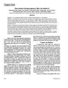
Pregnant women with fibroid evaluated for eligibility n = 64 Did not meet the inclusion criteria N = 10 Rallied with inclusion criteria N = 54 Missed follow-up N=7 Completed the study N = 47
Figure 1: Participants of the study in flow chart.
had previous myomectomy, one had uterine malformation, one had uterine adenomyosis, and 4 had medical disorder (two had history of diabetes mellitus and one had history of chronic hypertension). The follow-up of 7 cases was lost. So, ending data were included from 47 patients only (Figure 1). Ultrasonic examinations of the fibroid at 10–14 weeks, 20–24-weeks, and 28–34 weeks were recorded and studied statistically. The average of demographic data were as follows: for age 31.80 ± 3.27 years, body mass index (BMI) [calculated as weight in kilograms divided by the square of height in meters] 24.67 ± 2.46, gravidity 2.63 ± 1.21, parity 1.26 ± 1.03, duration of menstrual cycle/day 29.68 ± 3.10, and duration of menstrual period/day 6.46 ± 1.12. The percentage of spontaneous conception was 59.57% and 40.43% for using assisted reproductive technology (Table 1). Maternal outcome during antenatal period was represented in Table 2. 16 (34%) had threatened miscarriage (vaginal bleeding occurring at