Mar 25, 1992 - Conserved lysines of mouse ornithine decarboxylase were individually mutated to arginines. The mutations at amino acid residues 69, 115, ...
Vol. 267, No. 32, Issue of November 15, pp. 23057-23062,1992 Printed in U.S. A .
THEJOURNAL OF BIOLOGICAL CHEMISTRY 0 1992 by The American Society for Biochemistry and Molecular Biology, Inc.
Dominant Negative Mutants of Ornithine Decarboxylase* (Received for publication, March 25, 1992)
Stella TsirkaS andPhilip CoffinoSOll From the Departments of $Microbiology and Immunology and §Medicine, the University of California, San Francisco, California 94143
Conserved lysines of mouseornithine decarboxylase and structure of ODC. It would allow us to investigate interwere individually mutated to arginines. The mutations actions between the subunits ofODC. Such studies would at amino acid residues 69, 115, and 169 greatly re- reveal information aboutthe structureof the active site of the duced or abolished enzymaticactivity. Lysine 69 is the enzyme and would giveinsight into theorientation and folding site of Schiff base formation with the cofactor pyri- of the protein. doxal phosphate; the functional role of the other two Dominant negative mutants conditionally interfere with lysines essential for activity is not known. Coexpres- enzymatic function in vivo (6).The requirement for ODC is sion of wild type ornithine decarboxylase along with thought to be absolute, since mutants of Escherichia coli and the lysine 116 to arginine mutant reduced theactivity eukaryotic tissue culture cells in which polyamine biosynof the former without diminishingthe amount of wild thesis is disabled are notviable, unless exogenous polyamines type protein. This form of negative complementation was seen when wild typeandmutantprotein were are provided (1, 7). However, the amount ofODC activity coexpressed either by in vitro translation or in bacte- present in different tissues in vertebrates varies, suggesting ria. The data are consistent with the conclusion that a that the role it plays is more critical in some tissues than in wild type and mutant subunit form a heterodimer thatothers. Notsurprisingly, pregnant mice fed with potent inhibitors of ODC abort theirfetuses (8,9). Such evidence suggests is enzymatically inactive. that ubiquitous interference with ODC function, such as would arise from the use of homologous recombination to inactivate the ODC gene, results in earlyembryonic lethality, thus preventing study of the enzyme’s role during developOrnithine decarboxylase (ODC, EC 4.1.1.17)’ is the first ment. By judicious choice of tissue-specific promoters, howand rate-limiting enzyme in polyamine biosynthesis. It catalyzes the pyridoxal phosphate-dependent decarboxylation of ever, inhibitory consequences of dominant negative mutants ornithine toputrescine, the diamine precursor of polyamines. could be assessed in individual tissues in transgenic animals. We report here the identification of dominant negative Polyamines are ubiquitous in living organisms and areessenODC mutants. Our characterization of the dominant negative tial for growth and survival. Inhibition of their synthesis leads mutants suggests that 1) formation of an active site does not t o arrest of growth and later tocell death (1). The regulation of ODC activity is a complicated and elab- necessarily require participation of two active monomers; however, 2) since several of the mutants’ examined confer orate process, involving transcriptional (2), translational (3), and post-translational (4) control mechanisms. Mammalian partial or full dominant negative activity on the dimer comODC is a cytosolic protein which is rapidly degraded. How- plex, the subunitsof ODC do appear to interact. ever, truncation of the carboxyl-terminal 37 amino acids MATERIALS AND METHODS renders the protein more stable without affecting its activity Plasmids and Bacteria-The ODC bacterial expression plasmid (5). In addition, active mouse enzyme forms a homodimer of two 51-kDa subunits. It is not clear whether the subunits pSPODC7 (lo), which contains the BamHI-PvuII 900-base pair fragform a single catalytic unit orinstead function independently ment of pCQV2 (11) (the X p, promoter), linked to the TaqI-PuuII fragment of ODC cDNA in the EcoRI-SmaI sites of pSP64, was used of each other. for the expression of wild type or mutated ODC genes in E. coli strain We sought to address the question of how the active site 2205 (FM84 Asp& recA56), a strain with a deletion in the specgene functions. We wanted 1)to identify amino acids required for encoding ODC. Bacterial cultures without ODC activity, when grown activecatalysis by mutating individual residues conserved in M9 minimal medium, were supplemented with 100 p~ putrescine. For cotransformation experiments in E. coli, two different vectors among disparate species and 2) to determine whether the were used. The EcoRI-Sal1 fragment from pSPODC425, encoding a enzymatically inactive mutants foundexhibiteddominant truncated (but fully active) ODC protein under the X p, promoter, negative activity. was subcloned into the EcoRI-Sal1 sites of the lowcopy number The construction of dominant negative mutants would pro- kanamycin-resistant plasmid pK184 (Ref. 12, a generous gift from vide several novel and importantmeans to study the function Dr. Joblin, F. Edward H6bert School of Medicine) and transformed
* This work wassupported by National Institutes of Health Grants R01 GM45335 and R 0 1 CA29048. The costs of publication of this article were defrayed in part by the payment of page charges. This article must therefore be hereby marked “advertisement” in accordance with 18 U.S.C. Section 1734 solely to indicate this fact. II TOwhom correspondence should be addressed Box 0414, Dept. of Microbiology, University of California, San Francisco, CA 94143. Tel.: 415-476-1783; Fax: 415-476-0939. The abbreviations used are: ODC, ornithine decarboxylase; PCR, polymerase chain reaction; PAGE, polyacrylamide gel electrophoresis; DFMO, a-difluoromethylornithme.
in E. coli 2205 (pK184/425). The resulting kanamycin-resistant E. coli cells were transformed subsequently with variants of pSPODC7, in which individual lysines had been mutated to arginines. Positive colonies, selected in the presence of both kanamycin and ampicillin, were grown and assayed for ODC protein and activity. Site-directed Mutagenesis-Mutants converting conserved ODC lysines to arginines were generated using dUT-UNG mutagenesis (13). Mutants were isolated and identified using restriction analysis and sequencing. Wild type and mutant plasmids were used as DNA templates for the generation of PCR fragments used for expression studies in vitro. In Vitro Transcription and Translation-m7GpppG-capped RNAs
23057
Mutations That Inactivate ODC
23058
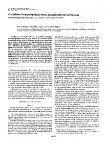
TABLE I were synthesized in vitro from linearized DNA templates (1.5 pg) or from PCR-generated DNA templates (0.5 pg) using T3 and T7RNA ODC activity of lysine to arginine mutants polymerase, respectively. Reaction mixtures were incubated a t 37 "C ODC activity in crude bacterial extracts for each mutant and for for 90 min, using the conditions recommended by the supplier. The the wild type control. Results are expressed as a percentage of the quality of the RNA synthesized was determined using gel electropho- activity in the wild type control. resis. For in vitro translations, -0.2-0.5 pg of RNA transcribed in Mutation ODC activity vitro was added to 14 pl of a reaction mixture containing rabbit reticulocyte lysate or wheat germ extract (Promega), according to the % supplier's specifications. To radiolabel the newly synthesized protein, 100 None (wild type) L-["S]methionine (ICN) (0.9 pCi) was included in the reaction mix99 K57R ture. To terminate the reaction, SDS-Laemmli sample buffer was 4 K69R added to thesamples, which were then boiled for 5 min and analyzed 0 K115R on SDS-PAGE (14). 98 K141R The in vitro transcription and translationefficiency of the systems K169R 8 used with PCR products was compared with that of the plasmid 100 K293R containing wild type ODC cDNA under control of the T3 promoter (15). This plasmid contains a full coding sequence for ODC, nucleotides 27-60 and 263-313 of the 5' leader (untranslated region), and 236 nucleotides of the 3"untranslated region. The PCR products none W.T. K169R K115R K69R lacked all 5'- and 3'-untranslated region sequences. However, the PCR products gave rise to comparable yields of translated products which had the correct ODC activities (data not shown), confirming that thecoding region is sufficient for adequate translation of ODC. Polymerase Chain Reaction-Using the polymerase chain reaction (PCR), we joined a T7 RNA polymerase promoter to the wild type and mutant ODC cDNAs. A 54-nucleotide oligomer that contained a T 7 RNA polymerase promoter sequence followed by 19 nucleotides corresponding to the 5' end of the ODC coding region was used as a 5' primer in PCRs to amplify wild type, truncated wild type, and mutant ODC cDNAs. The antisense oligomer used as a 3' PCR primer corresponded precisely to the sequence at the 3' end of the wild type ODC coding region. The program used was 92 "C for 1min, 58 "C for 1min, and 72 "C for 3 min for 30 cycles and a final extension I at 72"C for 7 min. After PCR, the unincorporated primers were removed by centrifugation through a Centricon 100 (Amicon) ultraFIG. 1. Synthesis of wild type and mutant mouse ODC in filtration unit. ODC Activity Assay-ODC activity assays were performed as de- bacteria. Bacteria carrying no plasmid or the plasmid pSPODC7 with wild type (W.T.) or mutant (K69R, K115R, or K169R) ODC scribed previously (16). Labeling with DFMO-After the invitro translation reactions were cDNAs were grown a t 32 "C. In the mid-log phase a portion of each completed using only nonradioactive amino acids, DL-[~,~-'H]DFMO culture was transferred to 42 "C to induce transcription of ODC and (a-difluoromethylornithine) was added to 10 p ~ The . labeling was incubated for 45 min. Both the 32 "Cand 42 "C cultures were labeled carried out a t room temperature for 30 min. The reactions were with ["S]methionine. Cold methionine was added after 1 min and terminated by the addition of Laemmli-sample buffer, boiled, and samples harvested and analyzed by SDS-PAGE and autoradiography. For each indicated strain, paired lanes represent cultures that were analyzed by SDS-PAGE and fluorography. Cross-linking-Wild type and mutant K69R cRNAs were trans- not induced (left) or induced (right). The arrows indicatethe position lated in the presence of ["S]methionine as described above. At the of the metabolically labeled mouse ODC. end of the translation reaction, the translation mixture was diluted 1 : l O in 20 mM Hepes buffer, pH 7.2, 0.1% bovine serum albumin, 0.1 each mutant and for the wild type control. The mutation to mM pyriodoxal phosphate and, where indicated, 1 ng of unlabeled arginine of three conserved lysines, K57R,K141R, and recombinant truncated mouse ODC protein. Unlabeled protein was K293R, had a negligible effect on ODC activity. The other added so that the concentration of specific ODC protein would not be limiting during the cross-linking reaction. The cross-linking re- three mutations, K69R, K115R, and K169R, resulted in significant loss of enzymatic activity. All three of these lysines agent bis(sulfosuccinimidy1) suberate (Pierce) was added to a final concentration of 1 mM, and the samples were incubated a t room are located within the amino-terminal half of the protein. temperature for 15 min. The reactions were quenched by the addition Synthesis of the three mutated proteins with reduced activof ethanolamine and were analyzed on 8% SDS-PAGE. ity was examined by brief labeling (1 min) of newly synthe" " "
" " "
" " "
" " "
" " "
sized bacterial proteins with [35S]methionine(Fig. 1).Similar amounts of a 51-kDa ODC protein were produced fromeach Identification of Negative Mutants-The amino acid se- of the mutant and wild type ODC cDNAs, demonstrating that quence of eukaryotic ornithine decarboxylase is highly con- the observed loss of activity for mutant proteins was not served. Among the conserved amino acids are the lysines 57, caused by a failure to synthesize a full-length protein. In 69, 115, 141, 169, and 293 (17). These lysines are of special addition, Western blot analysis confirmed that themutations interest, as at least one of them forms part of the active site did not significantly change the steady-state amounts of reand plays an important role in catalysis: ODC's cofactor, combinant ODC protein observed (results notshown). pyridoxal phosphate, necessarily forms a Schiff base with a In Vitro Transcription and Translation-To facilitate charlysine residue beforebinding to thesubstrate (ornithine) and acterization of the negative mutants, a cell-free system was decarboxylating it (18). used. For this purpose a T7 RNA polymerase promoter was To determine which of the lysines were required, we mu- joined by PCR to wild type or mutated ODC coding regions. tated each of them individually to arginine and transformed The ODC genes werethen transcribed in vitro using T7 RNA the resulting mutant cDNAs into E. coli strain 2205 (which polymerase. We haveshown previously that similar wild type has a deletion of the gene encoding bacterial ODC). Mutants constructs can be transcribed and translated in vitro to yield were confirmedby sequencing. The positive clones were then enzymatically active ODC (15).The in vitro transcribed RNAs assayed for ODC activity. Table I summarizes the ODC activ- were then translated in a rabbitreticulocyte lysate system. In ity in crude extracts, expressed as nmol/h/mg of protein, for vitro translation was performed either using [3sS]methionine RESULTS
Mutations ThatInactivate ODC to label the synthesized proteins (Fig. 2) or without labeled amino acids to produce translation products for determination ofODC activity. Wild type ODC and five mutants were studied the three found to have reduced activity when expressed in E. coli, K69R, K115R,and K169R, and two others, K141R and K293R. Noneof these mutations had a significant effect on in uitro synthesis of ODC (results notshown). Table I1 shows the ratio of the amount of ODC produced to enzymatic activity. The ratio of protein to activity provides a measure of relative specific activity and confirms that the lysine mutations that resulted in a profound loss of activity in a bacterial expression system did so as well when expressed in uitro. Similar results were seen when in uitro translation was done in a wheat germ extract system (results notshown). To test whether any of the three mutant products with reduced activity was dominant, themutant cDNAswere translated in the presence of wild type ODC cRNA. For this and subsequent experiments, the wild type ODC used was one that was truncated at amino acid 424. The truncated ODC is fully enzymatically active ( 5 ) and was made short so as to distinguish it by SDS-PAGE from the 461-amino acid fulllength mutant ODCs. Equal amounts of truncated wild type and full-length mutant ODC cRNAs were translated alone and in combination, and the products were assayed both for protein (Fig. 2) and for activity. A significant decrease in ODC activity was observed when any of the three mutants was cotranslated with wild type ODC: a 20,32, and 49% reduction compared with wild type alone for K169R, K69R, and K115R, respectively. However,there was no decrease in
23059
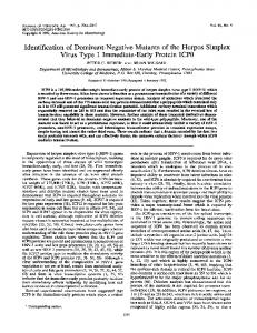
the synthesis of the truncated wild type ODC protein (Fig. 2), ruling out a simple and artifactual explanation for the decrease in enzymatic activity. Thus, we conclude that the observed inhibition of ODC activity was because of the presence of protein products of the mutated cRNAs. The significant decrease in enzymatic activity of wild type ODC in the presence of the mutant proteins suggested that all three of the negative mutants might act dominantly, implying that heterodimers could form but that the wild type subunits in them would exhibit reduced (or no) activity. To test this hypothesis further, we carried out cotranslation reactions using a fixed amount of wildtype cRNA and differing amounts of mutant cRNAs. As shown in Fig. 3,a reduction ofODC activity was observed as a function of increasing amounts of mutant cRNA. For K115R, the reduction was linearly dependent on the amount of mutant cRNA cotranslated with the wild type cRNA, resulting in -70% inhibition of the wild type ODC activity when K115R cRNAwas added in 2-fold excess over the wild type cRNA. The other two mutants also showed a progressive inhibition of wild type activity when increasing amounts of mutant were cotranslated with a constant amount of wild type ODC;however, the inhibition was less profound and not consistently cRNA concentration-dependent. When, in a converse manner, the amount of each mutant cRNA was held constant and the amount of wildtype cRNA increased, ODC activity increased (results not shown). This confirms that the inhibition observed using mutant cRNAs did not ensue from the addition of increasing quantities of total cRNA in the assay. In the inset of Fig. 3, the data from the same experiment are presented normalized for the amount of protein synthesized by KN W.Tf W.T.* W.T.r Kg(R K I W R K68F) K I I S Kt6PF) the different types of cRNAs used. Increasing K115R protein mqpmp” , -~ concentration correlates with gradual inhibition of wild type activity, as expected. We next examined the specificity of inhibition of wild type ODC activity after cotranslation with the mutantcRNAs. We cotranslated the same amounts as above of K293R or of an active S303A mutant ODC cRNA (19) with wild type ODC cRNA. No inhibition of ODC activity was observed.When cjun cRNA (used as a random, unrelated RNA) was cotranslated as above with wild type ODC cRNA, only a slight (68%)decrease of ODC activity was observed with the highest amount of c-jun cRNA used(data notshown). Thus, nonspecific inhibition does not appear to account for the excessive FIG. 2. In vitro translation of wild type and mutant mouse loss of activity observed when wild type ODC wascotranslated ODC cRNAs. Truncated wild type (W.T.)and full-length mutant with K69R, K115R,or K169R. (K69R, K115R, K169R) mouse ODC cRNAs (0.2 pg) were added We next determined whether the negative mutants could either individually or together to 14 pl of wheat germ extract translation mix (Promega) and were translated in the presence of [”SI complement each other by pairing to form active ODC hetmethionine a t 30 “C for 60 min. The reactions were terminated by erodimers. As reported previously foraspartate transcarbamthe addition of Laemmli-sample buffer and boiling and the labeled oylase (20), inactive mutantsubunits that have different proteins analyzed by SDS-PAGE. defects can assemble randomly and form inactive, partially active, and evenfully active enzyme. The three possible TABLE I1 pairwise combinations of K69R, K115R, and K169R cRNAs Activity of ODC mutants produced by translation in vitro were cotranslated, but no detectable activity was generated Specific activity is expressed as decarboxylase activity, assessed as (data not shown). This indicates that the mutants cannot 14C-labeledCO, generated from carboxyl-labeled ornithine, divided complement each other’s defects and is consistent with the by the amount of ODC produced, assessed by [%]methionine incorhypothesis that they display a dominant negative influence. poration into the in vitro translation product. Because the translation products were analyzed by denaActivity, Mutation Specific activity turing SDS-PAGE, no dimeric forms of ODC were observed 76 wild tvue directly. To establish that the mutant subunits of K69R, cpm “C/cpm %S K115R, and K169R associated with wild type subunits to 5.43 None100.00 (wild type) form heterodimers, we cross-linked the in uitro translation K69R 0.012 0.02 products, as described in Methods. As shown in Fig. 4, trunK115R 0.006 0.01 K141R 5.26 96.90 cated or full-length proteins translated individually can be K169R 0.04 0.024 cross-linked to produce dimeric products of expected and K293R 104.6 5.68 distinguishable sizes. When K69R was translated simultaneW.T.
~
Mutations That Inactivate ODC
23060
0 wt ODC (0.225 pg)
100.
80 -
FIG. 3. Effect
of increasing amounts of mutant ODCson wild type ODC activity. Truncated wild type (wt; (0.225 pg of cRNA) and increasingamounts (0.0625-0.45 pg) of full-length mutant cRNAs were translated together, as in Fig. 2, but in the presence of unlabeled methionine. ODC activity was then assayed. Synthesis of wild type and mutantproteins was tested by parallel translation in the presence of ['"S]methionine and analysis of the products by SDS-PAGE. In the inset, the results of the same experiment are displayed, showing the relationship between ODC activity and the ratio of mutant towild type protein synthesized.
h
E ..-+ Y
80-
60
m
40
i+l
-
K169R I wt
~
c)
60-
20
c1
u
n 0
0 0.5
40-
1
1.5
2
mut I wt
K69R. K115R. K169R(0.225pg) 0
0.1
0.2
0.3
0.4
0.5
ODC mutant RNA translated (pg) [wt RNA (0.225 pg) + increasing mutant RNA1
type/mutant heterodimers were recovered (Fig. 4,three rightmost lanes). The mutant proteins demonstrated differing affinities for wild type ODC in the following order: K69R > K169R K115R. This could reflect changes in the conformation of the mutant proteins which decrease heterodimer formation or stability, or differences in theefficiency of crosslinking of homo- and heterodimers. The above data, which demonstrated that K115R is the most potent inhibitor among the three mutants, suggested that heterodimers formed between K115R and wild type ODC are inactive. To gain further insight into the mechanism by which the wild type subunit was rendered inactive, we sought to determine whether it could be labeled by an ornithine substrate analog known as DFMO. DFMO, an ODC-activated suicide inhibitor, is thought tobind to and covalently modify active ODConly. Truncated wild type andmutant ODC cRNAs were translated individually or together in the presence of unlabeled methionine. [3H]DFM0 was then added, and the samples were incubated at room temperature for 15 FIG.4. Cross-linking of the in vitro translated wild type min and analyzed by SDS-PAGE. As shown in Fig. 5, a and mutant proteins. Wild type ( W.T.) and mutant cRNAs were DFMO-labeled band is observed in the sample containing in uitro translated as in Fig. 2 in the presence of [3sS]methionine. of the samples The protein-products of the translation reactions were cross-linked wild type proteinonly (firstl a n e ) but not in any containing mutant proteins only (lanes 24).Thus, as anticas described under "Materials and Methods." Cross-linking was also carriedout in the presence of an excess of unlabeled truncated ipated, DFMO labeled active protein only. Labeled bands recombinant wild type ODC (three rightmstlanes). The cross-linked corresponding to DFMO-modified wild type subunit were proteins were analyzed on an 8% SDS-PAGE. Arrows mark the observed in all of the samples in which wild type and mutant positions (from top to bottom) of cross-linked mutant-mutant dimer, protein were cotranslated (lanes 5-7). Quantitation of the mutant-wild type dimer, and wild type-wild type dimer. labeled bands revealed that the wild type ODC was labeled ously with truncated wild type protein, an intermediate sized half as much in the presence of mutant ODCs as when it was cross-linked species was formed which had the molecular translated by itself, suggesting that the labeling took place weight expected for a heterodimer between a full size and W.T.+ W.T.+ W.T.+ W.T. truncated subunit. Similar protein bands of the size of the K69R K115R K169R K69R K115R K169R heterodimeric species were obtained with the other two full+ length mutants, K115R and K169R, when cotranslated with + the truncated wild type protein. The intensity of the labeled dimeric bands obeyed the following order: K69R wild type > K115R K169R. This provides information about the relative ability of each of the FIG. 5. DFMO-labeling of the in vitro translated wild type polypeptides to form a dimer with itself. To determine the and mutant proteins. Truncated wild type ( W.T.) and mutant germ extract in the propensity of each mutant to associate with wild type mono- cRNAs were in uitro translated inawheat mers, we performed additional cross-linking studies on mix- presence of unlabeled methionine; each mutant cRNA was also cowith truncated wild type cRNA. The synthesized ODCs tures of labeled mutant ODC and unlabeled recombinant translated were then labeled with 10 p~ [3H]DFM0, as described under "Matruncated wild type ODC. Wild type protein (-1 ng) was terials and Methods." The labeled proteins were resolved by SDScross-linked to each of the mutant proteins (-0.3 ng), and PAGE. Arrows mark the positions of mutant (upper) and truncated approximately equal amounts of mutant homodimers and wild wild type (lower) ODCs.
-
-
-
-
*W:,.--.:r".~.:.
-
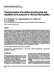
Mutations That Inactivate ODC predominantly on wild type homodimers. Unexpectedly,weak labeling of the K69R and K169R mutant ODC proteins was also detected. The intensities of the K69R- and K169Rlabeled bands were approximately 25% of that of the wild type ODC-labeled bands. This experiment demonstrates that wild type ODC protein translated in vitro foldssuch that itcan decarboxylate DFMO and subsequently become covalently modified by the nowreactive decarboxylated DFMO. In addition, the experiment confirms that inactive ODC is not labeled by [3H]DFM0, since K69R, K115R,and K169R werenot labeled when translated by themselves.Paradoxically, however, cotranslation of the K69R or K169R mutants with wild type cRNA resulted in formation of labeled mutant subunits. This suggests that the heterodimers formed by these mutants are at least partially active. Cotranslation of K115R with wild type protein did not result in the formation of a DFMO-labeled K115R subunit, suggesting that the dimerization of a wild type to a K115R subunit renders the wild type subunit inactive. This additionally supports the hypothesis that K115R exhibits dominant negative activity whencomplexed to wild type subunits. Alternatively, it might be argued that since the affinity of K115R forthe wild type subunit is relatively limited (Fig. 4), i t is possiblethat only a very low amount of heterodimer was formed, and consequently DFMO labeling of the mutant subunit in the heterodimer was too infrequent to be detected. However, the fact that wild type ODC activity is extensively inhibited when cotranslated in the presence of K115R protein suggests instead that inactive heterodimers of K115R and wild type ODC form quite readily and constitute asignificant fraction of the dimeric moleculespresent in the population. Negative Complementation in E. coli-To determine whether the dominant negative property observed for K115R could be reproduced in vivo,we transformed a kanamycinresistant plasmid encoding the wild type truncated mouse ODCcDNA(pK184/425) into 2205 E. coli cells and, as a control, transformed the same cells with an ampicillin-resistant plasmid encoding the inactive K69R mutant. To examine the interaction between wild type and mutant ODCs in cells expressing both, bacteria bearing the kanamycin-resistant plasmid encoding wild type truncated mouse ODC (pK184/ 425) were cotransformed with pSPODCK69R or pSPODCK115R or pSPODCK169R, all of which confer ampicillin resistance. Colonies resistant to both kanamycin and ampicillin were screened by restriction analysis and Western blotting. As shown in the upper panel of Fig. 6, almost equal quantities of the truncated wild type and full-length mutant proteins arepresent in the cells that express both. The lower panel of Fig. 6 depicts ODC activities determined in parallel. As expected, cellscarrying a single ODC, wildtype or mutant K69R, have, respectively, high activity and none. Of the three mutants coexpressed with wild type, only K115R inhibited wild type ODC activity significantly (- 60%). The reduction of activity observed was in close agreement with that predicted by the hypothesis that random formation of dimers occurred, i.e. half of the wild type subunits formed wildtype/wild type homodimers and the otherhalf formed inactive heterodimers with K115R subunits. The results in E. coli suggest 1)that no eukaryotic activity aside from ODC itself is required for dominance and 2) that the dominant negative activity is manifested adequately in vivo to use it in future experiments in eukaryotic organisms. DISCUSSION
We altered several lysine residues of the ODC protein that have been preserved in different species by conservative mu-
None
23061 'W.T.
W.T.' K69R K69R
W.T.' K l 1%
W.T.* K169R
I
I 0
15000
--i > "
10000
u
m U
n 0
5000
0
FIG.6. Coexpression of wild type andmutant ODC in bacteria. Bacteria carrying either the truncatedwild type ( W.T.)ODC cDNA alone or both the wild type and one of the three mutantODC cDNAs were grown a t 32 "C and thermally induced a t 42 "C, as in Fig. 1. Crude cell lysates were analyzed by Western blot(upper panel) and were tested for ODC activity (lower panel), as described under "Materials and Methods." Arrows mark the positions of truncated wild type (lower lane) and mutant(upper l a n e ) proteins. tations to arginines. Three of these mutations, K69R, K115R, and K169R, resulted in marked loss of activity by the altered ODCs. The loss of wild type ODC activity was expected for two of the three negative mutants. K69was recently identified2 (21) as the site a t which the cofactor pyridoxal phosphate binds. Mutation of this siteshould alter thebinding of pyridoxal phosphate to the enzyme and abolish its ability to form a Schiff base. We found recombinant K69R ODC to have residual activity, a few percent that of wild type (Table I). This is consistent with the properties of another pyridoxal phosphate enzyme, aspartate aminotransferase, in which a similar binding site mutation reduced but did not abolish activity (22). While this work wasin progress, Lu et al. (19) made several mutations in ODC in an effort to identify residues which participate in or affect the active site: K349A or the double mutant S303A/E308A had no effect on ODC activity, whereas the alteration of lysine 298 to alanine maintained 33% of ODC activity. Additionally, they reported that the mutation of histidine 197 or lysine 169 to alanine resulted inthe recovery of less than 1%of wild type ODC activity. We found also that the K169R mutant was inactive, even though our mutation to arginine is more conservative. Our final negative mutant, K115, altered aconserved lysine that is part of a stretch of conserved amino acids, Y-A-N-PC-K- (23). Recently, Jackson aligned the sequences of seven pyridoxal phosphate-dependent decarboxylases from five different organisms and derived a consensus sequence forthem. According to his alignment, the putative lysine required for Schiff base formation with pyridoxal phosphate is contained within the consensus sequence N-P-H-K (24). In mouse ODC this sequence is found at amino acids 112-115, although the residue immediately preceding lysine 115 is a cysteine instead of a histidine. The data of Lu et al. (19), however, are inconsistent with this conclusion: they identified lysine 69 as the residue with which pyridoxal phosphate forms a Schiff base. The datadescribed above demonstrate that lysines 69,115 and 169 are essential for ODC activity, and possibly participate in formation of the active site of the enzyme. We at-
's. Tsirka and P. Coffino, manuscript in preparation.
23062
Mutations That Inactivate ODC
tempted to purify the recombinant mutant enzymes, using our established ODC purification protocol (25). The K169R protein was purified to homogeneity using pyridoxamine phosphate-Affi-Gel and Mono Q column chromatography. Neither K69R nor K115R mutant protein bound securely to the Affi-Gel column, suggesting that both residues are important for the interaction ofODC with pyridoxal phosphate (results not shown). The three negative mutants, K69R, K115R, and K169R, were studied in a cell-free system and their interaction with wild type protein examined. Allof them were effective in inhibiting wild type ODC activity when coexpressed with it. Of the three, K115R had themost profound inhibitory effect when cotranslated with wild type protein (Fig. 3). Our results indicate that theinhibition proceeds via formation of inactive dimeric hybrids between mutant subunits andwild type subunits. The existence of heterodimers was confirmed by physically cross-linking mutant proteins to recombinant wild type proteins and observing the hybrid complexes on SDS-PAGE (Fig. 4). The results of this experiment also imply that wild type and mutantODC subunits exchange continuously. DFMO, the specific enzyme-activated suicide inhibitor of ODC, was used to provide information about the activity of the heterodimeric mutant proteins. Fig. 5 demonstrates that tritiated DFMO could not label the mutants when they were translated by themselves. DFMO was able, however, to modify and label K69R and K169R mutant polypeptides when they were synthesized along with wild type subunits by simultaneous translation of both cRNAs. The labeling efficiency of these heterodimers by DFMO apparently depends both on the ability of each mutant to form a heterodimer with the wild type subunit and on the activity of the heterodimer. The labeling results suggested thatthe heterodimers between K69R and wild type, and K169R and wild type might be partially active. The heterodimer between K115R and wild type does not label with DFMO. Given the ability of K115R to dimerize with wild typeprotein (Fig. 4), such a dimer apparently does form and must therefore be inactive since it does not becomes labeled. This result also suggests that ODC dimers are cross-modified by DFMO, i.e. that DFMO decarboxylated by one subunit can then modify the other member of the dimer pair. The reasoning for this is as follows. In thecotranslation lanes the K69R and K169R bands are labeled. Since modification by DFMO occurs after DFMO is decarboxylated, DFMO modification requires the presence of an active enzyme. Neither of the K69R or K169R mutants is active by itself, as demonstrated by the lack of modification in the lanes inwhich K69R and K169R are translated alone. We therefore conclude that the modification observed in cotranslation lanes was caused by theactivation of DFMO by the decarboxylating activity of the wild type subunits,and theactivated DFMO subsequently modified the neighboring inactive K69R or K169R subunits.
The data described above suggest that K115R acts as a negative mutant dominant to wild type ODC activity. We wanted also to determine whether dominance was observed in uiuo. Fig. 6 shows that when the three negative mutants were coexpressed with the wild type in E. coli, ODC activity was inhibited only in the bacteria cotransformed with the K115R mutant. Thisindicates that theinteraction of the wild type subunit with the inactive mutant subunit is sufficient for inhibition of ODC activity, and no other exogenous activity or factor is required. Thus the dominant negative mutant can be used in vivoto disrupt the activity of endogenous ODC. The identification of K115R as a dominantnegative mutant of ornithine decarboxylase provides two novel and important ways to study function and structureof the enzyme. First, it has and will allow us to investigate interactions between the subunits of active homodimeric mouse ODC. Second, we will now be able to use this dominant negative mutant tointerfere with ODC function in uiuo, by, for example, introducing it into transgenic mice in a tissue-specific manner. The dominant negative mutant will thus be used to ablate or modulate ODC activity in acontrolled and directed manner. Acknowledgment-Preliminary
experiments on mutagenesis of
ODC were carried out by Dan Medynski in this laboratory. REFERENCES 1. Tabor C. W., and Tabor H. (1984) Annu. Reu. Biochem. 63,749-790 and Kahana, C.'(1987) Mol. Cell. Biol. 7, 2641-2643 2. Katz, 3. White, M. W., Kameji, T., Pegg, A. E., and Morris, D. R. (1987) Eur. J. Biochem. 170,87-92 4. van Daalen Wetters, T., Macrae, M., Brabant, M., Sittler, A.,and Coffino, P. (1989) Mol. Cell. Biol. 9,5484-5490 5. Ghoda, L., van Daalen-Wetters, T., Macrae, M., Ascherman, D., and Coffino, P. (1989) Science 243,1493-1495 6. Herskowitz, I. (1987) Nature 3 2 9 , 219-222 7. Pilz, R., Steglich, C., and Scheffler, I. (1990) J. Biol. Chem. 2 6 6 , 8880-
x.,
8. FO;;;~ J., Part M., Prakash, N., Grove, J., Schechter, P., Sjoerdsma, A., and Koch-Wiser, J. (1980) Science 208,505-508 9. Fozard, J., Part, M., Prakash, N., and Grove, J. (1980) Eur. J. Phnrmacol. 66,379-391 10. Macrae, M., and Coffino, P. (1987) Mol. Cell. Biol. 7,564-567 11. Queen, C. (1983) J.Mol. Genet. 2,1-10 12. Joblin, M. G., and Holmes, R. K. (1990) Nucleic Acids Res. 18,5315-5316 13. Kunkel, T., Roberta, J., and Zakour, R. (1987) MethodsEnzymol. 1 6 4 , 367-382 14. Laemmli, U. K. (1970) Nature 227,680-685 15. Ghoda, L., Sidney, D., Macrae, M., Coffino, P. (1992) Mol. Cell. Biol. 1 2 , 2178-2185 16. Prichard, M. L., Seely, J. E., Poso, H., Jefferson, L. S., and Pegg, A. E. (1981) Biochem. Biophys. Res. Common. 100,1597-1603 17. Coffino, P. (1989) ,in Ornzthzv Decarboxylase: Biology, Enzymology, and Molecular Genetzcs (Hayashl, S., ed) pp. 135-144, Pergamon Press, New York 18. Bey, P., Danzin, C., and Jung, M. (1987) in Inhibition of Polyamine Metabolism: Biological S' niftcance and Basis for New Therapgs (McCann, P., Pegg, A., and gjoerdsma, A., eds) pp. 1-27, Academlc Press, San Diego 19. Lu, L., Stanley, B., and Pegg, A. (1991) Biochem. J. 277,671-675 20. Robey, E., and Schachman, H. (1985) Proc. Natl. Acad. Sci. U. S. A. 8 2 , 361-365 21. Poulin, R., Lu,L., Ackermann, B., Bey, P., and Pegg, A. E. (1992) J. Biol. Chem. 267,150-158 22. Kuramitsu, S., Inoue, Y., Tanase, S., Morino, Y., and Kagamiyama, H. (1987) Biochem. Biophys. Res. Commun. 146,416-421 23. Gupta, M., and Coffino, P. (1985) J. Bcol. Chem. 260,2941-2944 24. Jackson, F. R. (1990) J. Mol. Euol. 31,325-329 25. Phillips, M., Coffino, P., and Wang, C. (1987) J. Biol. Chem. 2 6 2 , 87218727