ã
Dentomaxillofacial Radiology (2002) 31, 218 ± 223 2002 Nature Publishing Group. All rights reserved 0250 ± 832X/02 $25.00 www.nature.com/dmfr
RESEARCH
3D volume rendering using multislice CT for dental implants MGP Cavalcanti*,1, A Ruprecht2,3,4 and MW Vannier3 1
Department of Radiology, Faculty of Odontology, University of SaÄo Paulo, Brazil; 2Department of Oral Pathology, Radiology, and Medicine, College of Dentistry, University of Iowa, Iowa, USA; 3Department of Radiology, College of Medicine, University of Iowa, Iowa, USA; 4Department of Anatomy and Cell Biology, College of Medicine, University of Iowa, Iowa, USA
Objectives: To determine the precision and accuracy of three-dimensional (3D) volume rendering spiral multislice computed tomography (CT)-based linear measurements of the mental foramen for dental implants, in vitro, and their precision, in vivo. Methods: Five cadaver heads were imaged by multislice spiral CT (Toshiba Aquilion) with 0.5 mm thick axial slices (0.5 mm/0.5 s of table feed) at 0.5 mm interval reconstructions. The image data sets were transferred to a networked computer workstation. Using computer graphics the data were analysed with a 3D volume rendering technique using Vitrea1 software. Two oral and maxillofacial radiologists, independently, made electronic linear measurements from the superior border of the mental foramen to the crest of the alveolar process. The soft tissues were removed and physical measurements made using a 3 SpaceTM (Polhemus, Colchester, VT, USA) electromagnetic digitizer with a personal computer running Windows1 98. The same linear measurements of 15 patients using the same imaging methodology were performed and the precision was analysed. Results: The ®ndings showed no statistically signi®cant inter- or intra-observer dierences in vitro and in vivo, or between imaging and physical measurements in vitro (P40.05). Conclusions: 3D multislice spiral CT imaging allows highly accurate measurements for dental implant placement in proximity to the mental foramen. Computer graphics software, using volume rendering is suitable for implant planning. Dentomaxillofacial Radiology (2002) 31, 218 ± 223. doi:10.1038/sj.dmfr.4600701 Keywords: tomography, X-ray computed; dental implantation; mandible; image processing, computer-assisted Introduction Implant placement is a rapidly growing component of dental practice. Accurate dental implant placement requires comprehensive pre-operative radiographic examination.1 ± 5 It has been demonstrated that computed tomography (CT) provides better visualization of the jaws than other radiographic methods.1 ± 5 Multislice spiral CT is a signi®cant advance in the technology of X-ray computed tomography, which results in rapid table speeds and the opportunity to greatly increase the speed of data acquisition. Previous studies demonstrated the precision and accuracy of 2D orthoradial multiplanar reformatted *Correspondence to: MGP Cavalcanti, University of SaÄo Paulo, Faculty of Odontology, Department of Radiology, Av. Prof. Lineu Prestes, 2227, SaÄo Paulo, SP, 05508-900, Brazil; E-mail:
[email protected]; 3D-Imaging Laboratory; www.usp.br/FO/labi3d Received 27 November 2001; revised 5 February 2002; accepted 7 February 2002
and 3D surface-rendered images of the mental foramen and inferior alveolar canal.5,6 Measurements of 3D surface-rendered images were found to be accurate, but with a high standard deviation, resulting in the conclusion that 3D surface-rendered images were not completely satisfactory for implant measurements.6 The process of 3D volume rendering involves: (1) navigation to ®nd an orientation that enables visualization of the structure of interest and (2) choice of opacity and color transfer function to suppress surrounding tissue and enhance the structure of interest for optimal view. Therefore, we believe it is important to apply computer graphics technology using the newer CT technology combined with 3D technique to test the accuracy and precision of linear measurements of dental implants. The aims of this work are to determine the precision (reproducibility) and the accuracy (validity) of 3D volume rendered images from a multislice spiral CT
3D CT of dental implants MGP Cavalcanti et al
219
A
B
C
Figure 1 Multiplanar reconstructed images with the crosshair lines exactly located at the same point (arrows) representing the superior border of the mental foramen in (A) axial, (B) coronal, and (C) sagittal views
scanner, in vivo and in vitro, using a desktop workstation for presurgical planning of dental implants related to the mental foramen.
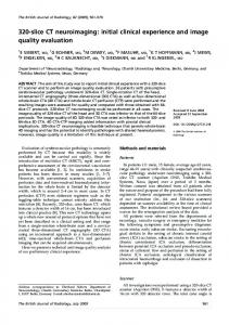
A
B
Materials and methods The current study was based on ®ve cadaver heads (aged between 55 and 65 years) provided by the Department of Anatomy and Cell Biology of the College of Medicine, The University of Iowa, following approval by the institutional review committee. All specimens were edentulous posterior to the mental foramen. The heads were imaged using a multislice spiral CT (Aquilion2 Toshiba-America Medical Systems Inc., Tustin, CA, USA) with 0.5 mm thick axial slices (0.5 mm/0.5 s of table feed and 0.5 mm interval reconstructions) beginning superior to the base of the skull and extending inferiorly to below the mandible, at 120 kVp and 150 mA. A low frequency ®lter cut-o was used in the reconstruction algorithm. The ®eld of view (FOV) was 20.3 cm, (5126512 matrix). The archived CT data were originally stored on optical disks (Imation Optical, 3M, rewritable optical disk, Imation Enterprises Corporation, Oakdale, MN, USA), and then transferred to a networked computer workstation (DELL Precision 420 Windows NT), to generate 3D volume rendered images. Subsequently the images were stored on the workstation desktop and in a CD-ROM to allow full retrospective review of any data and image processing. Using multiplanar reconstructed images, the crosshair tool lines were located at exactly the same point (arrows), namely the anterior border of the mental foramen (Figure 1). Next, the starting and endpoint of the segment of the mandibular arch to be traced in the axial slice were determined (Figure 2a). A line was created along the midpoints between the facial and lingual cortex of the mandible between these two points (Figure 2b). Another line (arrow) perpendicular to this line was generated from a point on the superior
Figure 2 (A) Lines perpendicular to the arch line traced on the axial view are generated. (B) The arch line is generated midway between the facial and lingual cortex of the mandible is shown. At the mental foramen a line (arrow) perpendicular to the arch line is created
most aspect of the mental foramen on the buccal surface (Figure 2b). A 3D model of the same tracing in MPR (multiplanar reconstructed) images was then generated automatically, with the tracing of the mandibular arch, the lines, and the same point in 3D, which corresponded to the superior border of the mental foramen, allowing to perform the measurement (Figure 3a). Using computer graphics the data were analysed with a 3D volume-rendering technique using Vitrea1 (Vital Images Inc., Plymouth, MN, USA) 2.1 software. In this 3D reconstructed image the inferior alveolar canal and mental foramen were depicted using the transparence and color tools (Figure 3b). The software tool protocol was used for subsequent image postprocessing and analysis. Two oral and maxillofacial radiologists processed the images and made electronic 3D linear measurements, independently, on two occasions separated by several weeks, from the superior border of the mental foramen to the crest of the alveolar process, bilaterally (Figure 3a,b,c). In total, 40 measurements were made. The soft tissues were subsequently removed, and the same measureDentomaxillofacial Radiology
3D CT of dental implants MGP Cavalcanti et al
220
A
B
Figure 4 3D-CT volume rendered reconstructed images of a patient using the same methodology as for the in vitro images in Figures 1 to 3, using color and transparence tools to show the inferior alveolar canal and the mental foramen. The distance from the superior border of the mental foramen to alveolar crest is 14.1 mm
SAS 6.09 (Statistical Analysis Systems, Institute Inc., Cary, NC, USA), with P-value less or equal to 0.05 for statistical dierence. Results C
Figure 3 (A) In the 3D reconstructed image lines are generated perpendicular to the line traced in Figure 2. The corresponding blue line (arrow) of the axial images is shown, as well as the measurement (11.8 mm) distance from the blue line to the crest of the alveolar process. (B) The 3D image is depicted in the transparence and color tool mode using the volume rendering technique, as is the mental foramen and the measurement from the superior part of the mental foramen to the corresponding alveolar crest: 11.8 mm. (C) The lines generated in A and B above are seen with the image of the bone in full transparence mode
ments were repeated using a 3 Space2 electromagnetic digitizer (Polhemus, Colchester, VT, USA) with a Pentium III personal computer (IBM, Armonk, NY, USA) and Windows1 98 (Microsoft, Redmond, WA, USA) used to display the physical measurements. The in vivo study was based on 15 patients who were referred for multislice CT for dental implant planning, where these 3D-CT methods and parameters were used (Figure 4). In this case 120 measurements were performed to analyse the reproducibility (precision) of the methodology in vivo. Statistical evaluation was performed using Analysis of Variance (ANOVA) with Dentomaxillofacial Radiology
There was no signi®cant dierence in either inter- or intra-observer measurements in vitro (P40.05). The mean dierence for inter- or intra-observer measurements was 0.10 mm each (Table 1), indicating a high level of precision. The measurements showed no signi®cant dierences between the image-based and physical measurements (P=0.35) (Table 2). The dierence between the mean actual and mean 3Dbased linear measurements was 0.25 mm (Table 2). The standard deviation (s.d.=0.51) and the standard error (s.e.=0.31) were also considered low in absolute values. This demonstrated the high accuracy of 3D linear measurements. In the in vivo study, there was no signi®cant dierence in inter- or intra-observer measurements (P40.05). The mean dierence in inter- or intraobserver measurements was 0.38 mm and 0.21 mm respectively (Table 3). This showed an acceptable level of reproducibility (precision) of those measurements. Discussion CT is considered an important imaging modality, because it is fast, reproducible, and reliable. Output has increased with the advent of the multislice CT scanner, which can acquire more slices in a shorter time, and cover more anatomy with thin slices than the previous spiral CT commonly used for dental implants. CT equipped with speci®c software designed for implantology can be used to determine the suitability of bone and speci®c sites for implant placement, the size of the implant that can be placed, and the need for pre-implantation ridge surgery. The osseous architecture and the positions of speci®c anatomical structures can be seen clearly. Using interactive software for
3D CT of dental implants MGP Cavalcanti et al
Table 1 Statistical analysis using ANOVA (analysis of variance), comparing inter- and intra-observer 3D-measurements in vitro (P=0.05 for significance) Source
Mean*
s.d.
s.e.
P
0.10 0.10
0.31 0.54
0.24 0.25
0.10 0.36
Observer Session
*Mean (mm) of the diernences between the inter- and intra-observer measurements, s.d. (standard deviation), s.e. (standard error), P (P value)
Table 2 Statistical analysis using ANOVA, comparing 3D-based linear measurements with physical measurements (P=0.05 for significance) 3D-CT vs physical
Mean*
s.d.
s.e.
P
0.25
0.51
0.31
0.35
*Mean (mm) of the dierences between the imaging and physical measurements; s.d. (standard deviation), s.e. (standard error), P (P value)
Table 3 Statistical analysis using ANOVA (analysis of variance), comparing inter- and intra-observer 3D-measurements in vivo (P=0.05 for significance) Source Observer Session
Mean*
s.d.
s.e.
P
0.35 0.21
0.51 0.54
0.36 0.29
0.18 0.41
*Mean (mm) of the dierences between the inter- and intra-observer measurements, s.d. (standard deviation), s.e. (standard error), P (P value)
reformatted CT, it is possible, in the mandible, to determine the distance between the alveolar crest and the mental foramen and inferior alveolar canal.5,6 With this technique, the radiologist can determine for the surgeon the exact amount of bone that must be removed or added, and the distance between the implant and vital structures.3 Several other authors have recommended that CT is the method of choice for dental implant planning.5,7 ± 9 Todd et al.10 compared linear tomography with CT for use with dental implants and found a magni®cation of 28% for linear tomography, in comparison with 4% for CT. In 1995 Luka et al.11 studied patients using a dental imaging program and spiral CT with a collimation of 1 mm, and a table feed of 1 mm/s. They stated that only in the 2D reformatted technique could the mandibular canal and mental foramen be identi®ed. It is generally agreed that spiral CT allows faster scanning, thereby decreasing the problem of patient movement during the data acquisition which has previously caused problems with image reconstruction and distortion in the ®nal results. In addition, spiral CT allows better resolution in the generated images.12 DelBalso et al.4 stated that 3D-CT surface rendered images, while not absolutely essential in pre-operative evaluation of dental implants, provide a gross overview of the alveolar ridge and adjacent osseous structures.
They also mentioned that important information can be obtained from 3D imaging of atrophic ridges, including the extent of ridge atrophy, areas where surgical augmentation must be performed to permit implant placement, and the spatial relationship of the maxilla and mandible. In previous studies we used Cemax ToothPix2 software, which allows 2 mm thick 2D orthoradial multiplanar reformatted images to be reconstructed.5,6 The plane of the images is perpendicular (orthogonal) to the curvature of the dental arch. We also demonstrated the accuracy of 3D measurements but with high standard deviations, and we stated that the standardization was not completely satisfactory, because of the limitation of the 3D technique for that purpose.6 3D volume rendering is used in an attempt to preserve, to the greatest extent possible, the information present in the image data set and in this regard has advantages over surface rendering, in which much of the original information in the image volume is implicitly discarded. Hopper and coworkers, using a phantom as an experimental material, found increased accuracy when using 3D volume rendered images, which re¯ects the ability to view the internal structures of the object, something not possible with 3D surface rendering. Also they found a low interobserver variability.13,14 Furthermore, 3D volume rendering had been shown to be accurate, within scanner resolution, for evaluation of smallcaliber vascular structures.15 Several papers have been published dealing with 3D volume rendering techniques for improved visualization of the liver, pancreas, abdominal aorta, and cerebral aneurysms, as well as for cardiac applications, and maxillofacial neoplasms with acceptable results.13,15 ± 19 The combination of subsecond spiral CT and 3D volume rendered reconstructed images allows rapid and detailed examination of the musculoskeletal system.12 Shimizu et al.18 reported that although surface rendered images provide good image quality with 3D displays, the selection of a single threshold and transparency of 0% results in less information than is available with volume rendered images. Lee et al.19 demonstrated the utility of volume-rendered 3D-CT in the head and neck region in radiation oncology. Applying this methodology to our research, we found high accuracy and precision in vitro and in vivo with 3D volume rendered images using color and transparence tools. Those results were better than in the previous study using 2D orthoradial and 3D surface rendered images.5,6 In our study, in order to assess the accuracy of the multislice scanner, which represents the newest technology in spiral CT, combined with 3D volume rendering, we used computer graphics systems to measure the images of cadaver heads in simulated implant situations. These measurements were validated on the dissected specimens by physical measurements of the same distance made with an electromagnetic
221
Dentomaxillofacial Radiology
3D CT of dental implants MGP Cavalcanti et al
222
digitizer. This device has previously been validated by Hildebolt and Vannier,20 as well as by several other researchers.5,6,21 Using a thinner protocol we found a mean dierence of less than 0.4 mm between the two sets of measurements (Tables 1 and 2). Localization of the points studied proved easy for both observers. This showed a high reproducibility for those measurements using this new 3D methodology in association with a submillimeter CT scanner. Furthermore, we believe that spiral 3D-CT reconstructed images oer the ability to obtain an accurate image of the body of the mandible and the alveolar process. Therefore, sectional models can be produced to show the location of the mental foramen and mandibular canal within the implantation ®eld. We think the combination of multislice CT and 3D volume rendering techniques using computer graphics systems allows several improvements over older technology, including patient data entry, study prescription, ®lming, archiving and image transmission. Our paper deals with this new technology showing its new application to dental implants, where we can greatly increase speed (0.5 s per slice thickness and interval of reconstruction) while improving both image quality and productivity, thus increasing the diagnostic and treatment planning eectiveness. We feel that CT in the oral and maxillofacial complex will continue to give rise to new applications based upon advances in the technology. Multislice 3D-CT provides excellent visualization and delineation of mandibular anatomy, which in turn permits establishment of the potential buccolingual position and inclination of the respective implants,
and allows the measurement of the amount of bone present. The latter is due to 3D volume rendering technique tools associated with thin slices and small intervals of reconstruction used (0.5 mm). Both observers clearly and easily identi®ed the superior and inferior borders of the mental foramen in the cross-sectional views, even in vivo and in vitro. Within approximately 10 min it was possible to generate images from the multislice spiral CT, send them to an independent workstation, reformat them, and perform 3D measurements. In conclusion, multislice spiral CT using a 3D volume rendering technique allows higher measurement accuracy and precision than a 3D surface rendering technique with respect to dental implantation in proximity to the mental foramen. The combination of multislice CT scanning and 3D volume rendering is established for dental implant applications and has provided valuable information for treatment planning. This makes it possible to formulate a more accurate diagnosis and treatment plan. Acknowledgements Research supported by the Grants from the Foundation of Research of SaÄ o Paulo State (FAPESP), 99/10276-4, Brazil. Department of Anatomy and Cell Biology of the College of Medicine at The University of Iowa that provided the cadaver heads. We acknowledge the capable technical assistance of Scot Daniel Thomas Henry, RTR (CT), Department of Radiology at The University of Iowa Hospitals and Clinics, who scanned the specimens. We also thank Ms Jane Jakobsen, MA from the Department of Preventive and Community Dentistry at The University of Iowa, for her assistance with the statistical analysis.
References 1. Shimura M, Babbush CA, Majima H, Yanagisawa SW, Sairenji E. Presurgical evaluation for dental implants using a reformatting program of computed tomography: maxilla/mandible shape pattern analysis (MSPA). Int J Oral Maxillofac Implants 1990; 5: 175 ± 181. 2. Abrahams JJ. Anatomy of the jaw revisited with a dental CT software program. Am J Neuroradiol 1993; 14: 979 ± 990. 3. James RA, Lozada JL, Truitt HP. Computer tomography (CT) application in implant dentistry. J Oral Implantol 1991; 17: 10 ± 15. 4. DelBalso AM, Greiner FG, Licata M. Role of diagnostic imaging in evaluation of dental implant patient. Radiographics 1994; 14: 699 ± 719. 5. Cavalcanti MGP, Yang J, Ruprecht A, Vannier MW. Validation of spiral computed tomography for dental implants. Dentomaxillofac Radiol 1998; 27: 329 ± 333. 6. Yang J, Cavalcanti MGP, Ruprecht A, Vannier MW. 2D and 3D reconstructions of spiral CT in localization of the inferior alveolar canal for dental implants. Oral Surg Oral Med Oral Pathol Oral Radiol Endod 1999; 87: 369 ± 374. 7. Yanagisawa K, Friedman C, Vinning E, Abrahams JJ. Denta Scan imaging of the mandible and maxilla. Head and Neck 1993; 15: 1 ± 7. 8. Weinberg LA. CT scan as a radiologic data base for optimum implant orientation. J Prosthet Dent 1993; 69: 381 ± 385. 9. Bessimo C, Lambrecht JT, Nidecker A. Dental implant planning with reformatted computed tomography. Dentomaxillofac Radiol 1995; 24: 264 ± 267. Dentomaxillofacial Radiology
10. Todd AD, Gher ME, Quintero G, Richardson AC. Interpretation of linear and computed tomograms in the assessment of implant recipient sites. J Periodontol 1993; 64: 1243 ± 1249. 11. Luka B, Brechtelsbauer D, Gellrich NC, Konig M. 2D and 3D CT reconstructions of the facial skeleton: an unnecessary option or a diagnostic pearl? Int J Oral Maxillofac Surg 1995; 24: 76 ± 83. 12. Pretorious ES, Fishman EK. Volume-rendered three-dimensional spiral CT: musculoskeletal applications. Radiographics 1999; 19: 1143 ± 1160. 13. Hopper KD, TuncË Lyriboz A, Wise SW, Neuman JD, Mauger DT, Kasales CJ. Mucosal detail at CT virtual reality: surface versus volume rendering. Radiology 2000; 214: 517 ± 522. 14. Jani AB, Pelizzari CA, Chen GTY, Grzeszczuk RP. Accuracy of object depiction and opacity transfer function optimization in CT volume-rendered images. J Comput Assist Tomogr 1998; 22: 459 ± 470. 15. Johnson PT, Fishman EK, Duckwall JR, Calhoum PS, Heath DG. Interactive three-dimensional volume rendering of spiral CT data: current applications in the thorax. Radiographics 1998; 18: 165 ± 187. 16. Cavalcanti, MGP, Vannier, MW. Measurement of the volume of oral tumors by three-dimensional spiral computed tomography. Dentomaxillofac Radiol 2000; 29: 35 ± 40.
3D CT of dental implants MGP Cavalcanti et al
17. Jani AB, Pelizzari CA, Chen GTY, Roeske J, Hamilton RJ, Macdonald RL, et al. Volume rendering quanti®cation algorithm for reconstruction of CT volume-rendered structures: Part I: Cerebral arteriovenous malformation. IEEE Trans Medical Imaging 2000; 19: 12 ± 24. 18. Shimizu T, Yoshikawa S, Uesugi Y. Three-dimensional computed tomography angiography and pulmonary vessels. Radiat Med 1999; 17: 151 ± 154. 19. Lee JS, Jani AB, Pellizari CA. Volumetric visualization of head and neck CT data for treatment planning. Int J Radiat Oncol Biol Phys 1999; 44: 693 ± 703.
20. Hildebolt CF, Vannier MW. Three-dimensional measurement accuracy of skull surface landmarks. Am J Phys Anthropol 1988; 76: 497 ± 503. 21. Cavalcanti MGP, Vannier MW. Quantitative analysis of spiral computed tomography for craniofacial clinical application. Dentomaxillofac Radiol 1998; 27: 344 ± 350.
223
Dentomaxillofacial Radiology