ies that bind to the heavy chain of Acanthamoeba myosin-IA. Eight of these antibodies bind to myosin-. IB and eight cross-react with Acanthamoeba myosin-II.
Characterization of Monoclonal Antibodies to Acanthamoeba Myosin-I that Cross-react with Both Myosin-II and Low Molecular Mass Nuclear Proteins S u s a n J. H a g e n , D a n i e l P. K i e h a r t , D o n a l d A. Kaiser, a n d T h o m a s D. Pollard Department of Cell Biology and Anatomy, Johns Hopkins Medical School, Baltimore, Maryland 21205. Dr. Hagen's present address is Division of Gastroenterology, Brigham and Women's Hospital, Boston, Massachusetts 02115. Dr. Kiehart's present address is Department of Cellular and Developmental Biology, Biological Laboratories, Harvard University, Cambridge, Massachusetts 02138.
Abstract. We characterized nine monoclonal antibodies that bind to the heavy chain of Acanthamoeba myosin-IA. Eight of these antibodies bind to myosinIB and eight cross-react with Acanthamoeba myosin-II. All but one of the antibodies bind to a 30-kD chymotryptic peptide of myosin-IA that derives from the COOH terminus of the molecule, and to tryptic peptides as small as 17 kD, hence these epitopes are clustered closely together on the heavy chain. None of the antibodies prevent heavy chain phosphorylation by myosin-I heavy chain kinase. One antibody inhibits the K+-EDTA ATPase activity and three antibodies inhibit
the actin-activated Mg÷÷-ATPase activity of myosin-I under the set of conditions that we tested. When fluorescent antibody staining of both whole cells and isolated nuclei is done, several of these monoclonal antibodies react strongly with nuclei. These antibodies also stain the cytoplasmic matrix, especially the cortex near the plasma membrane. All nine of the monoclonal antibodies bind to polypeptides of 30-34 kD that are highly enriched in nuclei isolated from Acanthamoeba. There is no myosin-I in the isolated nuclei, so the 30-34-kD polypeptides, not myosin-I, are responsible for the nuclear staining.
VER since it was discovered in Acanthamoeba (24), myosin-I has been a fascinating enigma. Unlike conventional myosins, including myosin-II from Acanthamoeba (21, 26), the myosin-I isozymes (IA and IB; reference 23) are globular proteins consisting of only single heavy chains and one or two light chains (4, 23, 24). The myosin-I isozymes have little (10, 17) or no (4) tail, so that they do not polymerize to form conventional bipolar filaments like other myosins (24, 25). On the other hand, myosin-I does have Ca++ and K+-EDTA ATPase activity similar to that of muscle myosin (24), binds reversibly to actin filaments (10, 22, 25) and, when phosphorylated on the heavy chain (22), possesses actin-activated ATPase activity (2, 22, 25). Myosin-I can propel plastic beads along actin filaments (4) and causes superprecipitation of actin filaments (9). Indirect fluorescent antibody staining with polyclonal antibodies to myosin-I showed that myosin-I is localized throughout the cytoplasm, but is especially abundant in the cortex of the amoeba (11). Until recently there was uncertainty regarding the existence of myosin-I in any other cell type, but this has been resolved with the isolation of a similar enzyme from the slime mold Dictyosteliura discoideum (6). In the present study, we characterized nine antibodies (eight originally described by Kiehart et al.; reference 17), including one that inhibits the K+-EDTA ATPase activity
and three that inhibit the actin-activated Mg++ ATPase of myosin-I. It is of equal interest that we show here that all of these antibodies to myosin-I also bind to polypeptides of 30-34 kD that are localized in the nucleoplasm. These nuclear polypeptides clearly bear some relationship to myosinI, since the two proteins share several epitopes. However, further studies (27; Rimm, D., and T. D. Pollard, manuscript in preparation) have shown that the nuclear proteins are not fragments of myosin-I. Adams and Pollard (1) have also used one of these antibodies to inhibit the movement of Acanthamoeba organelles in a model system. Part of this work was presented at the 1984 Annual Meeting of the American Society for Cell Biology (12).
© The Rockefeller University Press, 0021-9525/86/12/2121/8 $1.00 The/ournalofCellBiology, Volume 103(No. 6, Pt. 1),Dec. 1986 2121-2128
Materials and Methods Materials ATE dithiothreitol, imidazole (grade I), phenylmethylsulfonylfluoride (PMSF), sodium azide, polylysine, and benzamidine were purchased from Sigma Chemical Co. (St. Louis, MO). [32p]ATP was from Amersham Corp. (Arlington Heights, IL). Reagent grade chemicals were from J. T. Baker Chemical Co. (Phillipsburg, N J). Rhodamine-labeled goat antimouse Ig was from Cappel Laboratories (Cochranville, PA). Low gelling temperature Sea Plaque agarose was purchased from Marine Colloids Div., FMC Corp. (Rockland, ME). Alpha-chymotrypsin and L-l-p-tosylamino-2phenylethyl chloromethyl ketone (TPCK)-trypsin were purchased from
2121
Worthington Biochemical Corporation (Freehold, NJ). Immulon-1 and -2 Removawells (Dynatech Laboratories, Inc., Alexandria, Va) were used for solid-phase binding assays.
Cell Culture Acanthamoeba castellanii was grown in 15-liter aerated cultures (24) with a yield of about 15 g/liter.
Protein Purification Acanthamoeba myosin-I was purified from 1 kg of cells using Albanesi's modification (2) of earlier methods (21, 23, 24). Most of the experiments were performed on a pool of myosin-IA and -lB. Myosin-I was stored at 4°C in 10% glycerol, 100 mM KCI, 1 mM dithiothreitol, 3 mM sodium azide, and 20 mM imidazole (pH 7.5). Acanthamoeba myosiu-I heavy chain kinase was purified by the method of Hammer et al. (13) without final chromatography on histone-Sepharose. Actin was purified from rabbit skeletal muscle (20) by Dr. Masahiko Sato (Johns Hopkins Medical School).
Antibody Production Detailed methods for antibody production and purification are described by Kiehart et al. (17). Briefly, mice were immunized and boosted with 20 I.tg of native myosin-I. Hybridoma cells secreting antibodies to myosin-I were detected by a solid-phase binding assay, cloned twice, and grown as ascites tumors in mice. IgMs were purified by three cycles of low ionic strength precipitation and high ionic strength solubilization. IgGs were purified by chromatography on DEAE-cellulose.
(prepared fresh from paraformaldehyde), 1.5 % glucose, 50 mM NaCI, and 20 mM sodium phosphate (pH 7.0). After a brief rinse in buffer, the cells were permeabilized in acetone at - 2 0 " C for 30 s, then immediately rehydrated in a solution of 2 mg/ml Knox gelatin, 150 mM NaC1, 10 mM sodium phosphate, pH 7.5, (gelatin-PBS) containing 1 mM ethanolamine. The cells were rinsed twice in gelatin-PBS and then incubated for 1 h at 22°C with specific monoelonal antibody diluted to a concentration of 10 ttg/ml with gelatin-PBS. After a brief rinse in gelatin-PBS, the cells were incubated for 1 h at 22°C with rhodamine-labeled goat anti-mouse Ig diluted to 50 I~g/ml with gelatin-PBS. After three brief rinses, all coverslips were mounted with Mowel mounting agent and examined with a Leitz Orthoplan microscope. Controls for all antibody staining experiments included: (a) monoclonal antibody to myosin-H; (b) monoclonal antibody to chicken skeletal muscle myosin (17); and (c) rhodamine-labeled goat anti-mouse Ig alone. Antibody staining of isolated nuclei was similar to that described above for whole Acanthamoeba with these modifications: (a) nuclei were placed on clean glass coverslips (no polylysine); and (b) nuclei were permeabilized for 5 min in 0.1% Triton X-100 in gelatin-PBS rather than acetone at - 20°C. Frozen sections were prepared as described by Tokyuasu (28). Packed cells (200 p.l) were lightly fixed in 2% formaldehyde, 1.5% glucose, 50 mM NaCI, and 20 mM sodium phosphate (pH 7.0), washed, and then mixed with 1 ml of low gelling temperature Sea Plaque agarose at 37°C degrees. After the agar cooled to form a hard pellet containing the cells, it was cut into small pieces, washed in PBS, infiltrated with 2.3 M sucrose, and plunged into liquid nitrogen. Half-micron sections were made with glass knives using a microtome equipped with a cryokit (model MT-2B; Dupont Co., Sorvall Instruments Div., Newtown, CT). After sectioning, cells were treated in gelatin-PBS containing 1 mM ethanolamine, washed in gelatinPBS, stained, and observed as described above.
Antibody Characterization Methods for solid-phase binding assays, competitive binding assays with antibodies labeled with 35S or 1251, and reaction of antibodies ('~10 ttg/ml in culture supernatants) with polypeptides transferred to nitrocellulose paper are given by Kiehart et al. (17). We also determined the dependence of antibody binding to immobilized myosin-I on the concentration of 35S-labeled antibody (17). When plotted by the method of Scatehard, the data were linear and the slope was taken as an approximation of the relative dissociation constant of the antigen-antibody complexes, although we fully understand that this analysis is complicated by the multivalency of the IgM antibodies and the immobilization of the myosin on plastic.
Results Production of Monoclonal Antibodies to Myosin-I We cloned nine hybridoma cell lines secreting monoclonal antibodies to purified myosin-I and then designated each antibody as M1.X, where X is an integer from one to nine (Table I). Production of eight of these antibodies was initially described by Kiehart et al. (17). Most of the M1.X's are IgMs and all appear to have a high affinity for myosin-I (Table I)
Biochemical Methods Protein concentrations were measured by UV absorbance using these extinction coefficients: E 29° = 0.62 cm: • mg-1 for actin, E 2s° = 0.65 cm 2 • mg -1 for myosin-l, and E 2s° = 1.4 cm 2 • mg -l for mouse immunoglobulins. The Bradford assay (5) with ovalbumin as the standard was used to estimate the protein concentration in crude samples. Gel electruphoresis and blotting procedures are described by Kiehart et al. (17). K+-EDTA ATPase assays were done according to Pollard and Korn (24). The conditions for actin-activated ATPase assays were 4 mM MgC12, 1 mM ATE 10 mM KC1, 20 mM imidazole (pH 7.5), and 2 IXMactin at 30°C. Phosphorylation of the myosin-I heavy chain was carried out in 4 mM MgCI2, 1 m_M [~-32p]ATP, 50 mM KCI, 20 mM imidazole (pH 7.5), and evaluated by gel electrophoresis and autoradiography.
Table L Properties of Monoclonal Antibodies to Acanthamoeba Myosin-I Binding reactions Antibody Isotype MI.1 MI.2
IgG2B IgM
Isolation of Nuclei
MI.3
IgM
30 g of packed cells from Acanthamoeba were mixed with 2 vol of extraction buffer containing 8.5% sucrose, 25 mM KC1, 4 mM MgC12, 5 mM dithiothreitol, 18 ttg/ml benzamidine, 1 mM PMSF and 30 mM "Iris, (pH 7.5), homogenized in a glass Dounce homogenizer, and centrifuged at 2,000 rpm for 10 min at 4°C. The soft pellet was resuspended to a total volume of 60 ml with extraction buffer (above) and then homogenized again to separate vesicles from the nuclei. Buffer containing 78.2% sucrose (2.3 M), 25 mM KCl, 4 mM MgCI2 and 30 mM Tris, (pH 7.5) was added to a volume of 180 ml. Nuclei were collected by centrifugation at 20,000 rpm for 60 min in a rotor (model SW 28; Beckman Instruments, Inc., Fullerton, CA) at 4°C, through a 5-ml cushion of the 2.3-M sucrose buffer (above).
M1.4 M1.5 MI.6 M1.7 M1.8 M1.9
IgM IgM IgM IgG2B IgGl IgM
~
Myosin- Myosin- MyosinIA IB II
nM 0.05 0.02 0.08 0.24
+ + + +
+ + + +
*
+
0.08 0.22 0.20 0.13
+ + + +
Nuclear proteins
+ + +
+ + + +
+
+
+
+ + +
+ +* + +§
+ + + +
Acanthamoeba were allowed to spread for 1 h on polylysine-coated coverslips and then fixed for 15 min in a solution containing 2% formaldehyde
Binding to myosin-IA, myosin-IB, and myosin-II was assessed by solid-phase antibody binding assays with purified protein and by reaction with purified polypeptides on nitrocellulose blots of SDS gels. Binding to nuclear polypeptides was tested on blots of nuclei run on SDS gels. The apparent dissociation constant (/~*) was determined by Scatchard analysis of solid-phase binding data (17) using purified myosin-l. * MI.5 binds very weakly to denatured myosin-I and myosin-I that has been dried down on immulon wells, so we could not evaluate its affinity. * M1.7 hinds to myosin-l] on solid-phase assays but not on blots. § M1.9 hinds to myosin-II weakly on blots and not at all on solid-phase assays.
The Journal of Cell Biology, Volume 103, 1986
2122
Antibody Staining of Whole Cells and Isolated Nuclei
Antibody Staining of One-dimensional Tryptic Peptide Maps
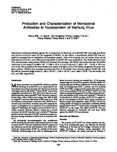
Figure 1. Reaction of monoclonal antibodies to myosin-I with the polypeptides of (A) whole cells, (B) a soluble protein fraction, and (C) isolated nuclei. Peptides were separated by SDS PAGE, transferred to nitrocellulose paper, and reacted with each antibody. Bound antibodies were detected with ~25I-goatanti-mouse Ig and autoradiography. Antibodies, (lane 1) M 1.1; (lane 2) M 1.2; (lane 3) M1.3; (lane 4) M1.4; (lane 5) Control IgG; and (lane 6) control IgM. The mobilities of myosin-I (130 kD), myosin-II (175 kD), and the cross-reactive nuclear protein (30 kD) are indicated on the left along with the molecular masses in kilodaltons of several standards. The heavy chains of myosin-IA and -IB were not resolved on these gels.
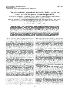
When reacted with a chymotryptic digest of myosin-IA consisting primarily of intact heavy chain (130 kD), a 100-kD peptide from the NH2-terminal region, and a 30-kD peptide from the COOH-terminal region (3, 10), all of the antibodies (except 1.5 that bound weakly) bound to the intact heavy chain and to the 30-kD peptide (Fig. 2). M1.7 bound only to these two peptides. The other antibodies bound weakly to peptides of 72 and 88 kD that do not appear on the stained gel. They also bound weakly to a peptide of 100 kD that we doubt, on the basis of the intensity of these bands relative to the 130-kD bands, is the major 100-kD peptide that appears on the stained gel. These results show that MI.1, M1.2, M1.3, M1.4, M1.6, M1.7, M1.8, and M1.9 all bind within 30 kD of the COOH terminus of the myosin-IA heavy chain. M1.5 bound only weakly to these blots, but reacted equally well with 130- and 100-kD bands. This is tentative evidence that M1.5 is the only antibody that binds to the major 100-kD
judging from the low concentrations of antibody required to achieve half maximal binding to myosin-I immobilized on plastic. Antibody M1.5 binds only weakly to denatured myosin-I or myosin-I that has been dried down on immulon wells, but it appears to have a high affinity for native myosinI, judging from its effects on ATPase activity described below.
Identification of Proteins that Bind M1.X Antibodies All of the M1.X antibodies bind to myosin-IA and all but M1.7 bind to myosin-IB in both solid-phase binding assays (not shown) and on electrophoretic transfers of SDS gels to nitrocellulose (Figs. 1 and 2). Even at high concentrations, no binding of M1.7 to myosin-IB was detected in either assay. When reacted with polypeptides from whole Acanthamoeba separated by gel electrophoresis, all nine M1.Xs bind to the 130-kD heavy chain of myosin-I and all react at least weakly with one or two polypeptides of 30 and 34 kD (Fig. 1 A). Eight of the nine M1.Xs also bind to the 175-kD heavy chains of myosin-II (Table I, Fig. 1; reference 17), although the strength of binding to myosin-II varies. MI.1 did not bind well on either blots or on solid-phase assays. M1.3 and M1.4 bound consistently on both blots and solid-phase assays. M1.7 bound on solid-phase assays but not on blots. M1.9 bound weakly on blots but not on solid-phase assays. Judging from quantitative solid-phase assays over a range of antibody concentrations, none of the M1.Xs bind to myosin-II as strongly as they bind to myosin-I. The heavy chains of both myosin-I and myosin-II are present in the soluble protein fraction (Fig. 1 B) while the low molecular mass polypeptides are highly enriched in a purified nuclear fraction (Fig. 1 C).
fragments of myosin-IA. Myosin-IA in 100 mM KCI, 10 mM Tris-HC1, pH 7.5, 1 mM dithiothreitol, and 12.5% glycerol was digested for 30 min at room temperature with alpha-chymotrypsin at an enzyme/substrate ratio of 1:100 (wt/wt). Proteolysis was terminated by the addition of an equal volume of 2X gel sample buffer at 100°C. After gel electrophoresis and electroblotting, individual strips of nitrocellulose containing alpha-chymotryptic peptides were reacted with 1 I.tg/ml monoclonal antibody. Bound antibodies were detected with ~zSI-goatanti-mouse Ig and autoradiography. The first lane is a gel stained with Coomassie Blue. Lanes 1-9 are the autoradiograms of strips stained with antibodies M I. I-M 1.9. (The authors are extremely grateful to Dr. Joseph P. Albanesi of the National Institutes of Health, who prepared the peptides.)
Hagen et al. Monoclonal Antibodies to Myosin-I
2123
Figure 2. Reaction of monoclonal antibodies with chymotryptic
that the cross-reactive site is on the proximal part of the tail of myosin-II (18). Identical results were obtained on blots and solid-phase assays. Second, high concentrations of M1.5 inhibit the binding of radiolabeled M2.1 to myosin-II. The M2.1 epitope is located at the junction of the heads with the tail of myosin-II (18). M1.2, M1.6, and M1.8 did not inhibit the binding of any of 21 radiolabeled M2.X antibodies to myosin-II, so we do not yet know exactly where any of these M1.X antibodies bind to myosin-II. None of 50 M2.X antibodies bind to myosin-I on blots of crude cell extracts (16, 17), nor do any of the M2.X antibodies specific for the proximal tail of myosin-II (M2.4, M2.6, M2.7, M2.10, M2.26) bind to purified myosin-I or compete with M1.4 for binding to myosin-I on blots or solid-phase assays.
Effects of Antibodies on Phosphorylation of the Myosin-I Heavy Chain None of the antibodies tested (MI.1, M1.2, M1.5, M1.6, M1.7, and M1.8) prevented phosphorylation of the myosin-I heavy chain by the heavy chain kinase when present in a 10-fold excess over the myosin-I and incubated with [7-32p]ATP for 10 rain (data not shown). Gel electrophoresis of the products of this incubation also showed that none of these antibodies were contaminated with enough protease activity to cleave detectable amounts of myosin-I heavy chain.
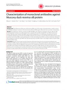
Figure 3. Reaction of monoclonal antibodies with tryptic peptides of myosin-IA. The experimental methods were identical to those in Fig. 2, except that TPCK-trypsin was used to digest the myosin. fragment, the part of the molecule with the nucleotide binding site and regulatory phosphorylation site (3). Similar experiments with tryptic peptides (Fig. 3) confirm that these antibodies bind near each other on the heavy chain, but are less informative regarding the location of this site along the heavy chain. With the exception of M1.5, all of the antibodies bind to a similar set of tryptic peptides of myosin-IA (Fig. 3). The major reactive peptides have molecular masses of 95, 27, and 17 kD, as seen particularly well with M1.7 (Fig. 3). Note that most of the antibodies also bind to several peptides of 80-100 kD and to another peptide of 50 kD. None of these peptides is a major band on the stained gel. None of the antibodies bind to a stable Coomassie Bluestained peptide of ,~41 kD (Fig. 3), shown by Albanesi et al. (3) to contain a uridine triphosphate binding site, or to another major peptide with a molecular mass of 73 kD.
Localization of Binding Sites by Competitive Binding Assays
Effect of Monoclonal Antibodies on ATPase Activity Antibody M1.5 inhibits the K+-EDTA ATPase activity of myosin-I in a concentration-dependent fashion (Fig. 5) with
1251.[abeled Antibodies M1.1 M1.2 M1.3 M1.4 M1.5 M1.6 M1,7 M1.8 M1.9
=5 o 1:3
-=
M1.1
7
9
54
6
7
39
60
M1,2
28
3
4
5
3
94
63
21
M1.3
28
5
1
5
7
15
48
14
M1,4
31
4
63
5
8
74
47
14
M1.5
88
74
111
76
89
121
94
77
M1,6
36
2
3
3
4
50
67
12
M1.7
57
11
38
7
61
50
41
M1.8
4
2
2
4
3
15
6
12
M1,9 104
3
2
6
5
36
54
10
Figure 4. Competitive binding assays. Radioiodinated antibodies
We used competitive binding studies to establish the relationship between antibody binding sites. Most of the M1.X antibodies can interfere with the binding of each other to myosinI (Fig. 4). The matrix is incomplete because M1.5 binds so weakly in these assays. Based on this assay, only MI.1 and M1.9 can bind simultaneously to myosin-I, and therefore occupy independent sites. It was previously established that M1.4 inhibits the binding of M2.4, M2.6, M2.7, and M2.26 to myosin-II (17) and new experiments confirm and extend this analysis of crossreactivity. First, antibodies M1.3, M2.4, M2.6, M2.7, and M2.26 strongly inhibit the binding of M1.4 to myosin-II, establishing that this competition is reciprocal and confirming
were competed with a 10-100-fold molar excess of unlabeled antibodies for binding to myosin-IA in the solid-phase assay (17). The fgures shown are counts bound corrected for nonspecific antibody binding and normalized to a value of 100 % established by samples with unlabeled control antibodies. The activity of M1.7 was lost during iodination, so competition between M 1.7 and an excess of the other antibodies for binding to myosin-IA was tested with an indirect assay. The amount of M1.7 bound to the wells was measured by ELISA using a goat anti-mouse IgG2Bcoupled to horseradish peroxidase. MI.1 is also an IgG2H, so it could not be tested. Similar results were obtained with ~25I-labeledprotein A at pH 6.0 where it binds to IgG2ss but not IgM or IgG~. In controls, the second reagents did not react with IgG~ or IgM monoclonal antibodies.
The Journal of Cell Biology, Volume 103, 1986
2124
50 % inhibition at a molar ratio of about four antibody sites per myosin molecule. Antibodies M1.2, M1.6, and a control IgM (Fig. 5) actually enhance the activity slightly, while MI.1, M1.7, and M1.9 have no effect. Since none of the other IgG-type or IgM-type antibodies inhibit the K+-EDTA ATPase activity of myosin-I, and since M1.5 does not degrade the myosin-I heavy chain, we think that the effect of M1.5 is due to an effect of antibody binding (rather than the action of a contaminant), in spite of the fact that an excess of antibody is required. Perhaps the binding of M1.5 to myosin-I is weak at the high ionic strength of the assay. We could not produce enough purified M1.3, M1.4, or M1.9 for definitive experiments. When tested at an actin concentration of 2 IxM, M1.5, M1.6, and M1.8 all inhibit the actin-activated Mg ++ ATPase of phosphorylated myosin-I in a concentration-dependent fashion (Fig. 6). This is the actin concentration that gives a peak of ATPase activity due to cross-linking of the actin illaments by myosin-I (2, 10). We did not test other aetin concentrations. M1.5 is the most potent inhibitor, with 100% inhibition at a molar ratio of less than one antibody site per myosin molecule. 50 % inhibition requires a molar ratio of ~0.6 for M1.6 and ~ 6 for M1.8. Even with a large excess of M1.8 there is some residual activity. MI.2 weakly inhibits activity at very high concentrations. Antibodies MI.1, M1.7, M2.2 (control IgG), and control IgM (referred to as antidynein in reference 17) did not inhibit actomyosin-I ATPase activity. M1.3, Mt.4, and M1.9 could not be tested.
Fluorescent Antibody Staining Monoclonal antibodies MI.1, M1.7, and M1.8 (all IgGs) stain fixed cells in the same way. There is intense staining of the nucleus and lighter staining throughout the cytoplasm (Fig. 7 A). In thin frozen sections (Fig. 7 B) it is clear that the nuclear staining is strongest in the nucleoplasm, is not especially intense at the nuclear envelope, and is weak in the nucleolus. The sections also show that the staining in the
@
~
~
-
x
~
@
~
con,.o,,0.
.a 3 ~ -
E
i
1.4
o
~
._> ~o....../Coot
ro I IqM
¢. "~ Lo
o
i=
o
< 0.6 0.4 -°
g_ o2
4+5°
u..
i
t
7 L
O
I
t
i
,
, ....
;
i
5 I0 15 Molar Rotio of Antibody to Myosin-~
20
Figure 6. Effect of M1.2, M1.5, M1.6, and M1.8 on the actinactivated ATPase activity of 61 nM phosphorylated myosin-I. The ratio of antibody to myosin is expressed as moles of antibody binding sites to moles of myosin. The actin concentration was 2 txM. The activity in the absence of antibody was 2.2 lxmol • min-~ • mg-]. Control IgM and IgG monoclonal antibodies did not inhibit the activity. cytoplasm is most intense very near the plasma membrane, confirming the observation of Gadashi and Korn (11) regarding polyclonal antibodies that stained only the cytoplasm. Two monoclonal IgMs (M1.6 and M1.9) and an immunoglobulin fraction from a rabbit immunized with myosin-I (rabbit JH-6; reference 17) also stain the nucleus more intensely than the cytoplasm (not shown). Monoclonal antibodies to myosin-II, all IgGs, give a completely different pattern of staining (Fig. 7 C). These antibodies stain the cytoplasmic matrix diffusely. Organelles, including the nucleus and contractile vacuole, are unstained, and the cortex is no more fluorescent than other parts of the matrix. Cells stained with monoclonal IgG against muscle myosin or second antibody alone are not fluorescent. Isolated nuclei stain the same as nuclei inside cells (Fig. 8). All three IgG monoclonal antibodies to myosin-I stain the nucleoplasm, while antibodies to myosin-II (Fig. 8, inset) and control antibodies do not stain nuclei.
Discussion
Our results provide new information about the relation of myosin-I to myosin-II and demonstrate a previously un• known structural homology between myosin-I and a protein located in the nucleus. The interpretation of these antibody n cross-reactions depends on location of the epitopes on myoI-sin-I, so we will consider that first. Eight of the MI.X antibodies characterized here bind to CI u.l sites on myosin-IA within 30 kD of the COOH terminus, as demonstrated by peptide maps (Fig. 2). These eight epitopes are within 17 kD of each other, since the antibodies all bind to a 17-kD tryptic peptide (Fig. 3). With the exception of z Y MI.1 versus M1.9 and MI.5 versus all others, all of the M1.Xs 0 I0 20 compete with each other for binding to myosin-I (Fig. 4). Molor Ratio of Antibody to M y o s i n - I This is not surprising since the antibodies, especially the six Figure 5. Effect of M1.5 on the K÷-EDTA ATPase activity of 61 IgMs, are considerably larger than the 17-kD peptide, so nM of myosin-I. The ratio of antibody to myosin is expressed as steric interference is to be expected. In our analysis of epimoles of antibody binding sites to moles of myosin. The control IgM is a monoclonal antibody that does not bind to myosin-I. topes on myosin-II (17), competitive binding was the most
Hagen et al. Monoclonal Antibodies to Myosin-I
2125
Figure 7. Localization of myosin-I and myosin-II in Acanthamoeba by indirect fluorescent antibody staining. Phase contrast and fluorescence of the same areas are illustrated. (A) Whole cells stained with M 1.8. This antibody stains both the nucleus (N) and the cytoplasm. (B) A 0.5-~m frozen section of Acanthamoeba stained with M1.8. The cytoplasmic staining is strongest near the plasma membrane (arrow). The nucleolus (Nu) is not stained as strongly as the nucleoplasm (N) (inset). (C) Whole cells stained with M2.5. Myosin-II is localized diffusely throughout the cytoplasm in Acanthamoeba. Note that myosin-II is not found in the nucleus (N) or contractile vacuole (CV). Bar, 10 ~m.
sensitive method for establishing that any two sites are distinct, but most o f the sites on myosin-I are so close.together that they are not distinguishable by this method. Fortunately, all of the M1.Xs have unique properties that are consistent with each binding to a distinct site on myosin-
The Journal of Cell Biology, Volume 103. 1986
I. All of the antibodies except M1.7 bind to both myosin-IA and myosin-IB, hence this unique MI.7 site on myosin-IA differs from all of the other epitopes. Only M1.5 inhibits the binding of M2.1 to myosin-II, so it must also be a unique epitope. M1.2, M1.3, and M1.4 all compete with each other for
2126
Figure 8. Isolated nuclei from Acanthamoeba stained indirectly with M1.8. Phase contrast and fluorescence of the same area are illustrated.
Inset shows isolated nuclei stained with M2.5. Bar, 10 p.m.
binding to myosin-l, but they can be distinguished because, of these three, only M1.4 competes with the M2.4 family for binding to myosin-II and only M1.2 fails to inhibit the binding of MI.4 to myosin-II. This is confirmed by reactions of the antibodies with tryptic peptides, since only M1.2 binds strongly to a 6-kD peptide (Fig. 3). One interpretation of these results is that each antibody binds to a unique site and that these sites are located close to each other. Higher resolution mapping of the sites will be necessary to confirm or refute this interpretation. Since myosin-IA and -IB share seven of these eight epitopes, the sequences of their COOH termini must be very similar, but not identical. The detailed peptide maps of A1banesi et al. (3) had previously established that the sequences of the 100-kD NH2-terminal regions of myosin-IA and -IB are different. Our work with antibodies (17; present report), and complementary work on primary structure (14, 15) and functional domains (10), shows that myosin-I is more closely related to myosin-II and to other myosins than was originally apparent (Fig. 9). The myosin-II molecule can be divided into a 100-kD NH2-terminal fragment and a 30-kD COOHterminal fragment. The NHz-terminal fragment has ATPase activity, an ATP-sensitive actin binding site (10), a phosphorylation site (3), and extensive sequence homology with the heads of other myosins (15). Like the heads of myosin-II, this part of myosin-I is not very antigenic; only six out of 50 M2.Xs bind to the heads of myosin-II (16, 18, 19) and no more than one of the nine M1.Xs binds to the corresponding 100kD fragment of myosin-I. In contrast, the region distal to the head-tail junction is the most antigenic part of myosin-II (16, 18) and the corresponding part of myosin-I has the binding sites for eight of the nine M1. Xs. The existence of antibodies that bind to both the proximal part of the tail of myosin-II and to the 30-kD COOH-terminal region of myosin-I raises the question of possible homology in these parts of the two
myosins. There are at least common epitopes, but close similarity is improbable, because the tails of all known conventional myosins (like myosin-II) are coiled coils of two parallel alpha-helical polypeptides, while myosin-I is composed of a single heavy chain. A single heavy chain is unlikely to form a parallel coiled coil. Furthermore, the COOH-terminal region of myosin-I has an unprecedented ATP-insensitive binding site for actin filaments (10). Our most unexpected result is the cross-reactivity of all nine M1.Xs with a low molecular mass polypeptide in the nucleus. In our previous publication (17), we speculated that the low molecular mass peptides are a breakdown product of myosin-I (17). While there is no doubt that fragments of myosin-I can be present in crude extracts, we are confident that the cross-reactive nuclear proteins are not fragments of myosin-I, because polyclonal antibodies raised against the
Hagen et al.
2127
Monoclonal
A n t i b o d i e s to M y o s i n - I
Head
II
Tail
AT P
N,
............
.C
i|
1
4
6 10 26
I
N
Chymotrypsin "~
ATP .
.
.
.
.
.
.
.
.
.
.
.
5 I 0
I 50
I 100 kDa
.
C
3 4 1 150
I 200
9. Comparison of myosin-I (I) and myosin-II (II) heavy chains. The polypeptides are aligned on the left at their NH2 terminus. Arrow indicates the site of the chymotryptic cleavage of myosin-I discovered by Fujisaki et al. (10). Shaded regions indicate the homologous, putative ATP binding regions sequenced by Hammer et al. (15). Stippled regions show the homologous regions that we have identified by antibody cross-reactivity, including the binding sites of specific antibodies indicated by numerals. Figure
34-kD protein purified from the nucleus react with the 30-34-kD nuclear proteins but not with purified myosin-I or other higher molecular mass polypeptides in whole cell samples (17; Rimm, D., and T. D. Pollard, manuscript in preparation). On the other hand, since the M1.X antibodies bind to several distinguishable sites on myosin-I, their crossreaction with the nuclear proteins suggests that there is substantial homology between myosin-I and the nuclear proteins, rather than some sort of spurious cross-reactivity as previously noted for other monoclonal antibodies (7, 8, 24). We would like to thank Dr. Joe Albanesi and Dr. John Hammer (National Institutes of Health) for helpful discussions on purification of myosin-I and myosin-I kinase, Dr. Albanesi for samples of myosin-I and myosin-I peptides, and Dr. Larry Gerace (Johns Hopkins Medical School) for his help in the preparation of isolated nuclei. We greatly appreciate the help of Ms. Pamela Maupin in preparing frozen sections, Dr. Mas Sato in contributing actin, and Ms. Toni Sahm in preparing the manuscript. This research was supported by a Muscular Dystrophy Association Postdoctoral Fellowship to Dr. Hagen, National Institutes of Health research grants GM-26388 and GM-26132 (awarded to Dr. Pollard), and CA-31460 and GM-33830 (awarded to Dr. Kiehart). Received for publication 13 January 1986, and in revised form 14 August 1986. References
1. Adams, R. J., and T. D. Pollard. 1986. Myosin-I propels organdies isolated from Acanthamoeba along actin filaments. Nature (Loud.). 322:754-756. 2. Albanesi, J. P., J. A. Hammer, and E. D. Korn. 1983. The interaction of F-actin with phosphorylated and unphosphorylated myosin-IA and myosinIB from Acanthamoeba-casteUanii. J. Biol. Chem. 258:10176-10181. 3. Albanesi, J. P., H. Fujisaki, and E. D. Korn. 1984. Localization of the active-site and phosphorylation site of Acanthamoeba myosins IA and IB. J. BioL Chem. 259:14184-14189. 4. Albanesi, J. P., H. Fujisaki, J. A. Hammer, Ill, E. D. Korn, R. Jones, and M. P. Sheetz. 1985. Monomeric Acanthamoeba myosins I support movement in vitro. J. Biol. Chem. 260:8649-8652. 5. Bradford, M. 1976. A rapid and sensitive method for the quantitation of microgram quantifies of protein utilizing the principle of protein-dye binding. Anal. Biochem. 72:248-254. 6. Cote, G. P., J. P. Albanesi, T. Ueno, J. A. Hammer, Ill, and E. D. Korn. 1985. Purification from Dictyostelium discoideum of a low-molecular-weight myosin that resembles myosin I from Acanthamoeba castellanii. J. Biol. Chem. 260:4543-4546. 7. Franklin, R. M., L. R. Emmons, R. P. Emmons, A. Oommen, J. R. Pink, A. M. Rijnbeek, M. Schnetzler, L. Tuderman, and E. Vainio, 1983. Monoclouai antibody which recognizes a common antigenic determinant or intermediate filament proteins, actin, and myosin. Hybridoma. 2:275-285. 8. Franklin, R. M., L. R. Emmons, R. P. Emmons, O. Kai, A. Oommen, J. R. Pink, A. M. Rijnbeek, M. Schnetzler, L. Tudermann, and E. Vainio. 1984. A monoclonal antibody recognizes an epitope common to an avian-
The Joumai of Cell Biology, Volume 103, 1986
specific nuclear antigen and to cytokeratins. J. Cell. Biochem. 24:1-14. 9. Fujisaki, H., J. P. Albanesi, T. J. Lynch, and E. D. Korn. 1985. Experimental evidence for the contractile activities of Acanthamoeba myosin IA and IB. J. Biol. Chem. 260:11183-11189. 10. Fujisaki, H., T. J. Lynch, J. P. Albanesi, and E. D. Korn. 1985. Actinbinding domains ofAcanthamoeba myosin-IA. J. CellBiol. 101 (5, pt. 2): 162a. (Abstr.) 11. Gadashi, H., and E. D. Koro. 1980. Evidence for differential intracellular localization of the Acanthamoeba myosin isozymes. Nature (Lond.). 286:452--456. 12. Hagen, S. C., D. P. Kiehart, D. A. Kaiser, and T. D. Pollard. 1984. Monoclonal antibodies as probes for the structure and function of myosin-I in Acanthamoeba. J. Cell Biol. 99 (4, pt. 2):35a. (Abstr.) 13. Hammer, J. A., J. P. Albanesi, and E. D. Korn. t983. Purification and characterization of a myosin-I heavy-chain kinase from Acanthamoeba-castellanii. J. Biol. Chem. 258:10168-10175. 14. Hammer, J. A., E. D. Kom, and B. M. Patterson. 1986. Isolation of a Non-muscle myosin heavy chain gene from Acanthamoeba. J. Biol. Chem. 261 : 1949-1956. 15. Hammer, J. A., G. Jung, and E. D. Korn. 1986. Genetic evidence that Acanthamoeba myosin-I is a true myosin. Proc. Natl. Acad. Sci. USA. 83: 4655--4659. 16. Kaiser, D. A., D. Bhandari, R. J. Adams, D. P. Kiehart, and T. D. Pollard. 1985. Evaluation of new monoclonal antibodies to Acanthamoeba myosinII. J. Cell Biol. 101 (5, pt. 2):158a. (Abstr.) 17. Kiehart, D. P., D. A. Kaiser, and T. D. Pollard. 1984. Monoclonal antibodies demonstrate limited structural homology between myosin isozymes from Acanthamoeba. J. Cell Biol. 99:1002-1014. 18. Kiehart, D. P., D. A. Kaiser, andT. D. Pollard. 1984. Direct locaiization of monoclonal antibody-binding sites on Acanthamoeba myosin-lI and inhibition of filament formation by antibodies that bind to specific sites on the myosinII tail. J. Cell Biol. 99:1015-1023. 19. Kiehart, D. P., and T. D. Pollard, 1984. Inhibition of Acanthamoeba actomyosin-II ATPase activity and mechanochernical function by specific monoclonal-antibodies. J. Cell Biol. 99:1024-1033. 20. MacLean-Fletcher, S., and T. D. Pollard. 1980. Identification of a factor in conventional muscle actin preparation which inhibits actin filament selfassociation. Biochem. Biophys. Res. Commun. 96:18-27. 21. Maruta, H., and E. D. Korn. 1977. Acanthamoeba myosin-II. J. Biol. Chem. 252:6501-6509. 22. Maruta, H., and E. D. Kom. 1977. Acanthamoeba cofactor protein is a heavy chain kinase required for actin activation of the Mg2+-ATPase activity of Acanthamoeba myosin-l. J. Biol. Chem. 252:8329-8332. 23. Maruta, H., H. Gadasi, J. H. Collins, and E. D. Korn. 1979. Multiple forms of Acanthamoeba myosin-l. J. Biol. Chem. 254:3624-3630. 24. Pollard, T. D., and E. D. Korn. 1973. Acanthamoeba myosin. I. Isolation from Acanthamoeba castellanii of an enzyme similar to muscle myosin. J. Biol. Chem. 248:4682-4690. 25. Pollard, T. D., and E. D. Korn. 1973. Acanthamoeba myosin. II. Interaction with actin and with a new cofactor protein required for actin activation of Mg ~ ATPase activity. J. Biol. Chem. 248:4691-4697. 26. Pollard, T. D., W. F, Stafford, and M. E. Porter. 1978. Characterization of a second myosin from Acanthamoeba castellanii. J. Biol. Chem. 253:47984808. 27. Rimm, D. L., and T. D. Pollard. 1985. Nuclear proteins that cross-react with antibodies to Acanthamoeba myosin-I. J. Cell Biol. 101 (5, pt. 2): 158a. (Abstr.) 28. Tokuyasu, K. T. 1980. Immunochemistry on ultrathin frozen sections. Histochem. J. 12:381--403.
2128