Disruption of the Lipid-Transporting LdMT-LdRos3 Complex in Leishmania donovani Affects Membrane Lipid Asymmetry but Not Host Cell Invasion Adrien Weinga¨rtner1, Bjo¨rn Drobot1, Andreas Herrmann1, Marı´a P. Sa´nchez-Can˜ete2, Francisco Gamarro2, Santiago Castanys2, Thomas Gu¨nther Pomorski3* 1 Institute of Biology and Biophysics, Humboldt-University Berlin, Berlin, Germany, 2 Instituto de Parasitologı´a y Biomedicina ‘Lo´pez-Neyra’, Consejo Superior de Investigaciones Cientı´ficas (CSIC), Armilla, Granada, Spain, 3 Department of Plant Biology and Biotechnology, University of Copenhagen, Copenhagen, Denmark
Abstract Maintenance and regulation of the asymmetric lipid distribution across eukaryotic plasma membranes is governed by the concerted action of specific membrane proteins controlling lipid movement across the bilayer. Here, we show that the miltefosine transporter (LdMT), a member of the P4-ATPase subfamily in Leishmania donovani, and the Cdc50-like protein LdRos3 form a stable complex that plays an essential role in maintaining phospholipid asymmetry in the parasite plasma membrane. Loss of either LdMT or LdRos3 abolishes ATP-dependent transport of NBD-labelled phosphatidylethanolamine (PE) and phosphatidylcholine from the outer to the inner plasma membrane leaflet and results in an increased cell surface exposure of endogenous PE. We also find that promastigotes of L. donovani lack any detectable amount of phosphatidylserine (PS) but retain their infectivity in THP-1-derived macrophages. Likewise, infectivity was unchanged for parasites without LdMT-LdRos3 complexes. We conclude that exposure of PS and PE to the exoplasmic leaflet is not crucial for the infectivity of L. donovani promastigotes. Citation: Weinga¨rtner A, Drobot B, Herrmann A, Sa´nchez-Can˜ete MP, Gamarro F, et al. (2010) Disruption of the Lipid-Transporting LdMT-LdRos3 Complex in Leishmania donovani Affects Membrane Lipid Asymmetry but Not Host Cell Invasion. PLoS ONE 5(8): e12443. doi:10.1371/journal.pone.0012443 Editor: Mauricio Martins Rodrigues, Federal University of Sa˜o Paulo, Brazil Received December 21, 2009; Accepted August 3, 2010; Published August 26, 2010 Copyright: ß 2010 Weinga¨rtner et al. This is an open-access article distributed under the terms of the Creative Commons Attribution License, which permits unrestricted use, distribution, and reproduction in any medium, provided the original author and source are credited. Funding: This work was supported by Research Training Group 1121 of the German Research Foundation (AW, AH and TGP), the Spanish grants SAF2005-01639 (SC), the ISCIII-Red de Investigacion Cooperativa en Enfermedades Tropicales (RICET) RD06/0021/0002 (FG) and by the Plan Andaluz de Investigacion (Cod. BIO130), the Marie Curie Flippase Network (AW, AH and FG), FAZIT (AW), the Carlsberg Foundation (TGP) and the Deutsche Forschungsgemeinschaft (TGP, grant PO748/9). The funders had no role in study design, data collection and analysis, decision to publish, or preparation of the manuscript. Competing Interests: The authors have declared that no competing interests exist. * E-mail:
[email protected]
exposure is mandatory for invasion of host cells and (ii) exposure of other lipids may facilitate infectivity as well. In fact, exposure of PS is typically accompanied by a loss of transbilayer asymmetry of other lipids. In particular, PE redistributes to the exoplasmic leaflet as well. Hence, this lipid may contribute to or even be critical for infectivity of parasites. Maintenance and regulation of membrane lipid asymmetry is governed by the concerted action of ATP-driven translocases that move specific lipids against a concentration gradient across the bilayer. Prime candidates for outward-directed lipid translocases are members of the ATP binding cassette (ABC) transporter family [7], whereas potential inward-directed lipid translocases belong to the P4 subfamily of P-type ATPases [8]. Members of both transporter families have also been implicated in the development of drug resistance in Leishmania, which includes resistance to alkyllysophospholipids [9–14]. This suggests that the mechanism by which the drugs are extruded from cells is closely related to the flippase mechanism by which lipids are translocated across membranes, and that these processes involve structurally similar, if not identical transporters. Studies aimed at identifying the molecular basis of miltefosine (hexadecylphosphocholine) resistance led to the identification of two Leishmania membrane proteins, LdMT, a member of the P4 subfamily of P-type ATPases, and LdRos3, a potential noncatalytic subunit of LdMT related to Cdc50 family [11,12,14]. Both
Introduction Most eukaryotic cells exhibit an asymmetric distribution of phospholipids in their plasma membrane, with aminophospholipids phosphatidylserine (PS) and phosphatidylethanolamine (PE) restricted to the cytosolic leaflet [1]. This asymmetric lipid distribution is maintained by ATP-dependent processes, suggesting that it is critical to normal cell function. Indeed, if cells fail to engage mechanisms to maintain lipid asymmetry, PS and PE appear at the cell surface, which leads to dramatic changes in cell function [2]. In multicellular organisms, one of the important consequences of altered membrane lipid asymmetry is the recognition and engulfment of PS exposing apoptotic cells by mononuclear macrophages. Recently, PS exposure has also been implicated in the infectivity of Leishmania, an obligate, intracellular parasite that infects cells of the mononuclear phagocyte lineage in their vertebrate hosts [3–5]. The parasite has a digenic life cycle, residing as flagellated extracellular promastigotes in the gut of the insect vector and as obligatory intracellular aflagellated amastigotes found in the parasitophorous vacuoles of mammalian macrophages. The observation of PS exposure on the cell surface of Leishmania parasites led to the hypothesis that the protozoan parasite might take advantage of a regulated loss of lipid asymmetry and PS presentation to invade and survive in host macrophages [6]. However, it remains open whether (i) PS PLoS ONE | www.plosone.org
1
August 2010 | Volume 5 | Issue 8 | e12443
Leishmania LdMT-LdRos3 Complex
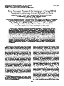
the miltefosin uptake defect in DLdMT parasites [12]. Nontransfected wild-type parasites served as a control. As shown in Figure 1C, the anti-GFP antibodies efficiently precipitated LdMTGFP and LdRos3 from detergent-solubilised membrane preparations obtained from parasites expressing LdMT-GFP. In contrast, an unrelated integral plasma membrane protein (metalloprotease gp63) was not present in the immunoprecipitate. As a control, a parallel immunoprecipitation from an extract obtained from a wild-type strain lacking the LdMT-GFP fusion did not precipitate LdRos3, indicating that the co-immunoprecipitation was specific. Analysis of the LdMT-GFP immunoprecipitates by mass spectrometry revealed that the preparations did not contain other Cdc50 members than LdRos3 (data not shown). Taken together, these results provide direct evidence that LdMT and LdRos3 reside in a stable complex in the membrane. We infer that the stoichiometry of both proteins in the complex is one because we did not detect corresponding fluorescent bands of retarded mobility for larger complexes.
proteins are primarily localized to the Leishmania plasma membrane and required for the rapid intracellular uptake of alkylphosphocholine drugs. In the budding yeast Saccharomyces cerevisiae, members of the two protein families have been found to form stable transporter complexes that function in the translocation of phospholipids from the exoplasmic to the cytoplasmic leaflet of cellular membranes [15,16]. Likewise, LdMT and LdRos3 have been implicated in the uptake of fluorescent 7nitrobenz-2-oxa-1,3-diazol-4-yl (NBD)-labelled phospholipids, suggesting a role for these proteins in controlling lipid asymmetry in the parasite plasma membrane [12,17]. However, lipid asymmetry of parasite plasma membranes is not well defined and the proteins involved in its regulation remain to be identified. In this work, we study the regulation of phospholipid asymmetry in promastigotes of Leishmania donovani and the relevance of asymmetry loss for infection. In particular, we show that LdMT and LdRos3 form a stable protein complex that is essential for inward translocation of NBD-PE and -PC across the parasite plasma membrane. Loss of either LdMT or LdRos3 leads to an increased cell surface exposure of endogenous PE. We therefore propose that the LdMT-LdRos3 complex plays an essential role in maintaining phospholipid asymmetry in the parasites plasma membrane. Since, to our surprise, promastigotes of Leishmania donovani lack any detectable amount of PS, we could study specifically the role of PS and PE exposure in host cell invasion. Control cells with functional LdMT-LdRos3 complexes were able to invade host cells showing that PS is not mandatory for infection. Furthermore, infection was not enhanced for parasites without LdMT-LdRos3 complexes indicating that redistribution of PE to the exoplasmic leaflet does not facilitate invasion.
LdMT and LdRos3 are required for the ATP-dependent inward translocation of NBD-PC and -PE To characterize the lipid transport activity of the LdMTLdRos3 complex in the plasma membrane we examined in wild type, DLdMT and DLdRos3 parasites the uptake of NBD-lipids. This can be monitored by flow cytometry and by fluorescence microscopy. Experiments were performed at 2uC to suppress endocytosis and suppress degradation of the NBD-lipids (consistently ,10%). Under these conditions, wild-type parasites efficiently internalized NBD-PC and NBD-PE, while NBD-PS and NBD-sphingomyelin (NBD-SM) were hardly taken up at all. The efficient uptake of NBD-PC and NBD-PE was significantly inhibited following ATP depletion by preincubation with sodium azide and 2-deoxyglucose or collapse of the proton electrochemical gradient with the protonophore carbonyl cyanide mchlorophenylhydrazone (CCCP) (Fig. 2A). Consistent with these results fluorescence microscopy of wild-type parasites labelled with NBD-lipids revealed an intensive labelling of intracellular membranes with NBD-PC and -PE but not with NBD-PS and SM (Fig. 2D). In contrast to wild-type parasites, DLdMT and DLdRos3 parasites were found defective in the low temperature uptake of NBD-PC and NBD-PE (Fig. 2B). These defects were due solely to the loss of LdMT and LdRos3, as lipid uptake was restored by re-expression of LdMT-GFP and LdRos3-GFP in DLdMT and DLdRos3 mutants, respectively (Fig. 2C). Based on these findings, we conclude that the LdMT-LdRos3 complex is essential to sustain an energy-dependent influx of NBD-PC and -PE across the parasite plasma membrane.
Results Leishmania LdMT and LdRos3 form a stable complex To investigate the ability of LdMT and LdRos3 to form a stable complex as their homologues in yeast, a C-terminally green fluorescent protein (GFP)-tagged version of LdRos3 (LdRos3GFP) was expressed in parasites lacking either LdRos3 (DLdRos3) or LdMT (DLdMT). Tagged LdRos3-GFP was functional, since it could suppress resistance of DLdRos3 parasites to the alkylphospholipid derivate miltefosine [12]. Total membrane preparations from the LdRos3-GFP expressing parasites were subjected to solubilisation in the presence of mild detergent and analysed by non-denaturing polyacrylamide gel electrophoresis (native-PAGE), in which complex formation between two proteins gives a new band with mobility different from that of either protein alone. Fluorimaging of the native gels identified one prominent fluorescent band of low molecular size for protein preparations obtained from DLdMT parasites expressing LdRos3-GFP (Fig. 1A). In protein samples obtained from DLdRos3 parasites expressing LdRos3-GFP, additional fluorescent bands of retarded mobility were detected (Fig. 1B, bands II–III). To test for the presence of LdMT in the GFP-labelled complexes, the fluorescent bands were excised from the native gel and subjected to SDSPAGE followed by western blot analysis. Immunoblotting with antibodies to LdMT and LdRos3 showed that the fluorescent band of low molecular size (band I) only contained LdRos3-GFP while the more slowly migrating bands (band II-III) corresponded to a complex of LdMT and Ros3-GFP. To corroborate that LdMT and LdRos3 form a stable complex, a C-terminally GFP-tagged version of LdMT (LdMT-GFP) was introduced into DLdMT parasites, and co-immunoprecipitation experiments were performed using anti-GFP-MicroBeads. Tagged LdMT-GFP has been shown to be functional, since it can rescue PLoS ONE | www.plosone.org
LdMT and LdRos3 sustain plasma membrane PE asymmetry in L. donovani We next tested whether the LdMT-LdRos3 complex helps to maintain a tight asymmetric distribution of aminophospholipids at the plasma membrane. Promastigote stages of Leishmania wild type, DLdMT and DLdRos3 lines were incubated with different concentrations of duramycin and papuamide B, cytolytic peptides that requires binding to cell surface-exposed PE and PS, respectively, to exert their cytotoxicity [18]. As shown in Figure 3A, both DLdMT and DLdRos3 lines were more sensitive to duramycin-induced cytolysis as compared to the wild-type line with an EC50 of duramycin that was approximately 1.8-fold lower than that for wild-type cells. Restoration of LdMT and Ros3 expression returned the duramycin sensitivity profile back to the wild-type pattern (Figure 3A). There was no significant 2
August 2010 | Volume 5 | Issue 8 | e12443
Leishmania LdMT-LdRos3 Complex
Figure 1. LdMT and LdRos3 form a stable complex in the Leishmania membrane. (A, B) Native- and SDS-PAGE analysis of LdMT and LdRos3GFP. LdRos3-GFP was expressed in DLdMT parasites (A) and in DLdRos3 parasites (B). Solubilised membrane proteins were separated by native PAGE and analyzed for GFP fluorescence. The membrane extract obtained from DLdRos3 parasites contains a prominent fast-migrating fluorescent band (band I). The extract obtained from DLdMT parasites also contains slow-migrating fluorescent protein bands (band II and III). Regions of the gel corresponding to the fluorescent bands were excised and loaded onto SDS-PAGE gels which were subsequently immunoblotted with polyclonal antibodies against LdRos3 (a-LdRos3) and LdMT (a-LdMT). Size markers indicate relative mobility of proteins in kDa. (C) Immunoblots from coimmunoprecipitation assays. LdMT-GFP was immunoprecipated from a detergent-solubilised membrane fraction (load) obtained from DLdMT parasites expressing LdMT-GFP as well as non-transfected wild-type parasites (wt) using anti-GFP-MicroBeads. Immunoprecipitates (IP) were subjected to immunoblot analysis using antibodies that recognize LdMT (a-LdMT), LdRos3 (a-LdRos3) and metalloprotease gp63 (a-gp63). doi:10.1371/journal.pone.0012443.g001
difference between the DLdMT and the DLdRos3 Leishmania lines as compared to wild-type cells with respect to their sensitivity to papuamide B and amphotericin B, a polyene macrolide antibiotic that binds to membrane ergosterol and induces cellular leakage [19]. To more directly visualize that deletion of LdMT or LdRos3 affects the lipid asymmetry in the plasma membrane, we analyzed the exposure of PE by labelling with biotinylated Ro09-0198, a peptide that specifically binds to PE [20,21]. As shown in Figure 3B, both the DLdMT and the DLdRos3 Leishmania lines bound more PE-sensing biotinylated Ro09-0198 peptide which was visualized with StreptavidinFITC. For a quantitative assessment of Ro09-0198 peptide binding, the FITC fluorescence associated with mutant and wildtype cells was measured by flow cytometry. As shown in Figure 3A, deletion of LdMT or LdRos3 caused a 10-fold increase in PE exposure when compared to wild type parasites based on Ro09-0198 peptide binding. We could, however, not detect in any of the lines labelling of propidium iodide (PI) PLoS ONE | www.plosone.org
negative parasites with annexin V-FITC, a protein that preferentially interacts with membranes containing PS [22]. To rule out the possibility that the observed differences in PE exposure resulted from substantial increase in the PE level in the DLdMT and the DLdRos3 lines as compared to wild-type parasites, we analyzed the total phospholipid composition in Leishmania parasites labelled to steady state with 32P-phosphate. Four major phospholipids could be identified with comparable levels in all of the strains: PC (44%–49%), PE (27%–29%), phosphatidylinositol (PI, 9%) and inositolphosphorylceramide (IPC, 4–5%) (Fig. 4A). Thus, the possibility that increased cell surface exposure of PE was caused by increased PE synthesis can be ruled out. Notably, we could not detect significant levels of PS by this method. When stained with ninhydrin, a reagent labelling lipids with free amino groups such as PS and PE, the chromatograms exhibited only one spot corresponding to PE (Fig. 4B). Likewise, lipid analysis by HPLC coupled ESI-MS did not reveal the presence of PS, suggesting that L. donovani does, if it 3
August 2010 | Volume 5 | Issue 8 | e12443
Leishmania LdMT-LdRos3 Complex
Figure 2. Inward translocation of NBD-PC and NBD-PE across the plasma membrane of Leishmania requires LdMT and LdRos3. Promastigotes of wild type (wt), DLdMT and DLdRos3 lines were labelled with NBD-lipids for 30 min at 2uC and than washed and analysed by flow cytometry (A–C) or visualised by fluorescence microscopy (D). ATP depletion was achieved by preincubation with 5 mM 2-deoxyglucose and 20 mM sodium azide. To abolish the proton electrochemical gradient 50 mM of the protonophore CCCP was used. As a control LdMT-GFP and LdRos3-GFP on episomal Leishmania expression vectors were reintroduced in DLdMT and DLdRos3 mutants, respectively; control, non-labelled cells showing the intrinsic fluorescence of the GFP fusion proteins. Data are normalized to NBD-PC internalization of wild-type parasites; 100% corresponds to 468696 a.u. NBD-PC. Data represent the means 6 SE of at least three independent experiments. For statistical analysis Welch’s test was performed. Significant differences in the lipid uptake of the mutants compared to the wild type are denoted by asterisks (** p = 0.05; * p = 0.01). doi:10.1371/journal.pone.0012443.g002
all, synthesize very low amounts of this phospholipid (Fig. 4C). The presence of only very low amounts of PS explains the equal sensitivity of the parasite lines to papuamide B and the lack of FITC-annexin V binding on intact parasites (see above).
logarithmic and stationary phase. Parasites and THP-1-derived macrophages were prelabelled with CellTrackerTM Green and CellTrackerTM Dil, respectively, and co-incubated at a ratio of 1:10 (cells:parasitetes). 16 h post infection, 30 to 70% of the THP1-derived macrophages were found to be infected. At this moment the number of intracellular parasites per infected macrophage ranged from 1 to 19 (Fig. 5A). The mean numbers of intracellular parasites per infected cell were similar: 2.6 for wt, 2.5 for DLdMT and 2.7 for DLdRos3 parasites, respectively. The total numbers of THP-1-derived macrophages infected by DLdMT and DLdRos3 parasites were not markedly different from that of wild-type parasites (Fig. 5B), indicating that at least in this infection model, changes in the plasma membrane PE arrangement of Leishmania parasites do not trigger specific recognition and removal of the parasites by macrophages.
Phagocytosis of DLdMT and DLdRos3 Leishmania lines in THP-1-derived macrophages is unchanged Exposure of PS on the exoplasmic leaflet of the Leishmania plasma membrane has been implicated in entry of the parasite into host cells [3–5]. The availability of two Leishmania lines strains displaying an altered plasma membrane lipid asymmetry and apparent lack of PS provided an opportunity to test whether other phospholipids are relevant for the recognition of Leishmania parasites by macrophages using the human monocytic cell line THP1. THP1 cells have many characteristics of human monocytes, including morphology, surface-membrane receptor, and the capacity to undergo maturational changes when induced with phorbol esters [23,24]. We used early log phase promastigotes to evade apoptotic subpopulations which increase during late PLoS ONE | www.plosone.org
Discussion Previous studies revealed a critical role for both the miltefosine transporter LdMT and the LdRos3 protein in the uptake and 4
August 2010 | Volume 5 | Issue 8 | e12443
Leishmania LdMT-LdRos3 Complex
Figure 3. LdMT and LdRos3 are required to sustain plasma membrane PE asymmetry in Leishmania. (A) Sensitivity of wild type (wt), DLdMT and DLdRos3 L. donovani parasites to the PE-binding peptide duramycin, the PS-binding peptide papuamide B and the sterol-binding amphotericin B. Parasites were diluted to 0.256106 cells/ml in medium containing duramycin, papuamide B or amphotericin B at the indicated concentrations. 72 h later viability was analysed as described in the Material and methods. As a control LdMT-GFP and LdRos3-GFP on episomal Leishmania expression vectors was reintroduced in DLdMT and DLdRos3 mutants, respectively. Means 6 S.E. of at least three independent experiments are shown as percentage of untreated control parasites. (B) Endogenous PS and PE at the exoplasmic leaflet of the plasma membrane of these strains was visualised with annexin V-FITC and Biotin-Rho/Streptavidin-FITC, respectively. Cells were analyzed by phase contrast (DIC) and fluorescence microscopy (PI, FITC). Bar, 10 mm. Fluorescence intensity histograms were obtained by flow cytometry as described under ‘‘Materials and Methods.’’ Cells incubated in the absence of biotinylated Ro09-0198 peptide served as controls (dash line). doi:10.1371/journal.pone.0012443.g003
CDC50A, for ER exit and delivery to the plasma membrane [25], and the Arabidopsis P4-ATPase ALA3 requires its Cdc50 binding partner ALIS1 to complement the lipid transport defect at the plasma membrane in Ddrs2Ddnf1Ddnf2 yeast mutant [26]. These findings hold also true for LdRos3 and the P4-ATPase LdMT. However, a closer inspection of the partially sequenced genome of L. infantum (http://www.genedb.org/genedb/linfantum/) reveals in total five genes encoding P4-ATPases (LinJ09_V3.0940, LinJ13_V3.1590, LinJ30_V3.2270, LinJ30_V3.2270, LinJ34_ V3.2460, LinJ34_V3.3000) and three genes encoding Cdc50 family members (LinJ09_V3.1080, LinJ32_V3.0540, LinJ35_V3.3450). Coming challenges are to identify whether all of these P4-ATPase require a Cdc50 binding partner for proper functioning and whether they can interact with several Cdc50 proteins as shown for the Arabidopsis P4-ATPase ALA3 [26]. In light of the results presented in this study, it seems that Leishmania LdMT does not associate with other Cdc50 members than LdRos3 and that the stoichiometry of both proteins in the complex is one.
potency of alkylphosphocholine type drugs and in phospholipid translocation at the plasma membrane of Leishmania parasites [11,12,14]. The present study provides direct evidence that both proteins form a stable complex that is essential for maintaining the normal asymmetric lipid distribution of at the parasite plasma membrane. Loss of plasma membrane PE asymmetry by disruption of the LdMT-LdRos3 complex does not alter the infectivity in macrophages. We conclude that in L. donovani loss of the normal asymmetric lipid distribution does not facilitate host cell invasion. Formation of a stable complex between P4-ATPases and Cdc50 family members seems to be indispensable for a proper targeting and functioning of P4-ATPases. In yeast, the Cdc50 family members Cdc50p, Lem3p and Crf1p can be co-immunoprecipitated with Drs2p, Dnf1p/Dnf2p and Dnf3p, respectively. Formation of these complexes is required for proper expression and endoplasmic reticulum (ER) export of either partner [15,16]. The human P4-ATPase ATP8B1 requires the Cdc50 member PLoS ONE | www.plosone.org
5
August 2010 | Volume 5 | Issue 8 | e12443
Leishmania LdMT-LdRos3 Complex
Figure 4. Total phospholipid composition of wild type, DLdMT and DLdRos3 L. donovani parasites. Promastigote stages of wild type (wt), DLdMT and DLdRos3 lines were labelled for 48 h with 32P-phosphate. Lipids were extracted, separated by two-dimensional thin layer chromatography, and then visualized by phosphorimager scanning (A) or ninhydrin staining (B). Representative two-dimensional TLC plates are shown. The location of individual species was verified by ESI-MS. Unidentified lipids are not marked. (C) Quantification of phospholipids in wild-type, DLdMT and DLdRos3 parasites. Data are expressed as the percentage of total phospholipids and represent the means 6 S.E. of three independent experiments. PC, phosphatidylcholine; PE, phosphatidylethanolamine; PI, phosphatidylinositol; IPC, inositolphosphorylceramide. doi:10.1371/journal.pone.0012443.g004
completely blocked the ATP-dependent uptake of NBD-PC and NBD-PE, indicating that the LdMT-LdRos3 complex facilitates the energy-coupled transport of natural PE and PC from the outer to the inner leaflet of the parasites plasma membrane. In support of this notion, loss of either LdMT or LdRos3 led to an increased cell surface exposure of natural PE, as evidenced by enhanced hypersensitivity to PE-binding peptide and labelling by biotinylated PE-binding peptide. These results are in line with a direct role of the LdMT-LdRos3 complex in pumping natural PE to the
Incubation of parasites with fluorescent phospholipid analogues (PC, PE, PS and SM) served as an approach to analyze lipid transport activity of the LdMT-LdRos3 complex at the plasma membrane. We found that L. donovani parasites efficiently internalize NBD-PC and NBD-PE even at 2uC while NBD-PS and NBD-SM are hardly taken at this temperature. Since endocytosis is blocked under this condition [27], uptake of NBD-PC and NBD-PE must involve a translocation step across the plasma membrane. Loss of either LdMT or LdRos3 PLoS ONE | www.plosone.org
6
August 2010 | Volume 5 | Issue 8 | e12443
Leishmania LdMT-LdRos3 Complex
reports [31,32] we could not detect endogenous PS in total lipid extracts prepared from L. donovani promastigotes, suggesting that this parasite does not synthesise considerable amounts of PS. Thus, an inward-directed transport activity for PS at the plasma membrane might not be required in L. donovani. However, all experiments undertaken in the present work were performed on the promastigote early stage. It will be interesting to determine whether L. donovani amastigotes synthesise PS and regulate its distribution in the parasite plasma membrane. A stage specific regulation of the plasma membrane asymmetry is conceivable since amastigotes up-regulate the expression of two P4-ATPases (LmjF30.2260, LmjF34.3220) and a plasma membrane ABC transporter associated with lipid export (LmjF11.1260) [33]. Leishmania amastigotes replicate within the mature phagolysosome compartment and have complex nutritional requirements which must be scavenged from the host cell [34]. It is therefore tempting to speculate that the LdMT-LdRos3 transporter complex or related P4-ATPases are involved in the acquisition of host phospholipids. The surface of the Leishmania parasite is a major point of interaction with the host throughout the infectious cycle. A number of surface glycoconjugates such as lipophosphoglycans, glycosylphosphatidylinositol (GPI)-anchored proteins (e.g. the metalloprotease gp63), and a heterogeneous group of small glycosylinositolphospho-lipids have been implicated in the ability of the parasite to infect and survive in host macrophages [35]. In addition, surface exposed PS has been associated with parasite infectivity [3–5]. However, these results must be interpreted with caution because PS exposure was concluded on basis of annexin V and anti-PS antibody binding in these studies. Despite being used extensively to label PS, neither annexin V nor anti-PS antibodies are specific for this lipid and also bind phosphatidic acid, phosphatidylglycerol and phosphatidylinositol-4,5-bisphosphate [36]. Thus, a direct proof for PS exposure by Leishmania parasites is currently lacking. In fact, we observed that L. donovani promastigotes lack significant amounts of PS, and annexin VFITC does not label living, PI negative L. donovani parasites but parasites positive for PI staining. Conceivably, the various Leishmania spp. might expose other lipids than PS that contribute to the infectivity. Our results on DLdMT and the DLdRos3 Leishmania mutant lines that display an altered PE asymmetry did not reveal major differences in terms of invasion efficiency into the human monocytic cell line THP-1. In line with these results, the DLdMT mutant remains infective and maintains virulence in cultures of primary isolated mouse peritoneal macrophages [37]. These results suggest that disruption of the LdMT-LdRos3 transporter complex and changes in the transbilayer distribution of PE, and probably PC, are not crucial for the infectivity of L. donovani promastigotes. Likewise, in the THP-1 infection model used here PS exposure is not mandatory for invasion of host cells. Future studies on other Leishmania species and mutants lacking the ability to synthesized PS may help to define in more detail the lipid types exposed on the cells surface and involved in parasite internalization and phagocyte inactivation.
Figure 5. Phagocytosis of DLdMT and DLdRos3 parasites by THP-1-derived macrophages is unchanged. CellTrackerTM Greenlabelled parasites were added (10:1) to THP-1-derived macrophages prelabelled with CellTrackerTM Dil (red) and co-incubated for 16 h at 37uC. (A) Micrograph of double-fluorescence labelling of THP-1-derived macrophages phagocytosing Leishmaina parasites. Bar, 10 mm. (B) Percentage of phagocytosing THP-1-derived macrophages was determined by flow cytometry. Values are means 6 SD of three independent experiments expressed as percentage of control (number of phagocytosed wild-type parasites by TPH-1-derived macrophages). The population of phagocytosing THP-1-derived macrophages ranged from 30 to 70% in control cultures. doi:10.1371/journal.pone.0012443.g005
cytosolic leaflet to generate an asymmetric membrane as recently suggested for P4-ATPases from other organisms [28,29]. The ability of wild-type parasites to take up NBD-PC suggests that natural PC is restricted to the inner leaflet of the parasites plasma membrane and that the outer leaflet is primarily composed of sphingolipids, as NBD-sphingomyelin is not taken up. The substrate specificity of the inward translocation machinery appears to vary between Leishmania species. In contrast to L. donovani, L. infantum displays not only an ATP-dependent internalization of NBD-PC and NBD-PE but also an active transport of NBD-PS across its plasma membrane [30]. Thus, this parasite might express one or more P4-ATPases of broader or different substrate specificity at the plasma membrane, respectively. The physiological relevance of these species-specific differences in substrate specificity remains to be established. Previous studies suggested that the LdMT-LdRos3 complex of L.donovani also recognizes NBD-PS, however, as a poor substrate [11,12]. Significant uptake of NBD-PS in this species was only observed when higher label concentrations were used as compared to NBDPC and NBD-PE. It is noteworthy that in agreement with former PLoS ONE | www.plosone.org
Materials and Methods Culture and transfection of Leishmania lines Promastigotes of L. donovani (MHOM/ET/67/HU3) and derivative lines LdMT knockout (DLdMT) and LdRos3 knockout (DLdRos3) [12] were maintained at 26uC in M-199 medium (Invitrogen, Karlsruhe, Germany) supplemented with 40 mM HEPES, 100 mM adenosine, 0.5 mg/ml hemin, 10 mM 6-biopterin and 10% heat inactivated fetal calf serum (Gibco, Invitrogen 7
August 2010 | Volume 5 | Issue 8 | e12443
Leishmania LdMT-LdRos3 Complex
GmbH, Karlsruhe, Germany). Parasites (36107 promastigotes) were transfected with the C-terminal fusion constructs LdMTGFP or LdRos3-GFP by electroporation (450 V, 500 mF) and then maintained in culture medium with 200 mg/ml G418 [11]. Unless indicated otherwise, all materials were purchase from SigmaAldrich (Taufkirchen, Germany).
(132 mM NaCl, 3.5 mM KCl, 0.5 mM MgCl2, 1 mM CaCl2, 5 mM glucose, 1 mM pyruvate, 20 mM HEPES, pH 7.4). Promastigotes in logarithmic phase of growth were harvested by centrifugation (10006g, 10 min, 4uC), washed twice with HPMI. To block the catabolism of NBD-lipids the cell suspension (107 parasites/ml) was pre-incubated with 5 mM 3-(4-octadecyl)benzoylacrylic acid (Biomol, Hamburg, Germany) and 1 mM phenylmethanesulphonylfluoride for 30 min at 28uC. To study the lipid uptake, the parasites were then incubated at 4uC with 5 mM NBD-lipid. After 30 minutes, cells were washed twice in HPMI containing 4% (w/v) fatty acid free bovine serum albumin to extract NBD lipids from the cell surface. Flow cytometry analysis was performed on a Becton Dickinson FACS (San Jose, CA) equipped with an argon laser (488 nm) using Cell Quest software. One ml of 1 mg/ml PI in H2O was added 200 ml cell suspension just before analysis. Ten thousand cells were analyzed at room temperature with gating during the acquisition. Live cells were selected based on forward/side-scatter gating and propidium iodide exclusion. The following fluorescence channels (log scale) were used: FL1 (530/30 nm, FITC, NBD, CellTrackerTM Green), FL2 (585/42 nm, PI, CellTrackerTM Dil). Data were analyzed by Cyflogic software.
Protein analysis and immunoprecipitation For preparation of membrane proteins, parasites were harvested, resuspended in ice-cold hypo-osmotic lysis buffer (5 mM TrisHCl pH 7.4) containing 1 mM phenylmethylsulfonyl fluoride, and then broken by vortexing with glass beads. The cell lysate was clarified by centrifugation at 5006g (10 min, 4uC). Subcellular membranes were collected by centrifugation at 100,0006g (1 h, 4uC) and resuspended in sample buffer (750 mM 6-aminocarproic acid, 50 mM Bistris pH 7.0, 20% glycerol) to a protein concentration of 5 mg/ml. Protein concentration was measured using the BCA protein assay kit (Pierce Chemical Company, Rockford IL, USA). Membranes were solubilised by adding 20 ml of ndodecyl-b-D-maltopyranoside (DDM, 10%) to 100 ml suspended membranes, corresponding to a DDM/protein ratio of 4 (w/w). After incubation for 60 min on ice, insoluble material was removed by centrifugation (100,0006g, 1 h, 4uC). For Clear Native-PAGE, solubilised membrane proteins (1 mg) were loaded directly onto a 6%–15% gradient gel (6%–15% acrylamid/bisacrylamid in 50 mM Bis Tris-HCl pH 7.0, 500 mM 6-aminocarproic acid). Electrophoresis was carried out at 4uC at 500 V for 16 h (electrophoresis buffer = 50 mM Tricine, 15 mM Bis Tris-HCl pH 7.0, 0.05% Na-taurodeoxycholat, pH 7.0) and bovine serum albumin (monomer, 66 kDa; dimer, 132 kDa; trimer, 198 kDa; tetramer, 264 kDa) was used as molecular weight marker. Gels were scanned for GFP fluorescence using a Fuji FLA3000 phosphoimager (Raytest GmbH, Straubenhardt, Germany) equipped with a 473 nm argon laser and 510 nm long pass filter. Fluorescent bands were excised from the native gel and subjected to SDS-PAGE followed by western blot analysis. GFP-co-immunoprecipitation assays were performed using antiGFP microBeads (Miltenyi Biotec, Bergisch Gladbach, Germany). Per immuno-isolation, a 1 ml reaction was prepared in lysis buffer containing 150 ml of anti-GFP microBeads slurry, 250 ml of detergent-solubilised protein. The reactions were rotated gently at 4uC for 30 min. Beads were separated from the supernatants using mColumns with a mMACS separator (Miltenyi Biotec) and washed three times with lysis buffer containing 0.05% DDM. Membrane protein extracts and immuno-isolated membrane proteins were subjected to western blot analysis. Immunoblots were probed with polyclonal antibodies against LdMT and LdRos3 [14], and gp63 (kindly provided by Robert McMaster). Horseradish peroxidase-conjugated secondary antibodies were from Bio-Rad (Hercules, CA). Blots were developed using enhance chemiluminescence (ECL plus Kit, GE-Healthcare, Freiburg, Germany). Protein mass spectrometry of the immunoprecipitates was performed as described elsewhere [38].
Drug Sensitivity Assays in L. donovani lines To determine parasite sensitivity to amphotericin B, papuamide B (Flintbox, Lynsey Huxham) and duramycin, 2,56104 parasites were incubated in 96well plates (100 ml) for three days at different drug concentrations before determining cell proliferation by the 3(4,5-dimethyl-2-thiazolyl)-2,5-diphenyl-2H-tetrazolium bromide (MTT) colorimetric assay as previously described [11]. The 50% effective concentration (EC50) was defined as the drug concentration required for half-maximal inhibition of the cellular growth rate. The EC50 for each line was calculated by nonlinear regression analysis using SigmaPlot 2000 for Windows (SPSS Inc., Chicago, IL, USA).
Annexin V und Bio-Rho Assay To visualize endogenous PE on the cell surface, 56106 promastigotes in logarithmic phase of growth were incubated in 20 ml HPMI containing 38 mM biotinylated Ro09-0198 (provided by Kazuma Tanaka). After 1 h at 4uC, cells were washed with phosphate buffered saline (PBS) containing 0.5% (w/v) bovine serum albumin and then fixed with PBS containing 5% (w/v) formaldehyde for 1 h at 30uC. Promastigotes were then washed in PBS, resuspended in 250 ml PBS containing 5 mg/ml StreptavidinFITC and incubated for 30 min at 25uC prior to microscopy analysis. To measure exposure of endogenous PS on the cell surface, about 56105 parasites were incubated on ice for 10 min in the dark with 125 ng annexin V-FITC and 1 mg PI in 0.5 ml of binding buffer (10 mM HEPES pH 7.4, 140 mM NaCl, 2.5 mM CaCl2). Cells were washed, resuspended in 0.5 ml of binding buffer and subjected to microscopy.
Metabolic labelling and lipid fractions analysis
NBD-lipid uptake and flow cytometry analysis in L. donovani lines
Parasites (56107) were inoculated in 100 ml of supplemented M-199 containing 100 mCi [32P]H3PO4 (1 mCi, HartmannAnalytic GmbH, Braunschweig, Germany) and grown for 48 h at 26uC. Cells were harvested by centrifugation, washed twice with PBS. Total cellular lipids were extracted by the method of Bligh and Dyer [39] and separated by two-dimensional thin layer chromatography (I: chloroform/methanol/25% aqueous ammonium hydroxide, 90:54:7; II: chloroform/acetone/methanol/ acetic acid/water, 50:20:10:10:5). The 32P-containing radiolabelled spots were imaged on a 32P-sensitive screen and quantified
Palmitoyl-(NBD-hexanoyl)-phosphatidylserine (NBD-PS), -phosphatidylethanolamine (NBD-PE), -phosphatidylcholine (NBDPC) and 6-NBD-hexanoyl-sphingosine-1-phosphocholine (NBDsphingomyelin; NBD-SM) were from Avanti Polar Lipids (Birmingham, AL). Appropriate amounts of analogues (5 nmol of NBD-lipids for 107 cells) in chloroform/methanol (1:1) were transferred to a glass tube, dried under nitrogen, dissolved in 5 ml absolute ethanol and vortexed with the desired volume of HPMI PLoS ONE | www.plosone.org
8
August 2010 | Volume 5 | Issue 8 | e12443
Leishmania LdMT-LdRos3 Complex
on a Fuji Imaging System imager (FLA3000, Raytest, Straubenhardt, Germany). Lipids were also visualized with common lipidlocating agents such as iodine or ninhydrin (0.25% ninhydrin w/v in acetone). For lipid determination by HPLC (Agilent 1200 series) coupled to ESI-MS (Finnigan LTQ Fourier transform ion cyclotron resonance) lipid spots from iodine stained 2D-TCL were scraped of, extracted as described above and dried. The HPLC column (BioBasic-4 C4 5 m 15061 mm) was rinsed with 70% Buffer B (70% acetonitril, 25% 2-propanol, 5% H2O; 10 mM triethylammonium acetat, 1 mM acetic acid) and 30% Buffer A (95% H2O, 5% acetonitril; 10 mM triethylammonium acetat, 1 mM acetic acid) first. Next, the lipids (dissolved in acetonitril/methanol (1:1)) were loaded to the column and the concentration of Buffer A was increased stepwise to 100%. Eluted fractions were directly injected to the ESI-MS and analysed in negative ion mode.
microscopy. For flow cytometry, macrophages were rinsed intensively with PBS, trypsinised for 20 min at 37uC and finally suspended in supplemented RPMI-1640.
Fluorescence Microscopy Epifluorescence microscopy and image acquisition were carried out using an inverse Axiovert 100 standard fluorescence microscope (Carl Zeiss, Oberkochen, Germany) equipped with a cooled CCD camera (Coolsnap, visitron systems, Puchheim, Germany) driven by Metamorph software (Universal Imaging, Downingtown, USA). NBD fluorescence was viewed using a PlanAPO 1006/1.3 NA oil objective with the following filter set: BP 450–490, FT 510, and BP 512–542. Confocal laser scanning microscopy was performed using an inverted Fluoview 1000 microscope (Olympus, Tokio, Japan) and a 606 (N.A. 1.35) oilimmersion objective. Fluorescence of FITC and CellTracker Green was excited with a 488 nm argon laser and recorded between 500 and 530 nm. Fluorescence of PI and CellTracker CM-Dil was excited with a 559 nm argon laser and recorded between 570 and 600 nm. Images with a frame size of 2566256 pixels were acquired.
Phagocytosis of L. donovani lines by macrophages THP-1 cells were cultured at 37uC with 5% CO2 in RPMI1640 medium (Gibco) supplemented with 2 mM glucose, 50 mM b-mercaptoethanol, 1 mM Na-pyruvate, and 10% fetal calf serum. To stimulate differentiation into macrophages, 100 ng/ml phorbol 12-myristate 13-acetate (Biomol, Hamburg) was added to THP-1 cells (26105/ml). After 12 h, adherent cells were washed 3 times with PBS and cultured as described above. Three days postdifferentiation, THP-1 cells (106/flask) were labelled for 40 min in 3 ml RPMI-1640 medium containing 0.5 mM CellTrackerTM Dil (Invitrogen). Non-accumulated dye was removed by washing the cells 5 times with RPMI-1640 medium. Promastigotes (107) from early log phase were labelled for 40 min in 2.5 ml M-199 medium containing 0.5 mM CellTrackerTM Green (Invitrogen). Then, parasites were washed five times with M-199 and finally resuspended in RPMI-1640. To study phagocytosis of promastigotes by macrophages, about 107 CellTrackerTM-labeled parasites were added to 106 CellTrackerTM Dil-labeled adherent macrophages (macrophage:parasite ratio, 1:10). After 16 h, cultures were analysed by flow cytometry and fluorescence
Acknowledgments We gratefully acknowledge Frederic Mu¨ller for helping with lipid mass spectrometry, Michael Linscheid for giving the opportunity to perform the lipid mass spectrometry measurements, Gunnar Dittmar for protein mass spectrometry, Robert McMaster for generously providing the gp63 antibody, Juan Patron for the THP-1 cell line, Kazuma Tanaka for biotinylated Ro09-0198, and Stephen M. Beverly for pX, pXG-GFP+2/ and pXG-9GFP+ vectors.
Author Contributions Conceived and designed the experiments: AW TGP. Performed the experiments: AW BD. Analyzed the data: AW BD TGP. Contributed reagents/materials/analysis tools: AH MPSC FG SC. Wrote the paper: AW AH TGP.
References 1. Devaux PF (1991) Static and dynamic lipid asymmetry in cell membranes. Biochemistry 30: 1163–1173. 2. Fadeel B, Xue D (2009) The ins and outs of phospholipid asymmetry in the plasma membrane: roles in health and disease. Crit Rev Biochem Mol Biol 44: 264–277. Available: http://dx.doi.org/10.1080/10409230903193307 via the Internet. Accessed 7 Aug 2010. 3. de Freitas Balanco JM, Moreira ME, Bonomo A, Bozza PT, AmaranteMendes G, et al. (2001) Apoptotic mimicry by an obligate intracellular parasite downregulates macrophage microbicidal activity. Curr Biol 11: 1870–1873. 4. Wanderley JLM, da Silva LHP, Deolindo P, Soong L, Borges VM, et al. (2009) Cooperation between apoptotic and viable metacyclics enhances the pathogenesis of Leishmaniasis. PLoS One 4: e5733. Available: http://dx.doi.org/10. 1371/journal.pone.0005733 via the Internet Accessed 7 Aug 2010. 5. Wanderley JLM, Moreira MEC, Benjamin A, Bonomo AC, Barcinski MA (2006) Mimicry of apoptotic cells by exposing phosphatidylserine participates in the establishment of amastigotes of Leishmania (L) amazonensis in mammalian hosts. J Immunol 176: 1834–1839. 6. Wanderley JLM, Benjamin A, Real F, Bonomo A, Moreira MEC, et al. (2005) Apoptotic mimicry: an altruistic behavior in host/Leishmania interplay. Braz J Med Biol Res 38: 807–812. 7. Pomorski T, Holthuis JCM, Herrmann A, van Meer G (2004) Tracking down lipid flippases and their biological functions. J Cell Sci 117: 805–813. Available: http://dx.doi.org/10.1242/jcs.01055 via the Internet Accessed 7 Aug 2010. 8. Tang X, Halleck MS, Schlegel RA, Williamson P (1996) A subfamily of P-type ATPases with aminophospholipid transporting activity. Science 272: 1495–1497. 9. Castanys-Mun˜oz E, Alder-Baerens N, Pomorski T, Gamarro F, Castanys S (2007) A novel ATP-binding cassette transporter from Leishmania is involved in transport of phosphatidylcholine analogues and resistance to alkyl-phospholipids. Mol Microbiol 64: 1141–1153. 10. Castanys-Mun˜ oz E, Pe´rez-Victoria JM, Gamarro F, Castanys S (2008) Characterization of an ABCG-like transporter from the protozoan parasite
PLoS ONE | www.plosone.org
11.
12.
13.
14.
15.
16.
9
Leishmania with a role in drug resistance and transbilayer lipid movement. Antimicrob Agents Chemother 52: 3573–3579. Available: http://dx.doi.org/10. 1128/AAC.00587-08 via the Internet. Accessed 7 Aug 2010. Pe´rez-Victoria FJ, Gamarro F, Ouellette M, Castanys S (2003) Functional cloning of the miltefosine transporter. A novel P-type phospholipid translocase from Leishmania involved in drug resistance. J Biol Chem 278: 49965–49971. Available: http://dx.doi.org/10.1074/jbc.M308352200 via the Internet. Accessed 7 Aug 2010. Pe´rez-Victoria FJ, Sa´nchez-Can˜ete MP, Castanys S, Gamarro F (2006) Phospholipid translocation and miltefosine potency require both L. donovani miltefosine transporter and the new protein LdRos3 in Leishmania parasites. J Biol Chem 281: 23766–23775. Available: http://dx.doi.org/10.1074/jbc. M605214200 via the Internet. Accessed 7 Aug 2010. Pe´rez-Victoria FJ, Sa´nchez-Can˜ete MP, Seifert K, Croft SL, Sundar S, et al. (2006) Mechanisms of experimental resistance of Leishmania to miltefosine: Implications for clinical use. Drug Resist Updat 9: 26–39. Available: http://dx. doi.org/10.1016/j.drup.2006.04.001 via the Internet. Accessed 7 Aug 2010. Sa´nchez-Can˜ete MP, Carvalho L, Pe´rez-Victoria FJ, Gamarro F, Castanys S (2009) Low plasma membrane expression of the miltefosine transport complex renders Leishmania braziliensis refractory to the drug. Antimicrob Agents Chemother 53: 1305–1313. Available: http://dx.doi.org/10.1128/AAC.01694-08 via the Internet. Accessed 7 Aug 2010. Furuta N, Fujimura-Kamada K, Mol Biol CellSaito K, Yamamoto T, Tanaka K (2007) Endocytic recycling in yeast is regulated by putative phospholipid translocases and the Ypt31p/32p-Rcy1p pathway. Mol Biol Cell 18: 295–312. Available: http://dx.doi.org/10.1091/mbc.E06-05-0461 via the Internet. Accessed 7 Aug 2010. Saito K, Fujimura-Kamada K, Furuta N, Kato U, Umeda M, et al. (2004) Cdc50p, a protein required for polarized growth, associates with the Drs2p Ptype ATPase implicated in phospholipid translocation in Saccharomyces cerevisiae. Mol Biol Cell 15: 3418–3432. Available: http://dx.doi.org/10. 1091/mbc.E03-11-0829 via the Internet. Accessed 7 Aug 2010.
August 2010 | Volume 5 | Issue 8 | e12443
Leishmania LdMT-LdRos3 Complex
29. Zhou X, Graham TR (2009) Reconstitution of phospholipid translocase activity with purified Drs2p, a type-IV P-type ATPase from budding yeast. PNAS 106: 16586–16591. 30. Arau´jo-Santos JM, Gamarro F, Castanys S, Herrmann A, Pomorski T (2003) Rapid transport of phospholipids across the plasma membrane of Leishmania infantum. Biochem Biophys Res Commun 306: 250–255. 31. Zhang K, Pompey JM, Hsu FF, Key P, Bandhuvula P, et al. (2007) Redirection of sphingolipid metabolism toward de novo synthesis of ethanolamine in Leishmania. EMBO J 26: . pp 1094–1104. Available: http://dx.doi.org/10. 1038/sj.emboj.7601565 via the Internet. Accessed 7 Aug 2010. 32. Zufferey R, Allen S, Barron T, Sullivan DR, Denny PW, et al. (2003) Ether phospholipids and glycosylinositolphospholipids are not required for amastigote virulence or for inhibition of macrophage activation by Leishmania major. J Biol Chem 278: 44708–44718. Available: http://dx.doi.org/10.1074/jbc. M308063200 via the Internet. Accessed 7 Aug 2010. 33. Saxena A, Lahav T, Holland N, Aggarwal G, Anupama A, et al. (2007) Analysis of the Leishmania donovani transcriptome reveals an ordered progression of transient and permanent changes in gene expression during differentiation. Mol Biochem Parasitol 152: 53–65. Available: http://dx.doi.org/10.1016/j.molbiopara. 2006.11.011 via the Internet. Accessed 7 Aug 2010. 34. Naderer T, McConville MJ (2008) The Leishmania-macrophage interaction: a metabolic perspective. Cell Microbiol 10: 301–308. 35. Naderer T, Vince JE, McConville MJ (2004) Surface determinants of Leishmania parasites and their role in infectivity in the mammalian host. Curr Mol Med 4: 649–665. 36. Yeung T, Heit B, Dubuisson JF, Fairn GD, Chiu B, et al. (2009) Contribution of phosphatidylserine to membrane surface charge and protein targeting during phagosome maturation. J Cell Biol 185: 917–928. Available from: http://dx.doi. org/10.1083/jcb.200903020. 37. Seifert K, Pe´rez-Victoria FJ, Stettler M, Sa´nchez-Can˜ete MP, Castanys S, et al. (2007) Inactivation of the miltefosine transporter, LdMT, causes miltefosine resistance that is conferred to the amastigote stage of Leishmania donovani and persists in vivo. Int J Antimicrob Agents 30: 229–235. Available: http://dx.doi. org/10.1016/j.ijantimicag.2007.05.007 via the Internet. Accessed 7 Aug 2010. 38. Guenther UP, Handoko L, Varon R, Stephani U, Tsao CY, et al. (2009) Clinical variability in distal spinal muscular atrophy type 1 (DSMA1): determination of steady-state IGHMBP2 protein levels in five patients with infantile and juvenile disease. J Mol Med 87: 31–41. Available: http://dx.doi.org/10.1007/s00109008-0402-7 via the Internet. Accessed 7 Aug 2010. 39. Bligh EG, Dyer WJ (1959) A rapid method of total lipid extraction and purification. Can J Biochem Physiol 37: 911–917.
17. Pe´rez-Victoria FJ, Castanys S (2003) Gamarro F Leishmania donovani resistance to miltefosine involves a defective inward translocation of the drug. Antimicrob Agents Chemother 47: 2397–2403. 18. Parsons AB, Lopez A, Givoni IE, Williams DE, Gray CA, et al. (2006) Exploring the mode-of-action of bioactive compounds by chemical-genetic profiling in yeast. Cell 126: 611–625. Available: http://dx.doi.org/10.1016/j.cell.2006.06. 040 via the Internet. Accessed 7 Aug 2010. 19. Brajtburg J, Powderly WG, Kobayashi GS, Medoff G (1990) Amphotericin B: current understanding of mechanisms of action. Antimicrob Agents Chemother 34: 183–188. 20. Emoto K, Kobayashi T, Yamaji A, Aizawa H, Yahara I, et al. (1996) Redistribution of phosphatidylethanolamine at the cleavage furrow of dividing cells during cytokinesis. Proc Natl Acad Sci U S A 93: 12867–12872. 21. Iwamoto K, Kobayashi S, Fukuda R, Umeda M, Kobayashi T, et al. (2004) Local exposure of phosphatidylethanolamine on the yeast plasma membrane is implicated in cell polarity. Genes Cells 9: 891–903. 22. Gerke V, Moss SE (2002) Annexins: from structure to function. Physiol Rev 82: 331–371. 23. Anttila HS, Reitamo S, Ceska M, Hurme M (1992) Signal transduction pathways leading to the production of IL-8 by human monocytes are differentially regulated by dexamethasone. Clin Exp Immunol 89: 509–512. 24. Dasgupta B, Roychoudhury K, Ganguly S, Akbar MA, Das P, et al. (2003) Infection of human mononuclear phagocytes and macrophage-like THP1 cells with Leishmania donovani results in modulation of expression of a subset of chemokines and a chemokine receptor. Scand J Immunol 57: 366–374. 25. Paulusma CC, Folmer DE, Ho-Mok KS, de Waart DR, Hilarius PM, et al. (2008) ATP8B1 requires an accessory protein for endoplasmic reticulum exit and plasma membrane lipid flippase activity. Hepatology 47: 268–278. 26. Poulsen LR, Lo´pez-Marque´s RL, McDowell SC, Okkeri J, Licht D, et al. (2008) The Arabidopsis P4-ATPase ALA3 localizes to the golgi and requires a betasubunit to function in lipid translocation and secretory vesicle formation. Plant Cell 20: 658–676. Available: http://dx.doi.org/10.1105/tpc.107.054767 via the Internet. Accessed 7 Aug 2010. 27. Parodi-Talice A, Arau´jo JM, Torres C, Pe´rez-Victoria JM, Gamarro F, et al. (2003) The overexpression of a new ABC transporter in Leishmania is related to phospholipid trafficking and reduced infectivity. Biochim Biophys Acta 1612: 195–207. 28. Coleman JA, Kwok MCM, Molday RS (2009) Localization, purification and functional reconstitution of the P4-ATPASE, ATP8A2, a phosphatidylserine flippase in photoreceptor disc membranes. J Biol Chem Available from: http:// dx.doi.org/10.1074/jbc.M109.047415 via the Internet. Accessed 7 Aug 2010.
PLoS ONE | www.plosone.org
10
August 2010 | Volume 5 | Issue 8 | e12443