●
Cutting Edge: Anti-Inflammatory Properties of Low Levels of IFN-␥1 Liat Flaishon,2* Ian Topilski,2*† David Shoseyov,* Rami Hershkoviz,* Elizabeth Fireman,† Yoram Levo,† Sylvia Marmor,† and Idit Shachar3*
Activation of naive T and B cells occurs only within the context of organized lymphoid tissue. Thus, the continuous recirculation of mature lymphocytes is crucial for the development of primary immune response to foreign Ags. We have previously shown that low levels of IFN-␥ inhibit homing of B cells to the secondary lymphoid organs. In this study, we demonstrate that similarly low doses of IFN-␥ down-regulate integrin-mediated adhesion and migration of naive T and Th2 cells, and have a profound effect on the in vivo homing of naive T cells to the lymph nodes. Moreover, we show that these low doses of IFN-␥ have anti-inflammatory effects in an in vivo asthma model. Thus, in contrast to the proinflammatory effects of IFN-␥ at relatively high concentrations, low dose IFN-␥ appears to exert global suppressory effects on T cell trafficking and may have clinical application as an anti-inflammatory agent. The Journal of Immunology, 2002, 168: 3707–3711.
T
he surveillance of the body for foreign Ags is a critical function of the immune system. Lymphocytes migrate from the blood into tissues and secondary lymphoid organs, and return to the blood via lymph vessels and the thoracic duct (1). The majority of lymphocytes are capable of tissue selective trafficking (homing), recognizing organ-specific adhesion molecules on specialized endothelial cells (2). Leukocytes are specifically recruited by a combination of molecular events: bloodborne cells use primary adhesion molecules to tether to, and roll on the luminal wall of postcapillary venules, and thereby encounter and rapidly transduce signals to up-regulate secondary adhesion molecules, which then mediate firm arrest (3, 4). Naive T lymphocytes traffic through the T cell areas of secondary lymphoid organs in search of Ag presented by dendritic cells *Department of Immunology, Weizmann Institute of Science, and †Sourasky Medical Center, Tel-Aviv, Israel Received for publication January 15, 2002. Accepted for publication February 25, 2002. The costs of publication of this article were defrayed in part by the payment of page charges. This article must therefore be hereby marked advertisement in accordance with 18 U.S.C. Section 1734 solely to indicate this fact.
(5, 6). Upon Ag recognition, specific T cells proliferate and, in the presence of polarizing cytokines, differentiate into Th1 or Th2 cells, which produce distinct patterns of cytokines and mediate various types of protective and pathological responses (7, 8). After T cell differentiation into Th1 or Th2 phenotypes, the lymph node (LN)4 homing receptors are down-regulated, while expression of tissue homing receptors is acquired (9, 10). Recently, we demonstrated that to prevent premature encounter with Ag, immature B cells down-regulate their own integrin-mediated adhesion to the extracellular matrix (ECM). This inhibition is mediated by IFN-␥, which is transcribed and secreted by immature B cells to down-regulate their migration to the LNs in an autocrine manner. In addition, these low levels of IFN-␥ were found to dramatically affect homing of mature B cells to secondary lymphoid organs (11). The powerful inhibitory effect of IFN-␥ on adhesion of B lymphocytes both in vitro and in vivo led us to suggest that this cytokine might serve as a more general modulator of the immune response and have an influence on other subsets of the immune system. Therefore, in this study, we analyzed the ability of low levels of IFN-␥ to regulate migration of T cells in vitro and in vivo. Our findings demonstrate that low levels of IFN-␥ prevent adhesion and migration of naive T and Th2 cells in vitro and have anti-inflammatory effects in an in vivo asthma model.
Materials and Methods Animals C57BL/6 or BALB/c mice were used at 6 – 8 wk of age. All animal procedures were approved by the Animal Research Committee at the Weizmann Institute (Rehovot, Israel).
Cells Spleen cells were obtained from mice as previously described (12). T cells were enriched by using the MACS system (Miltenyi Biotec, Auburn, CA). Spleen cells were incubated with anti-CD45R (B220) magnetic beads, and the CD45 negative cells were collected. CD4⫹ T cells were enriched using anti-CD4 magnetic beads. To obtain Th1 and Th2 cells, the T cell-enriched population was incubated with Con A (250 g/ml; Roche, Basel, Switzerland) and IL-12 (3.5 ng/ml; Life Technologies, Rockville, MD) or IL-4 (103 U/ml; Life Technologies) for 96 h. Th1 supernatant was collected from the IL-12-treated cells.
Adhesion assay Adhesion assays were performed as described previously (11).
1
This research was supported by the Israel Science Foundation founded by the Academy of Sciences and Humanities; and the German-Israeli Foundation for Scientific Research and Development. I.S. is an incumbent of the Alvin and Gertrude Levine Career Development Chair of Cancer Research. L.F. was supported by the Israeli Health Ministry. 2
Transwell migration Chemotaxis was assayed by using transwell chambers as was previously described (13).
L.F. and I.T. contributed equally to this study.
3
Address correspondence and reprint requests to Dr. Idit Shachar, Department of Immunology, Weizmann Institute of Science, Rehovot 76100, Israel. E-mail address:
[email protected] Copyright © 2002 by The American Association of Immunologists
●
4 Abbreviations used in this paper: LN, lymph node; ECM, extracellular matrix; AHR, airway hyperactivity; BAL, bronchoalveolar lavage; Penn, enhanced expiratory pause; SDF-1, stromal-cell-derived factor 1.
0022-1767/02/$02.00
3708
CUTTING EDGE: IFN-␥ INHIBITS INFLAMMATORY RESPONSE IN AN ASTHMA MODEL
Tracking of cells in vivo The assay, based on labeling cells with BCECF-AM (Molecular Probes, Eugene, OR), was performed as described previously (11).
OVA sensitization and challenge BALB/c mice were immunized i.p. on days 0, 7, and 14 as previously described (14). The OVA/OVA group was i.p. injected with 300 l 0.9% NaCl from day 15 (first day of challenge) for 5 days, 5 min before each inhalation. The IFN-␥ group was i.p. injected with murine IFN-␥ (6 U in 300 l 0.9% NaCl; Genentech, South San Francisco, CA) from day 15 (first day of challenge) for 5 days, 5 min before each inhalation.
Measurement of airway hyperactivity (AHR) AHR was determined as previously described (15) by calculating the enhanced expiratory pause (Penn) measured in a plethysmographic box during expiration (Buxco Electronics, Sharon, CT).
bronchoalveolar lavage (BAL) BAL was performed on day 19, 4 h after the last AHR evaluation as was previously described (14).
Lung histology Lungs were inflated with 1 ml of 10% formalin until distended. Samples for paraffin sectioning were prepared as described previously (14). Immunohistochemistry was performed as previously described (16). The sections were stained with anti-CD3 (Serotec, Oxford, U.K.) followed by biotinylated anti-rat IgG Ab. The slides were developed using 3,3-diaminobenzidine as a substrate.
Cytoskeleton rearrangement The cells were analyzed as previously described (13).
Polyclonal stimulation CD4⫹ T cells were cultured in the presence of Con A (2.5 g/ml; Roche) in the presence or absence of elevated concentrations of IFN-␥. Proliferation was determined after 4 days by incorporation of [3H]thymidine.
Results
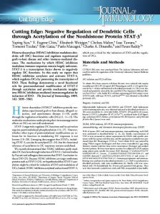
Low levels of IFN-␥ inhibit integrin-mediated adhesion and migration of T cells in vitro To migrate to LNs or to sites of inflammation, T cells must interact with components of the ECM (5). To determine whether low levels of IFN-␥ negatively regulate homing of naive T cells, we studied their integrin-mediated adhesion to the major ECM component, fibronectin. 51Cr-labeled naive T cells were activated with or without PMA, a potent agonist of integrin-mediated adhesion (17), or stromal-cell-derived factor 1 (SDF-1), a potent T cell chemoattractant and integrin agonist (18) in the presence or absence of low levels of IFN-␥ (0.1 U/ml). Upon activation, T cell adhesion to fibronectin was dramatically augmented, while in the presence of low levels of IFN-␥ (Fig. 1A) the integrin-mediated adhesion response to PMA or chemoattractant of these T cells was abolished. Because IFN-␥ is secreted at high levels by Th1 (⬃350 ng/ml, equivalent to ⬃35,000 U/ml), T cells might encounter high levels of IFN-␥ in vivo. To investigate whether these high levels of IFN-␥ have any influence on the homing of naive T cells, we examined the adhesion of naive T cells to fibronectin in the presence of supernatant collected from cultured Th1 cells. As can be seen in Fig. 1A, Th1 cell supernatant did not alter the adhesion response of PMA- or SDF-1-stimulated T cells, and they were able to increase their adhesion response in a manner similar to untreated cells. Moreover, in the presence of high levels of rIFN-␥ (50 u/ml), the adhesion response was hardly affected (Fig. 1B). Because Th1 cells secrete ⬃104 times higher levels of IFN-␥ than used in our assays (19, 20), our results suggest that while low levels of IFN-␥ regulate adhesion and migration of cells, high levels of IFN-␥ do not similarly affect these processes.
We further analyzed the inhibitory effect of low levels of IFN-␥ on the transwell chemotactic migration of naive T cells. Naive T cells were suspended with or without low (0.1 U/ml) and higher (1 and 10 U/ml) levels of IFN-␥, placed in the upper chamber of a transwell, and were allowed to migrate toward SDF-1 placed in the lower chamber. Low levels of IFN-␥ inhibited the migration of T cells toward SDF-1 by ⬃30% (Fig. 1C), while higher doses of this cytokine (1 and 10 U/ml) had no inhibitory effect on the migration of the cells. In B cells, the inhibitory signal of IFN-␥ is transmitted through the IFN-␥ receptor, whose engagement leads to inhibition of cytoskeleton rearrangement (13). To determine whether IFN-␥ similarly regulates cytoskeletal rearrangement in T cells, we followed actin polymerization of SDF-1-activated naive T cells, which were preincubated in the presence or absence of lower (0.1 U/ml) and higher (1 U/ml) levels of IFN-␥. As can be seen in Fig. 1D, SDF-1 stimulation induced actin polymerization, which was inhibited by pretreatment with 0.1 U/ml of IFN-␥. This inhibition was lost in the presence of higher levels of this cytokine. Thus, the inhibition of adhesion and migration of T cells by low levels of IFN-␥ correlate with reduced actin polymerization. To directly show that low levels of IFN-␥ are not toxic for the cells, we analyzed CD4⫹ T cell proliferation in the presence or absence of elevated concentrations of IFN-␥. As can be seen in Fig. 1E, IFN-␥ did not inhibit the proliferative response of the cells, showing that the lack of adhesion and migration of T cells do not result from the cell death. When naive T cells find their specific Ag, they proliferate and differentiate into effector and memory T cells. Effector CD4⫹ T cells can be subdivided into two functionally distinct subsets, Th1 and Th2 cells. Because Th2 cells do not secrete IFN-␥, we determined whether the migration of Th2 cells is nevertheless affected similarly by low levels of IFN-␥. Th2 cells pretreated with or without low levels of IFN-␥ were placed in the upper chamber of a transwell and were allowed to migrate toward SDF-1 placed in the lower chamber. As can be seen in Fig. 1F, low dose IFN-␥ suppressed the migration of SDF-1-activated Th2 cells. However, Th2 cells had a broader dose response to IFN-␥, and 1 U/ml of IFN-␥ inhibited the chemotactic-dependent migration of the cells, suggesting that naive and effector T cell population respond differently to various IFN-␥ concentrations. Low levels of IFN-␥ have an anti-inflammatory function in vivo The in vitro powerful inhibitory effect of IFN-␥ on adhesion and migration of T lymphocytes suggests that this cytokine might regulate homing of T cells in vivo and serve as an anti-inflammatory compound. To determine the in vivo effect of IFN-␥ on homing of naive T cells, we analyzed their entry into the LNs. T cells preincubated in the presence or absence of IFN-␥ (0.1 or 1 U/ml) were washed, labeled with the fluorescent dye, and an equal number of IFN-treated and -untreated cells were injected i.v. into mice. The proportion of labeled cells recovered in the LNs was determined 3.5 h after injection. As can be seen in Fig. 1G, compared with the control (medium-treated) population, homing of IFN-␥-treated cells into the LNs was almost abolished. Therefore, IFN-␥ inhibits the migration of naive T cells to the LNs. To further study the in vivo effect of IFN-␥, we analyzed its influence on the effector Th2 cells in an asthma model. Asthma is a chronic inflammatory disorder of the airways that is characterized by intermittent episodes of airway obstruction and wheezing. Asthma is characterized by variable airflow obstruction, AHR, and airway inflammation. Although asthma is multifactorial in origin, it has been suggested that T lymphocytes, and in particular CD4⫹ T cells producing a Th2 pattern of cytokines, have a prominent
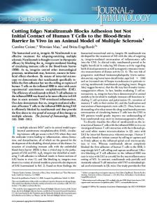
The Journal of Immunology effect in the pathogenesis of this disease (16, 21). To examine the possible anti-inflammatory effect of low dose IFN-␥ on allergic airway reactivity, we used a treatment protocol of i.p. injection of 6 U/day (0.25 U/g) of IFN-␥, beginning on the first day of OVA inhalation. As can be seen in Fig. 2A, the inhalation of aerosolized OVA caused the infiltration of eosinophils (48.9% OVA/OVA vs 0.3% control; p ⫽ 0.0026) and lymphocytes (14.5% OVA/OVA vs 3.4% control) into the BAL of OVA-sensitized mice, while a low dose of IFN-␥ dramatically inhibited the Ag-induced eosinophil (16.8% IFN-␥ vs 48.9% OVA/OVA; p ⫽ 0.01) and lymphocyte (8.7% IFN-␥ vs 14.5% OVA/OVA) recruitment into the BAL. Moreover, low levels of IFN-␥ dramatically inhibited homing of lymphocytes to the lung LNs (Fig. 2B). The AHR of conscious spontaneously breathing mice were evaluated at baseline (on day 0) immediately after the OVA inhalation (early reaction), and 24 h after OVA inhalation (late reaction). OVA/OVA-challenged mice had a significantly increased airway responsiveness to the early Ag challenge (⌬ Penn for the early reaction 0.93; baseline 0.092 vs OVA/OVA early 0.185; p ⫽ 0.0002). Furthermore, the airway responsiveness of the OVA/ OVA-challenged mice was significantly augmented to the late Ag challenge (⌬ Penn 0.52). However, no AHR was observed in the
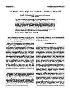
3709 early reaction (⌬ Penn 0.04) or late reaction (⌬ Penn 0.02) of control (nonchallenged) mice. Treatment of the OVA/OVA-challenged mice with low dose IFN-␥ significantly reduced AHR (OVA/OVA 0.185 vs IFN-␥ 0.113 early; p ⫽ 0.000028). ⌬ Penn for early reaction was significantly reduced by 79% (⌬ Penn early reaction OVA 0.93 vs ⌬ Penn early reaction IFN-␥ 0.2). Moreover, IFN-␥ reduced the late AHR in OVA-challenged mice (OVA/ OVA 0.14 vs IFN-␥ 0.11; p ⫽ 0.03). ⌬ Penn for late reaction was significantly reduced by 59% (⌬ Penn late reaction OVA 0.48 vs ⌬ Penn late reaction IFN-␥ 0.2) (Fig. 2C). Histopathologic examination of lung tissue from OVA mice revealed a pleomorphic peribronchial and perivascular infiltrate consisting of eosinophils, lymphocytes, macrophages, and neutrophils that was not seen in control mice (Fig. 3). The peribronchial and perivascular inflammatory infiltrates were given an inflammatory score of between 1 and 4 by two different viewers. The inflammatory infiltrate in the OVA/OVA-treated mice was significantly increased as compared with the control animals (OVA/OVA 3 ⫾ 0.86 vs control 0.11 ⫾ 0.33; p ⫽ 7 ⫻ 10⫺8). Lung histological changes in low dose IFN-␥-treated mice were barely detectable, with evidence of only slight accumulation of inflammatory cells around the bronchioles. The peribronchial inflammatory infiltrate
FIGURE 1. Low levels of IFN-␥ inhibit adhesion migration and homing of T cells. A, 51Cr-labeled T cells were resuspended in the presence or absence of SDF-1 or PMA stimulation in the presence or absence of IFN-␥, or supernatant collected from Th1 cells and adhesion was analyzed. One experiment representative of five is depicted. B, 51Cr-labeled T cells were resuspended in the presence or absence of PMA stimulation in the presence or absence of elevated concentrations of IFN-␥ and adhesion was analyzed. C, T cells were placed on transwell plates in the presence or absence of IFN-␥. Migration was measured after 3 h of incubation and was analyzed by FACS. One experiment representative of three is depicted. D, T cells were pretreated with IFN-␥ for 30 min. The cells were then stimulated with SDF-1. Next, the cells were fixed, permeabilized, their intracellular F-actin stained with FITC-phalloidin, and analyzed by FACS. The results obtained for the untreated cells were considered as 100% polymerization of F-actin. One experiment representative of five is depicted. E, CD4⫹ T cells purified from C57BL/6 mice were cultured in the presence of Con A (2.5 g/ml) in the presence or absence of elevated concentrations of IFN-␥. Proliferation was determined by addition of 1 Ci of [3H]thymidine for the last 18 h of a 4-day culture. F, Th2 cells were incubated in the presence or absence of IFN-␥ on transwell plates. Migration was measured after 3 h of incubation, and cell number was analyzed by FACS. One experiment representative of three is depicted. G, T cells labeled with BCECF AM were incubated in the presence or absence of IFN-␥ that were injected to mice. After 3.5 h, the LN were collected and the FITC-positive population was analyzed by FACS.
3710
CUTTING EDGE: IFN-␥ INHIBITS INFLAMMATORY RESPONSE IN AN ASTHMA MODEL
FIGURE 2. Low levels of IFN-␥ dramatically inhibit an allergic response. Control (untreated) OVAprimed mice (OVA/OVA) and OVA-primed mice i.p. injected with IFN-␥ were analyzed for (A) BAL cell recovery in mice after treatment with IFN-␥. Percentage of eosinophils and lymphocytes recovery in BAL fluids from control. Values represent the mean of nine animals. B, Number of lymphocytes arrived to the lung LN. Values represent the mean of four animals. C, Airway responsiveness was assessed on day 15. Values shown represent ⌬ Penn, which was calculated by subtracting control Penn measurements before Ag challenge from the Penn measurements after late or early Ag challenge. Baseline Penn levels were comparable among untreated control, OVA-primed mice, and OVA-primed mice treated with IFN-␥. Values represent the mean of nine animals.
was significantly reduced in these IFN-␥-treated mice (IFN-␥ 0.88 ⫾ 0.54 vs OVA/OVA 3 ⫾ 0.86; p ⫽ 0.000013). Immunohistochemical staining of lung tissue demonstrated an increased number of T cells (CD3-positive cells) in OVA/OVA mice. Treatment of mice with low doses of IFN-␥ dramatically reduced this T cell infiltration (Fig. 3).
Discussion IFNs were originally characterized as a group of related molecules induced to mediate an immediate defensive response to viral in-
FIGURE 3. Lung histology in mice that received IFN-␥ treatment. Histologic features of control OVA-primed (OVA/OVA) and OVA-primed treated with IFN-␥ (OVA/OVA ⫹ IFN). A1–3, ⫻10; B1–3, ⫻60; C1–3, BAL ⫻100; D1–3 ⫻20 CD3 cells.
fections. Type II IFN or IFN-␥ was initially identified based on its antiviral activities, and is now known to play a much more important role as a proinflammatory molecule (22). Previously, we showed that low levels of IFN-␥ down-regulate homing of B cells to the LNs but not to the spleen (11). The data presented in this study demonstrate that low levels of IFN-␥ inhibit similar processes of T cells and as a consequence, down-regulate the Th2 inflammatory response. Low dose IFN-␥ inhibits adhesion and transwell migration of naive T cells and of Th2 cells in vitro. The effect of IFN-␥ on naive T cells is concentration-dependent with a maximum inhibition of migration and cytoskeleton rearrangement at 0.1 U/ml. Moreover, our results demonstrate an inhibitory in vivo effect of low levels of IFN-␥. Low levels of this cytokine inhibit the migration of labeled naive cells to the LNs and decreases the AHR, airway inflammation, and eosinophil recruitment to the bronchial airways in the asthma Th2 model. Thus, IFN-␥, in addition to acting as a local proinflammatory cytokine, may under specific conditions exert counterinflammatory effects on T cells. It is possible that exposure of circulating effector cells in the blood or the spleen to low levels of IFN-␥ serves to suppress their migratory capacities. We suggest that the profound inhibition observed in the in vivo model might result from the combined effect of low levels of IFN-␥ on various T cell populations. These low levels of IFN-␥ down-regulate homing of naive T cells to the LNs and thereby reduce the exposure of these T cells to Ag, and as a consequence their activation is prevented. In addition, these low levels of IFN-␥ dramatically inhibit homing of effector Th2 cells to sites of inflammation where they exert their function. Thus, IFN-␥ has a dual function in the asthma model, both at sensitization and effector stages, which results in a dramatic inhibition on the inflammatory response. Several studies have previously analyzed the effect of IFN-␥ on asthma. Targeted disruption of the IFN-␥ receptor gene results in a prolonged airway eosinophilia in response to allergen (23). In addition, i.p. administration of rIFN-␥ decreases the OVA-induced eosinophil infiltration in a dose-dependent manner with a significant
The Journal of Immunology minimum effect at 600 U/day and almost complete inhibition at doses higher than 20,000 U/day (24). These relatively high doses of IFN-␥, which were considered to function in the transition from Th2 to Th1 cells, can potentiate the effects of other proinflammatory mediators. Our results show that low levels of IFN-␥, if properly targeted, might have a clinical application as an antiinflammatory agent.
Acknowledgments We thank Ofer Lider, Ronen Alon, and Michal Schwartz; and members of the Shachar lab for helpful discussions and ideas and review of this manuscript.
References 1. Gowans, J. L., and E. J. Knight. 1964. The route of re-circulation of lymphocytes in the rat. Proc. R. Soc. Lond. B. Biol. 159:257. 2. Butcher, E. C., R. G. Scollay, and I. L. Weissman. 1980. Organ specificity of lymphocyte migration: mediation by highly selective lymphocyte interaction with organ-specific determinants on high endothelial venules. Eur. J. Immunol. 10: 556. 3. Butcher, E. C. 1991. Leukocyte-endothelial cell recognition: three (or more) steps to specificity and diversity. Cell 67:1033. 4. Springer, T. A. 1994. Traffic signals for lymphocyte recirculation and leukocyte emigration: the multistep paradigm. Cell 76:301. 5. Butcher, E. C., and L. J. Picker. 1996. Lymphocyte homing and homeostasis. Science 272:60. 6. Banchereau, J., and R. M. Steinman. 1998. Dendritic cells and the control of immunity. Nature 392:245. 7. Abbas, A. K., K. M. Murphy, and A. Sher. 1996. Functional diversity of helper T lymphocytes. Nature 383:787. 8. O’Garra, A. 1998. Cytokines induce the development of functionally heterogeneous T helper cell subsets. Immunity 8:275. 9. Campbell, J. J., and E. C. Butcher. 2000. Chemokines in tissue-specific and microenvironment-specific lymphocyte homing. Curr. Opin. Immunol. 12:336.
3711 10. Sallusto, F., C. R. Mackay, and A. Lanzavecchia. 2000. The role of chemokine receptors in primary, effector, and memory immune responses. Annu. Rev. Immunol. 18:593. 11. Flaishon, L., R. Hershkovitz, F. Lantner, O. Lider, R. Alon, Y. Levo, R. A. Flavell, and I. Shachar. 2000. Autocrine secretion of interferon ␥ negatively regulates homing of immature B cells. J. Exp. Med. 192:1381. 12. Shachar, I., E. A. Elliot, B. Chasnoff, I. S. Grewal, and R. A. Flavell. 1995. Reconstitution of invariant chain function in transgenic mice in vivo by individual p31 and p41 isoforms. Immunity 3:373. 13. Flaishon, L., F. Lantner, R. Hershkovitz, Y. Levo, and I. Shachar. 2001. Low levels of IFN-␥ down-regulate the integrin-dependent adhesion of B cells by inhibition of cytoskeleton rearrangement. J. Biol. Chem. 276:46701. 14. Topilski, I., A. Harmelin, R. A. Flavell, Y. Levo, and I. Shachar. 2002. Preferential Th1 immune response in invariant chain-deficient mice. J. Immunol. 168:1610. 15. Djukanovic, R., W. R. Roche, J. W. Wilson, C. R. Beasley, O. P. Twentyman, R. H. Howarth, and S. T. Holgate. 1990. Mucosal inflammation in asthma. Am. Rev. Respir. Dis. 142:434. 16. Cohn, L., R. J. Homer, N. Niu, and K. Bottomly. 1999. T helper 1 cells and interferon ␥ regulate allergic airway inflammation and mucus production. J. Exp. Med. 190:1309. 17. Shimizu, Y., G. A. van Seventer, E. Ennis, W. Newman, K. J. Horgan, and S. Shaw. 1992. Crosslinking of the T cell-specific accessory molecules CD7 and CD28 modulates T cell adhesion. J. Exp. Med. 175:577. 18. Bleul, C. C., R. C. Fuhlbrigge, J. M. Casasnovas, A. Aiuti, and T. A. Springer. 1996. A highly efficacious lymphocyte chemoattractant, stromal cell-derived factor 1 (SDF-1). J. Exp. Med. 184:1101. 19. Tau, G. Z., T. von der Weid, B. Lu, S. Cowan, M. Kvatyuk, A. Pernis, G. Cattoretti, N. S. Braunstein, R. L. Coffman, and P. B. Rothman. 2000. Interferon ␥ signaling alters the function of T helper type 1 cells. J. Exp. Med. 192: 977. 20. Harris, D. P., L. Haynes, P. C. Sayles, D. K. Duso, S. M. Eaton, N. M. Lepak, L. L. Johnson, S. L. Swain, and F. E. Lund. 2000. Reciprocal regulation of polarized cytokine production by effector B and T cells. Nat. Immunol. 1:475. 21. Huang, S. K., H. Q. Xiao, J. Kleine-Tebbe, G. Paciotti, D. G. Marsh, L. M. Lichtenstein, and M. C. Liu. 1995. IL-13 expression at the sites of allergen challenge in patients with asthma. J. Immunol. 155:2688. 22. Farrar, M. A., and R. D. Schreiber. 1993. The molecular cell biology of interferon-␥ and its receptor. Annu. Rev. Immunol. 11:571. 23. Coyle, A. J., S. Tsuyuki, C. Bertrand, S. Huang, M. Aguet, S. S. Alkan, and G. P. Anderson. 1996. Mice lacking the IFN-␥ receptor have impaired ability to resolve a lung eosinophilic inflammatory response associated with a prolonged capacity of T cells to exhibit a Th2 cytokine profile. J. Immunol. 156:2680. 24. Iwamoto, I., H. Nakajima, H. Endo, and S. Yoshida. 1993. Interferon ␥ regulates antigen-induced eosinophil recruitment into the mouse airways by inhibiting the infiltration of CD4⫹ T cells. J. Exp. Med. 177:573.