www.ajbrui.org Afr. J. Biomed. Res. Vol. 21 (January, 2018); 61- 63 Short communication
Dental Formula and Dental Abnormalities Observed in the Eidolon helvum (Fruit Bat) Captured from the Wild Igado O1; *Femi-Akinlosotu O2; Omobowale T3; Ajadi R4 and Nottidge H3 1
Department of Veterinary Anatomy, Faculty of Veterinary Medicine, University of Ibadan, Nigeria. Department of Anatomy, Faculty of Basic Medical Sciences, College of Medicine, University of Ibadan, Nigeria. 3 Department of Veterinary Medicine, Faculty of Veterinary Medicine, University of Ibadan, Nigeria. 4 Department of Veterinary Medicine and Surgery, College of Veterinary Medicine, Federal University of Abeokuta, Ogun State, Nigeria. 2
ABSTRACT Bats are known to be the only flying mammals. Eidolon helvum is a species of fruit bats that is reported to have great economic and health importance, while also having an unsavoury reputation in many cultures in Nigeria. This study investigated the dental formula and dental pathologies of Eidolon helvum captured from the wild. The dental profile of 35 adult bats (20 males, 15 females) was assessed. The animals were observed to have a heterodont dentition. The males were observed to have a total of 30 to 36 teeth, while the females had 30 to 34 teeth. A missing lower incisor was observed in some of the females while the males showed a varying number of upper premolar. Severe attrition and edentulousness were observed in some bats examined. Results obtained from this work may shed more light on the different dental pathologies observed in the adult Eidolon helvum in Nigeria. Keywords: Eidolon helvum, dental formula, dental abnormalities, attrition *Author for correspondence: E-mail:
[email protected];
[email protected]; Tel:+234-7031163053 Received: August 2017; Accepted: November, 2017 Abstracted by: Bioline International, African Journals online (AJOL), Index Copernicus, African Index Medicus (WHO), Excerpta medica (EMBASE), CAB Abstracts, SCOPUS, Global Health Abstracts, Asian Science Index, Index Veterinarius
INTRODUCTION A healthy dentition is important to both quality of life and nutrition. However, oral disease has been shown to affect systemic health (Moynihan and Petersen, 2004; Ali, 2015). Dentition is the characteristic arrangement of teeth, the type, shape and number in a particular species at a certain age. They are important for aesthetics, phonation and protection (Holt et al., 2000). In Veterinary Medicine, dentition is an essential tool for aging (Getty, 1975; Dyce et al., 2002). Broadly, dentition can be divided into deciduous and permanent. The dental formula is the numerical representation of the different types of teeth in the oral cavity (Zengingul et al., 2007). Alterations in the dentition may have widespread influences, including the feeding habit of the animal. Tooth wear is an allembracing term used to describe the combined processes of erosion, attrition and abrasion and the diagnosis of dental pathology and also age determination (Zengingul et al., 2007). The African straw-coloured bat, Eidolon helvum, is a megachiroptera, and the second largest fruit eating bat in Africa, with unique morphological, ecological and
reproductive characteristics (Juste et al., 2000). It is found in Africa, Asia (the Arab Peninsula) and the southwest of the Arab Peninsula (Odukoya et al., 2009). Weight and size have been generally used to determine or estimate the age (juvenile, adolescent and adult) in the Eidolon helvum (Odukoya et al., 2009). The males have been reported to be larger than the females (Odukoya et al., 2009; Igado et al., 2015). Of public health importance is the fact that these bats have been reported to be reservoirs of the rabies virus and other viruses that can infect humans and other domestic animals and wildlife (Aghomo et al., 1990; Calisher et al., 2006). The economic importance lies in the fact that as unsavoury as their reputation may be, they have been found to be useful in the control of insects, reseeding cut forests, and in pollinating plants that provide food for humans and other species (Calisher et al., 2006). In spite of the health and economic importance of bats, and the fact that various works have been done describing the anatomy of the Eidolon helvum, there is currently a dearth of information on the dental pathologies in the bats captured from the wild, and specifically on the dental abnormalities and
Dental abnormalities in Eidolon helvum dentition of the E helvum, which is a very common bat in Nigeria. This study hopes to bridge that gap by providing baseline data, while results obtained may also find application in the field of comparative dental and forensic anatomy. MATERIALS AND METHODS Ethical approval for this study was obtained from the Faculty of Veterinary Medicine, University of Ibadan, Nigeria (Ethical code number – 12/13/03). In handling the bats and carrying out all procedures, guidelines for the care and use of experimental animals (National Institute of Health (NIH), USA and the Faculty of Veterinary Medicine, University of Ibadan, Nigeria) were strictly adhered to. All the bats used for this study were assessed to be adults using the weight index according to Odukoya et al., (2009). A total of 35 bats (20 males, 15 females) were used in this study. They were captured in Abeokuta, Ogun State, Nigeria, using mist nets set up at roosting sites. They were transported to the laboratory in metal cages and euthanized using chloroform chamber. The weights were determined with the aid of a digital weighing scale (Electronic Kitchen Scale, Camry® EK5350) and recorded in grams. The temporomandibular joint was dislocated to allow for maximum opening of the mouth, so as to gain full access to the oral cavity. Linear measurements when employed were carried out with a digital vernier calliper (Neiko®, sensitivity of 0.01mm). RESULTS All animals examined were observed to have heterodont dentition. Generalised attrition ranging from mild to severe was observed on all the teeth in all bats examined. Attrition was more pronounced on the premolars and molars.
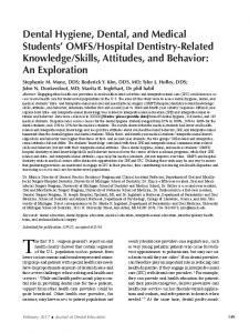
In the males, the dental formula was I 2/2 C1/1 PM 2-3/3 M 2-3/2-3, total 30 to 36. Two males (10%) had only 2 upper premolars, while the others had 3 (90%) per quadrant. Three males had 3 upper molars (15%), while ten (50%) had 3 lower molars per quadrant. One male (5%) was observed to have only one lower incisor, while another one had no lower incisor (5%). The number of incisors, canine and lower premolars (left and right) were observed to be consistent in all specimens examined, except one that recorded asymmetry of the incisor with the left side being 1/1 and the right 2/2. There was generalized attrition of all the teeth especially the lower molars (Figure 1B). This pathology was however more prominent on the palatal surface of the teeth. The specimens also showed a space between the upper incisors measuring 0.40cm which was a little bit different when compared to the lower incisors’ space (0.35cm). In the females, the dental formula was I 2/1-2 C1/1 PM 3/3 M 2/2-3, total 30 to 34. In one female, there was extreme severe attrition while the visible teeth were I 2/0 C1/1 with partial edentulous of the upper and lower jaw (Figure 1C). Partial edentulous was also observed in another female specimen, both in the upper and lower jaws. The visible teeth in this particular specimen were lower canine (right), 1st premolar (right & left), the last 2 molars (right & left) and upper incisors (right & left), canines (right & left), last 2 molars (left) and last molar (right). The female bats however had generalized attrition of the molars which was milder when compared to the male bats. This attrition was symmetrical both buccally and palatally. A particular female specimen having a dental formula of I2/0; C1/1; PM3/3; M2/3 showed a very severe generalized attrition of all the teeth. There was also a space between the upper incisors measuring 0.42cm and between the lower incisors measuring 0.30cm.
Plate 1: Oral cavity of bats, with the tongues removed, showing increasing levels of attrition, from left (A) to right (C). Compare the degree of attrition on a canine in A (arrow) to a canine in B (arrow) and C (arrow). Note the generalized severe attrition of the teeth in B and partial edentulousness of the upper and lower jaws in C (arrow heads).
62
Afr. J. Biomed. Res. Vol. 21, No.1 (January) 2018
Igado, Akinlosotu, Omobowale et al
Dental abnormalities in Eidolon helvum
DISCUSSION The primary function of the teeth is to prepare food for swallowing and also to facilitate digestion. Teeth have their respective forms to facilitate prehension, incision and trituration (process of reduction of food substance into smaller particles to form an homogenous material) of food (Nelson and Ash, 2010). The dentition of heterodont mammals is characteristically known to be symmetric bilaterally (Juste and Ibáñez, 1993). This is consistent with what was seen in most of the bats examined. The reason for one subject recording asymmetry of the incisors is not clear but may be due to developmental instability as previously reported by Juste and Ibáñez (1993), where some frugivorous bats, Myonycteris brachycephala, from the São Tomé Island in West Africa showed an asymmetrical absence of a lower incisor. This asymmetry reported in São Tomé Island occurred invariably in the left or right mandible. Incisors are employed mainly in nibbling, grooming and also for breaking food before it is taken into the oral cavity (Dyce et al., 2002). The attrition observed on the incisors was not as severe as that observed in the premolar and molar. This is probably due to the food-dividing function of the incisors. The space between the left and right upper incisors remained relatively constant in all specimens examined, irrespective of gender. The reason for this lack of gender disparity is currently not clear, especially since in a previous experiment on the E helvum, the males recorded higher values for width of the oral cavity, between left and right upper canines (Igado et al., 2015). Some of the females in this study recorded a missing lower incisor, although symmetrical, unlike previous asymmetrical reports (Juste and Ibáñez, 1993). In mammals, the canine teeth have been known to function as tools for killing, grasping, opening and dividing prey/food (Mellet, 1985; Freeman, 1992). The E. helvum is a fruit eating bat, and so may probably not use the canine teeth as much as the flesh-eating bat. In addition, canines are used to initially tear up food, which are then processed by postcanine teeth (the premolars and molars (Freeman, 1992). This may account for the canines generally showing the least attrition in all the bats examined in the current study. The shapes of the incisal and occlusal surfaces of the teeth are related to the function they perform and also the movements of the mandible required in carrying out in chewing a variety of foods (Nelson and Ash, 2010). The cheek teeth (premolars and molars) showed the greatest attrition due to their function as grinding teeth, which is essential in frugivorous bats. The males showed a tendency to have greater number of teeth, evidenced by an extra upper molar in some males. Dental formula recorded in this current study appeared to have similarities to the report in Euderma maculatum (spotted bat), where dental formula is I 2/3 C1/1 PM 2/2 M3/3, total 34 (Watkins, 1977). Even though the weights of the bats used were similar, the degree of attrition showed that there were young adults as well as old adults. The fact that the males showed more severe attrition and edentulousness may probably imply that they eat more,
63
relative to the females, as observed in their higher body weight in previous studies (Odukoya et al., 2009; Igado et al., 2015). In conclusion, our findings revealed sexual dimorphism in the number of teeth, with the males having a larger number; and also revealed that the males had the greater tendency to develop attrition relative to the females. The findings herein suggest its usefulness in the field of comparative dental and forensic anatomy. REFERENCES Aghomo HO, Ako-Nai AK, Oduye OO, Tomori O, Rupprecht CE (1990). Detection of rabies virus antibodies in Fruit Bats (Eidolon helvum) from Nigeria. Journal of Wildlife Diseases, 26 (2): 259-261. Calisher CH, Childs JE, Field HE, Holmes KV, Schountz T (2006). Bats: Important Reservoir Hosts of Emerging Viruses. Clinical Microbiology Reviews, 19(3): 531–545 Ali DA (2015). Knowledge of the Relationships between Oral Health, Diabetes, Body Mass Index and Lifestyle among Students at the Kuwait University Health Sciences Center, Kuwait. Med Princ Pract; 25:176–180 Dyce KM, Sack WO, Wensing CJG (2002). Textbook of Veterinary Anatomy. 3rd edn. Philadelphia: Saunders Company. Freeman PW (1992). Canine teeth of bats (Microchiroptera): size, shape and role in crack propagation. Biological Journal of the Linnean Sociely (1992), 45: 97-1 15. Holt R, Roberts G, Scully C (2000). ABC of oral health and disease. British Medical Journal, 320: 1652 – 1655. Igado OO, Omobowale TO, Ajadi RA, Nottidge HO (2015). Gross morphometric studies on the tongue, buccal cavity and hard palate of the fruit bat (Eidolon helvum). Anatomia Histologia Embryologia, 44 (4): 283–287. Juste J, Ibáñez C (1993). An asymmetric dental formula in a mammal, the São Tomé Island fruit bat Myonycteris brachycephala (Mammalia: Megachiroptera). Canadian Journal of Zoology, 71(1): 221 – 224. Juste J, Machordom A, Ibáñez C (2000). Morphologic and allozyme variation in Eidolon helvum in the Gulf of Guinea (West-Central Africa). Biol. J. Linn. Soc. 71: 359-378. Mellett JS (1985). Autocclusal mechanisms in the carnivore dentition. Australian Mammalogy, 8: 233-238. Moynihan P, Petersen PE (2004). Diet, nutrition and the prevention of dental diseases. Public Health Nutr: 7(1A), 201– 226 Nelson SJ, Ash MM (2010). Wheeler’s Dental Anatomy, Physiology, and Occlusion. 9th edition, Saunders Elsevier, pg 75. Odukoya SA, Ofusori DA, Adeeyo OA, Ayoka OA, Abayomi TA, Ajayi SA, Falana BA (2009). Comparative biochemical studies of the pregnant and non pregnant uterine limbs of the frugivorous bat, Eidolon helvum. J Cell Anim Biol, 3: 175-178. Getty R (1975) Sisson and Grossman’s Anatomy of the Domestic Animals. (5th edition), WB Saunders Company, Philadelphia. ISBN 0-7216-4107-5. Watkins LC (1977). Euderma maculatum. Mammalian Species, 77: 1 – 4. Zengingul A, Eskimez S, Değer Y, Kama J (2007). Tooth Wears and Dentoalveolar Compensation of Vertical Height. Biotechnol. & Biotechnol. Eq. pg. 362-365.
Afr. J. Biomed. Res. Vol. 21, No.1 (January) 2018
Igado, Akinlosotu, Omobowale et al