Neuroendocrinology Letters ISSN 0172–780X Copyright © 2002 Neuroendocrinology Letters
Effects of morphine on tumour growth
1. Department of Anaesthesia and Intensive Care, Aarhus University Hospital, 8000 Aarhus C, Denmark. 2. Neuroscience Research Institute, State University of New York/College at Old Westbury, Old Westbury, New York, 11568 USA. Correspondence to: Mads Rasmussen M.D., Department of Anaesthesia and Intensive Care, Aarhus University Hospital, 8000 Aarhus C, DENMARK . PHONE : +45 89493507; FA X : +45 89493502 E-MAIL :
[email protected] May 1, 2002 May 2, 2002
Key words:
endogenous opioids; morphine; neoplasia; cancer; apoptosis; nitric oxide
Neuroendocrinology Letters 2002; 23:193–198
Abstract
pii: NEL230302R01
Copyright © Neuroendocrinology Letters 2002
Introduction Endogenous opiate alkaloids, such as morphine, and their peptide counterparts have been implicated in a wide variety of pharmacological and physiological functions [1]. Opiate alkaloids appear to represent one of the immune and vascular inhibitory/ anti-inflammatory systems in an organism whereas opioid peptides appear to have proinflammatory capabilities [1–3]. Thus, from an immune perspective, these signalling molecules are potential candidates as tumour growth modifiers. This article will focus on morphine’s influence on the regulatory mechanisms involved in tumour growth.
Endogenous opioid peptides influence tumour growth Since 1983, numerous studies have demonstrated that endogenous opioid peptides are involved in the growth regulation of tumour cells. In several studies, Zagon et al demonstrated that [MET5] enkephalin, also termed opioid growth factor (OGF), through interaction with the OGF receptor (OGFr) [4], inhibits cell proliferation in a variety of cancer cell lines in vitro [5] [6–8] and in vivo [9,10]. Using immunocytochemistry they demonstrated that OGF and the OGF receptor were present in a variety of tumour cells [6,10], suggesting that growth regulation by endogenous opioid peptides is auto-
R EV IEW
Endogenous opiate alkaloids, such as morphine, and their peptide counterparts have been implicated in a wide variety of pharmacological and physiological functions. In addition to their use in the treatment of pain, opioids, appears to be important in the growth regulation of normal and neoplastic tissue. This review will focus on the influence of endogenous and exogenous opioids on tumour growth, with emphasis on immunoregulatory and antiproliferative mechanisms.
NEL
Submitted: Accepted:
I N V IT ED
Mads Rasmussen1, Wei Zhu2, Jan Tønnesen2, Patrick Cadet2, Else Tønnesen1 & George B. Stefano 2
193
Mads Rasmussen, Wei Zhu, Jan Tønnesen, Patrick Cadet, Else Tønnesen & George B. Stefano
crine. Further experiments by this group revealed that OGF was tonally active because persistent blockade of opioid-receptor interaction with the potent opioid antagonist, naltrexone, or the removal of OGF, using antibodies to this peptide, resulted in an increase in the number of tumour cells [5,8]. It appears that the mechanism of OGF’s action on tumour growth is related to a strong influence on cell proliferative events, since previous studies have demonstrated that binding of OGF to the OGF receptor depressed both DNA synthesis and mitosis within hours [11,12]. Recently Bisignani et al raised the question whether one mechanism contributing to the proliferation of cancer cells is a defect in the machinery producing this tumoursuppressing element [5]. In this regard, in 1989 Zagon et al characterized this opioid receptor, designated at that time Zeta and now OGFr, which binds [MET5] enkephalin and unlike other opioid receptors is directly involved in the proliferation of cells [4]. Receptor displacement studies demonstrated that the binding of [MET5] enkephalin was not influenced by ligands selective for µ-, δ-, and κreceptors, suggesting a specific interaction of [MET5] enkephalin and its receptor. Furthermore, they demonstrated that sodium and guanine nucleotides inhibited binding of [ MET5] enkephalin, and suggested that this opioid binding site may have some similarities in molecular organization to other opioid sites in brain tissues, neoplastic tissues and cells. In earlier studies of opioid agonist binding, they argued that both sodium and guanine nucleotides have been found to be necessary for functional coupling of the opioid binding site to regulatory units such as adenylate cyclase. In addition, their study demonstrated that the binding site for [MET5] enkephalin is proteinaceous in character because protein inhibitors were necessary for optimal binding reactions, and binding was reduced by proteolysis of the preparation with trypsin.
Exogenous morphine influences tumour growth Morphine is widely used as an analgesic to treat pain in a variety of patients including those with cancer. However, there is evidence that morphine has extra-analgesic actions and significantly alters tumour growth. Ishikawa et al demonstrated that morphine (10 mg/kg) given daily for 10 days, enhanced the growth of several different tumour cell lines in vivo [13]. However, other studies suggest that the analgesic qualities of morphine contribute to the control of metastasis following surgery. Page et al demonstrated that pre- and postoperative administration of morphine significantly attenuated the metastatic-enhancing effects of surgery [14–16]. In addition, intermittent bolus of morphine administration to animals was associated with a reduction in the growth of tumour cells that gained access to the circulation during the surgical procedure [17]. The authors proposed that the most likely
194
explanation was a direct morphine effect on host resistance to metastatic tumour growth. Hatzoglou et al demonstrated that morphine decreases the cell growth of human breast cancer cells in vitro, despite the lack of µ receptors in the cancer cells as determined by a limited screening with pharmacological agents [18]. They raised the question whether this antiproliferative effect of morphine could be mediated through interaction with other receptor systems. In a further study they showed, that morphine may exert its antiproliferative effect on breast cancer cells through interaction with the somatostatin receptor SSTR2, suggesting a functional interaction of morphine with the inhibitory somatostatinergic system [19]. Previous studies have demonstrated a direct inhibitory effect of somotostatin analogues on the growth of human cancer cells [20–22]. The possible interaction of morphine with other receptor systems is supported by the findings of Maneckjee and Minna. They found that the inhibitory effects of morphine on the growth of lung cancer could be reversed by nicotine, suggesting an interaction between the opioid system and acetylcholine receptors [23]. In addition, Zagon et al demonstrated in receptor binding studies that only a few tumours express µ opioid binding sites [24]. Thus, given the knowledge at that time, it seems that µ opioid receptors do not play a significant role in opioid mediated regulation of tumour growth. However, new evidence from our group suggests that other physiologically active µ opioid receptor splice variants may be present and operational in immune and vascular tissues that had gone previously undetected [25].
Morphine and apoptosis The molecular mechanism by which morphine influence tumour growth in vivo and in vitro is not clear. One hypothesis is that morphine promotes apoptosis in tumour cells. Maneckjee and Minna demonstrated that treatment of human lung cancer cells with 0.1–1 µM morphine or methadone resulted in morphological changes and cleavage of DNA into nucleosome-sized fragments characteristic of apoptosis [26]. Sueoka et al demonstrated that morphine attenuated the growth of various cancer cell lines in vitro through inhibition of tumour necrosis factor (TNF)-α mRNA expression and TNF-α release [27]. Transcription of TNF-α gene is in part regulated by the transcription factor, nuclear factor κB (NFκB) [28]. In a further study, they demonstrated that the anticancer activity of morphine and the five times more potent morphine derivatives, (–)-3-Acetyl-6βacetylthio-N-cyclopropyl-methylnormorphine (KT-90), (–)-6β-acetylthiomorphine (KT 87) was mediated through apoptosis associated with inhibition of nuclear factor κB (NFκB) in human cancer cell lines [29]. NFκB is a DNA binding protein that induces expression of genes for several inflammatory mediators such as TNF-α and augments transcription of various
Morphine and tumour growth
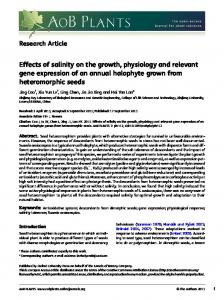
genes involved in cell proliferation. Recently it was demonstrated that inhibition of NFκB attenuates apoptosis resistance in lymphoid cells [30]. Furthermore, we demonstrated that morphine can directly inhibit NFκB actions via the liberation of nitric oxide [31,32], introducing another variable in the morphine signalling cascade that may explain morphine’s antiproliferative and apoptotic actions. Figure 1.
Morphine influences natural killer (NK) cell activity The role of NK cells in both the metastasis enhancing effects of surgery and the attenuation of these effects by morphine was investigated by Page et al [15]. Surgery induced a suppression of whole blood NK cytotoxic activity and a decreased number of circulating Large Granular Lymphocytes (LGL)/NK cells measured 4 h postoperatively [15]. The authors observed that in LGL/NK depleted animals, morphine had no impact on tumour cell retention. They suggested that LGL/NK cells play a critical role in morphine’s antimetastatic effects. In addition, Provincialli et al reported that “chronic” morphine treatment in cancer patients was accompanied by a decrease in the in vitro NK cell cytotoxic activity [33] while the treatment significantly increased Lymphokine Activated Killer (LAK) cell activity when compared to healthy controls [33]. Morphine influence’s on DNA synthesis It has previously been demonstrated that µ-, δ-, and κ- opioid agonists attenuate thymidine incorporation into DNA in glial [34] and developing neural cells
[35–38]. Furthermore, Barg et al showed that morphine inhibited DNA synthesis through inhibition of thymidine incorporation in C6 rat glioma cells that express opioid receptors [39]. Their results imply that inhibition of phosphoinositol signal transduction and CA 2+ mobilization is responsible for reduction in thymidine incorporation [40]. This finding is in accordance with previous reports demonstrating that opioid peptides and opiate alkaloids can inhibit phosphoinositol turnover [34,41–43]
Endogenous morphine, nitric oxide and the regulation of tumour growth Morphine has been shown to be involved in immunomodulation. Studies have demonstrated that morphine, not opioid peptides, via the µ3 opiate receptor is coupled to constitutive nitric oxide release in endothelial and immunocompetent cells [44,45]. Recently, expression of this opiate receptor subtype was demonstrated for the first time in human specimens of cancer tissue (non-small-cell lung carcinoma) [46]. The authors demonstrated that activation of the µ3 opiate receptor by opiate alkaloids in tumour cells leads to a rapid and substantial release of NO [46]. They suggested that the anti-cancer effects of morphine were mediated by nitric oxide through the µ3 receptor. However, they also speculated that increased nitric oxide production in lung carcinoma may indicate that tumours use endogenous opiates that bind to the µ3 receptor and thereby down regulate the host immune response to tumour growth. Several studies have demonstrated that morphine is produced endogenously in various human and mamFigure 1. How morphine might alter tumour growth. Morphine stimulates intracellular Ca2+ transients, that, in turn activates constitutive nitric oxide synthase (cNOS) and liberate nitric oxide (NO). NO may have a direct effect on tumour growth. Furthermore, NO inhibits disassociation of the IκBα inhibitor omplex, NFκB binding to the respective DNA promotor region and the subsequent expression of proinflammatory cytokines. Thereby stimulating apoptosis and downregulation of the carcinogenic effects of TNFα.
Neuroendocrinology Letters ISSN 0172–780X Copyright © 2002 Neuroendocrinology Letters
195
Mads Rasmussen, Wei Zhu, Jan Tønnesen, Patrick Cadet, Else Tønnesen & George B. Stefano
Figure 2. The mechanisms by which L-arginine metabolites influence cell proliferation. L-arginine is the common substrate for two enzymes, arginase and nitric oxide synthase (NOS). Arginase conversts L-arginine to L-ornithine. L-ornithine can be subsequently used by ODC to form polyamines which is essential for cell proliferation. NO is a potent inhibitor of ODC, suggesting
malian tissues. It binds to the opioid-peptide insensitive µ3 receptor and functions as a signalling molecule involved in immune down regulation [3,47–49]. It is demonstrated that morphine through activation of constitutive nitric oxide synthase (cNOS) liberates nitric oxide. Nitric oxide, in turn, inhibits binding of the DNA transcription factor NFκB to DNA and thereby down regulating the expression of genes for several inflammatory cytokines [3,31,32]. Figure 1. Furthermore, NO stabilizes the NFκB inhibitor, IκBα, which prevents its degradation. In addition, inhibition of NFκB attenuates apoptosis resistance as described previously and with that promotes down regulation of tumour growth. Figure 1. Regarding the effect of nitric oxide on tumour growth regulation, previous studies have shown that nitric oxide produced by immune and endothelial cells is tumouricidal possibly by inducing apoptosis [50]. Other studies have demonstrated that upregulation of the expression of inducible NO synthase (iNOS) in hepatic metastases and metastatic melanoma cells is associated with apoptosis, suppression of tumourigenicity, and abrogation of metastasis [51,52]. However, other studies have demonstrated tumour promoting effects of nitric oxide, and overall, NO seems to play a variety of contradictory roles in tumour growth regulation [53,54]. Contradicting results have also been obtained regarding the functional interaction between the opioidergic and the nitric oxide system. Kampa et al demonstrated that opioid agonists, active on κ-opioid receptors decrease NO2– /NO3-release and NOS activity in vitro [55]. Based on their results and reports demonstrating nitric oxide involvement in tumour progression and metastasis, the authors suggested opiates as potential in cancer treatment. Recently, Gobert et al demonstrated that the vigorous host response, i.e. up regulation of iNOS, to the
196
that the antiproliferative role of NO is attributed to inhibition of polyamine formation. L-arginine is also metabolized by NOS to L- citrulline and NO via an intermediate, NOHA. NOHA interferes with cell proliferation by inhibition of arginase and arginine-derived polyamines. Arginase promotes polyamine formation and suppress tumour cytotoxicity by reducing NO production.
human gastric pathogen Helicobacter pylori failed to eradicate the organism [56]. This was due to bacterial arginase down-regulating eukaryotic nitric oxide production. We surmise a similar process may be occurring with tumours, explaining the contradictory results. That is, certain tumours have a process to neutralize nitric oxide tumouricidal actions. In this regard the generation of NO through activation of nitric oxide synthase (NOS) has been shown to be antiproliferative [57]. However, the mechanism by which increased NOS activity and the production of nitric oxide causing cytostasis is not clear. The intermediate NG-hydroxyL-arginine (NOHA) in the oxidation of arginine is a strong inhibitor of the enzyme arginase. One of the products of arginase catalyzing L-arginine is L-ornithine. L-ornithine can be subsequently used by the enzyme ornithine decarboxylase (ODC) to form polyamines, which are essential components of cell proliferation. Figure 2. Cells that have been activated with cytokines show a significant increase in NOHA and nitric oxide or its oxidized metabolites [57]. Therefore, NOHA may be playing a role as a biological inhibitor of endogenous arginase activity, thereby promoting down regulation of tumour cell proliferation. Figure 2. This may explain the mechanism by which activation of NOS is involved in cytostasis. In addition, it has been demonstrated that NO is a potent inhibitor of ODC, thereby suggesting that the antiproliferative action of NO is attributed to inhibition of polyamine formation [57]. Figure 2. Few breast cancer cells lines were shown to have high arginase activity and very low NOS activity [58]. Nitric oxide derived from macrophages is known to have tumouricidal activity and polyamines may promote the growth of tumour cells [59]. Therefore, it appears that arginase may be playing a role in promoting tumour growth by inhibiting the production of
Morphine and tumour growth
nitric oxide. The significance of this phenomenon is enhanced by our early discussion concerning morphine’s ability to stimulate cNOS derived NO release, since this action may not be observed in the presence of arginase. In this regard, a biomedical strategy recognizing this process may be designed, strengthening a role for opiate as a new tumouricidal agent. Acknowledgements: This work was supported by the following grants: NIDA 09010; NIMH 47392 and the NIH Fogarty INT 00045. REFERENCES 1 Stefano GB, Scharrer B, Smith EM, Hughes TK, Jr., Magazine HI, Bilfinger TV et al. Opioid and opiate immunoregulatory processes. Crit Rev Immunol 1996; 16:109–144. 2 Stefano GB, Salzet B, Fricchione GL. Enkelytin and opioid peptide association in invertebrates and vertebrates: immune activation and pain. Immunol Today 1998; 19:265–268. 3 Stefano GB, Goumon Y, Casares F, Cadet P, Fricchione GL, Rialas C et al. Endogenous morphine. Trends Neurosci 2000; 23:436–442. 4 Zagon IS, Goodman SR, McLaughlin PJ. Characterization of zeta (zeta): a new opioid receptor involved in growth. Brain Res 1989; 482:297–305. 5 Bisignani GJ, McLaughlin PJ, Ordille SD, Beltz MS, Jarowenko MV, Zagon IS. Human renal cell cancer proliferation in tissue culture is tonically inhibited by opioid growth factor. J Urol 1999; 162:2186–2191. 6 McLaughlin PJ, Levin RJ, Zagon IS. Regulation of human head and neck squamous cell carcinoma growth in tissue culture by opioid growth factor. Int J Oncol 1999; 14:991–998. 7 McLaughlin PJ, Zagon IS, Skitzki J. Human neuroblastoma cell growth in tissue culture is regulated by opioid growth factor. Int J Oncol 1999; 14:373–380. 8 Zagon IS, Smith JP, McLaughlin PJ. Human pancreatic cancer cell proliferation in tissue culture is tonically inhibited by opioid growth factor. Int J Oncol 1999; 14:577–584. 9 Zagon IS, Hytrek SD, Lang CM, Smith JP, McGarrity TJ, Wu Y et al. Opioid growth factor ([Met5]enkephalin) prevents the incidence and retards the growth of human colon cancer. Am J Physiol 1996; 271:R780–R786. 10 Zagon IS, Hytrek SD, Smith JP, McLaughlin PJ. Opioid growth factor (OGF) inhibits human pancreatic cancer transplanted into nude mice. Cancer Lett 1997; 112:167–175. 11 Zagon IS, McLaughlin PJ. Endogenous opioid systems regulate growth of neural tumor cells in culture. Brain Res 1989; 490:14–25. 12 Zagon IS, Roesener CD, Verderame MF, Ohlsson-Wilhelm BM, Levin RJ, McLaughlin PJ. Opioid growth factor regulates the cell cycle of human neoplasias. Int J Oncol 2000; 17:1053–1061. 13 Ishikawa M, Tanno K, Kamo A, Takayanagi Y, Sasaki K. Enhancement of tumor growth by morphine and its possible mechanism in mice. Biol Pharm Bull 1993; 16:762–766. 14 Page GG, Ben Eliyahu S, Yirmiya R, Liebeskind JC. Morphine attenuates surgery-induced enhancement of metastatic colonization in rats. Pain 1993; 54:21–28. 15 Page GG, Ben Eliyahu S, Liebeskind JC. The role of LGL/NK cells in surgery-induced promotion of metastasis and its attenuation by morphine. Brain Behav Immun 1994; 8:241–250. 16 Page GG, McDonald JS, Ben Eliyahu S. Pre-operative versus
postoperative administration of morphine: impact on the neuroendocrine, behavioural, and metastatic-enhancing effects of surgery. Br J Anaesth 1998; 81:216–223. 17 Yeager MP, Colacchio TA. Effect of morphine on growth of metastatic colon cancer in vivo. Arch Surg 1991; 126:454–456. 18 Hatzoglou A, Bakogeorgou E, Castanas E. The antiproliferative effect of opioid receptor agonists on the T47D human breast cancer cell line, is partially mediated through opioid receptors. Eur J Pharmacol 1996; 296:199–207. 19 Hatzoglou A, Ouafik L, Bakogeorgou E, Thermos K, Castanas E. Morphine cross-reacts with somatostatin receptor SSTR2 in the T47D human breast cancer cell line and decreases cell growth. Cancer Res 1995; 55:5632–5636. 20 Bogden AE, Taylor JE, Moreau JP, Coy DH, LePage DJ. Response of human lung tumor xenografts to treatment with a somatostatin analogue (Somatuline). Cancer Res 1990; 50:4360–4365. 21 Prevost G, Foehrle E, Thomas F, Pihan I, Veber N, Starzec A et al. Growth of human breast cancer cell lines is inhibited by the somatostatin analog BIM23014. Endocrinology 1991; 129:323–329. 22 Setyono-Han B, Henkelman MS, Foekens JA, Klijn GM. Direct inhibitory effects of somatostatin (analogues) on the growth of human breast cancer cells. Cancer Res 1987; 47:1566–1570. 23 Maneckjee R, Minna JD. Opioid and nicotine receptors affect growth regulation of human lung cancer cell lines. Proc Natl Acad Sci U S A 1990; 87:3294–3298. 24 Zagon IS, McLaughlin PJ, Goodman SR, Rhodes RE. Opioid receptors and endogenous opioids in diverse human and animal cancers. J Natl Cancer Inst 1987; 79:1059–1065. 25 Cadet P, Bilfinger TV, Fimiani C, Peter D, Stefano GB. Human vascular and cardiac endothelia express mu opiate receptor transcripts. Endothelium 2000; 7:185–191. 26 Maneckjee R, Minna JD. Opioids induce while nicotine suppresses apoptosis in human lung cancer cells. Cell Growth Differ 1994; 5:1033–1040. 27 Sueoka N, Sueoka E, Okabe S, Fujiki H. Anti-cancer effects of morphine through inhibition of tumour necrosis factoralpha release and mRNA expression. Carcinogenesis 1996; 17:2337–2341. 28 Shakhov AN, Collart MA, Vassalli P, Nedospasov SA, Jongeneel CV. Kappa B-type enhancers are involved in lipopolysaccharide-mediated transcriptional activation of the tumor necrosis factor alpha gene in primary macrophages. J Exp Med 1990; 171:35–47. 29 Sueoka E, Sueoka N, Kai Y, Okabe S, Suganuma M, Kanematsu K et al. Anticancer activity of morphine and its synthetic derivative, KT-90, mediated through apoptosis and inhibition of NF-kappaB activation. Biochem Biophys Res Commun 1998; 252:566–570. 30 Jeremias I, Kupatt C, Baumann B, Herr I, Wirth T, Debatin KM. Inhibition of nuclear factor kappaB activation attenuates apoptosis resistance in lymphoid cells. Blood 1998; 91:4624–4631. 31 Welters ID, Fimiani C, Bilfinger TV, Stefano GB. NF-kappaB, nitric oxide and opiate signaling. Med Hypotheses 2000; 54:263–268. 32 Welters ID, Menzebach A, Goumon Y, Cadet P, Menges T, Hughes TK et al. Morphine inhibits NF-kappaB nuclear binding in human neutrophils and monocytes by a nitric oxide-dependent mechanism. Anesthesiology 2000; 92:1677–1684. 33 Provinciali M, Di Stefano G, Raffaeli W, Pari G, Desiderio F, Fabris N. Evaluation of NK and LAK cell activities in neoplastic patients during treatment with morphine. Int J Neurosci 1991; 59:127–133. 34 Barg J, Belcheva MM, Coscia CJ. Evidence for the implication of phosphoinositol signal transduction in mu-opioid inhibition of
Neuroendocrinology Letters ISSN 0172–780X Copyright © 2002 Neuroendocrinology Letters
197
Mads Rasmussen, Wei Zhu, Jan Tønnesen, Patrick Cadet, Else Tønnesen & George B. Stefano DNA synthesis. J Neurochem 1992; 59:1145–1152. 35 Bartolome JV, Bartolome MB, Lorber BA, Dileo SJ, Schanberg SM. Effects of central administration of beta-endorphin on brain and liver DNA synthesis in preweanling rats. Neuroscience 1991; 40:289–294. 36 Kornblum HI, Loughlin SE, Leslie FM. Effects of morphine on DNA synthesis in neonatal rat brain. Brain Res 1987; 428:45–52. 37 Stiene-Martin A, Hauser KF. Opioid-dependent growth of glial cultures: suppression of astrocyte DNA synthesis by met-enkephalin. Life Sci 1990; 46:91–98. 38 Zagon IS, McLaughlin PJ. Endogenous opioid systems regulate cell proliferation in the developing rat brain. Brain Res 1987; 412:68–72. 39 Barg J, Belcheva MM, Levy R, McHale RJ, McLachlan JA, Johnson FE et al. A monoclonal anti-idiotypic antibody to opioid receptors labels desipramine-induced opioid binding sites on rat C6 glioma cells and attenuates thymidine incorporation into DNA. Glia 1994; 10:10–15. 40 Barg J, Belcheva MM, Zimlichman R, Levy R, Saya D, McHale RJ et al. Opioids inhibit endothelin-mediated DNA synthesis, phosphoinositide turnover, and Ca2+ mobilization in rat C6 glioma cells. J Neurosci 1994; 14:5858–5864. 41 Barg J, Belcheva MM, Rowinski J, Coscia CJ. kappa-Opioid agonist modulation of [3H]thymidine incorporation into DNA: evidence for the involvement of pertussis toxin-sensitive G protein-coupled phosphoinositide turnover. J Neurochem 1993; 60:1505–1511. 42 Mangoura D, Dawson G. Chronic opioid treatment attenuates carbachol-mediated polyphosphoinositide hydrolysis in chick embryo neuronal cultures. Brain Res 1991; 548:273–278. 43 Periyasamy S, Hoss W. Inhibition of carbachol-stimulated phosphoinositide turnover by U-50,488H in rat hippocampus – involvement of GTP-binding protein. Eur J Pharmacol 1991; 207:101–109. 44 Magazine HI, Liu Y, Bilfinger TV, Fricchione GL, Stefano GB. Morphine-induced conformational changes in human monocytes, granulocytes, and endothelial cells and in invertebrate immunocytes and microglia are mediated by nitric oxide. J Immunol 1996; 156:4845–4850. 45 Stefano GB, Hartman A, Bilfinger TV, Magazine HI, Liu Y, Casares F et al. Presence of the mu3 opiate receptor in endothelial cells. Coupling to nitric oxide production and vasodilation. J Biol Chem 1995; 270:30290–30293. 46 Fimiani C, Arcuri E, Santoni A, Rialas CM, Bilfinger TV, Peter D et al. Mu3 opiate receptor expression in lung and lung carcinoma: ligand binding and coupling to nitric oxide release. Cancer Lett 1999; 146:45–51. 47 Gintzler AR, Levy A, Spector S. Antibodies as a means of isolating and characterizing biologically active substances: presence of a non-peptide, morphine-like compound in the central nervous system. Proc Natl Acad Sci U S A 1976; 73:2132–2136. 48 Goumon Y, Bouret S, Casares F, Zhu W, Beauvillain JC, Stefano GB. Lipopolysaccharide increases endogenous morphine levels in rat brain. Neurosci Lett 2000; 293:135–138. 49 Goumon Y, Stefano GB. Identification of morphine in the rat adrenal gland. Brain Res Mol Brain Res 2000; 77:267–269. 50 Nicotera P, Bonfoco E, Brune B. Mechanisms for nitric oxideinduced cell death: involvement of apoptosis. Adv Neuroimmunol 1995; 5:411–420. 51 Xie K, Huang S, Dong Z, Gutman M, Fidler IJ. Direct correlation between expression of endogenous inducible nitric oxide synthase and regression of M5076 reticulum cell sarcoma hepatic metastases in mice treated with liposomes containing lipopeptide CGP 31362. Cancer Res 1995; 55:3123–3131. 52 Xie K, Huang S, Dong Z, Juang SH, Gutman M, Xie QW et al.
198
Transfection with the inducible nitric oxide synthase gene suppresses tumorigenicity and abrogates metastasis by K-1735 murine melanoma cells. J Exp Med 1995; 181:1333–1343. 53 Lejeune P, Lagadec P, Onier N, Pinard D, Ohshima H, Jeannin JF. Nitric oxide involvement in tumor-induced immunosuppression. J Immunol 1994; 152:5077–5083. 54 Wink DA, Vodovotz Y, Laval J, Laval F, Dewhirst MW, Mitchell JB. The multifaceted roles of nitric oxide in cancer. Carcinogenesis 1998; 19:711–721. 55 Kampa M, Hatzoglou A, Notas G, Niniraki M, Kouroumalis E, Castanas E. Opioids are non-competitive inhibitors of nitric oxide synthase in T47D human breast cancer cells. Cell Death Differ 2001; 8:943–952. 56 Gobert AP, McGee DJ, Akhtar M, Mendz GL, Newton JC, Cheng Y et al. Helicobacter pylori arginase inhibits nitric oxide production by eukaryotic cells: a strategy for bacterial survival. Proc Natl Acad Sci U S A 2001; 98:13844–13849. 57 Buga GM, Wei LH, Bauer PM, Fukuto JM, Ignarro LJ. NG-hydroxy-L-arginine and nitric oxide inhibit Caco-2 tumor cell proliferation by distinct mechanisms. Am J Physiol 1998; 275:R1256–R1264. 58 Singh R, Pervin S, Karimi A, Cederbaum S, Chaudhuri G. Arginase activity in human breast cancer cell lines: N(omega)hydroxy-L-arginine selectively inhibits cell proliferation and induces apoptosis in MDA-MB-468 cells. Cancer Res 2000; 60:3305–3312. 59 Chang CI, Liao JC, Kuo L. Macrophage arginase promotes tumor cell growth and suppresses nitric oxide-mediated tumor cytotoxicity. Cancer Res 2001; 61:1100–1106.