mal fat, and this was independent of fat pad weight. To test the hypothesis that 3-adrenergic-receptor stimulation was involved in the downregulation of leptin ...
Does b3-adrenoreceptor blockade attenuate acute exercise-induced reductions in leptin mRNA? S. BROOKE BRAMLETT,1 JUN ZHOU,2 RUTH B. S. HARRIS,2 STEPHEN L. HENDRY,1 TRUDY L. WITT,1 AND JEFFREY J. ZACHWIEJA1 1Exercise and Nutrition Program and 2Neurosciences Laboratory, Pennington Biomedical Research Center, Louisiana State University, Baton Rouge, Louisiana 70808 Bramlett, S. Brooke, Jun Zhou, Ruth B. S. Harris, Stephen L. Hendry, Trudy L. Witt, and Jeffrey J. Zachwieja. Does b3-adrenoreceptor blockade attenuate acute exercise-induced reductions in leptin mRNA? J. Appl. Physiol. 87(5): 1678–1683, 1999.—We investigated the effect of a single bout of exercise on leptin mRNA levels in rat white adipose tissue. Male Sprague-Dawley rats were randomly assigned to an exercise or control group. Acute exercise was performed on a rodent treadmill and was carried out to exhaustion, lasting an average of 85.5 6 1.5 min. At the end of exercise, soleus muscle and liver glycogen were reduced by 88% (P , 0.001). Acutely exercised animals had lower (P , 0.05) leptin mRNA levels in retroperitoneal but not epididymal fat, and this was independent of fat pad weight. To test the hypothesis that b3-adrenergic-receptor stimulation was involved in the downregulation of leptin mRNA in retroperitoneal fat, a second experiment was performed in which rats were randomized into one of four groups: control, control 1 b3-antagonist, exercise, and exercise 1 b3-antagonist. A highly selective b3-antagonist (SR-59230A) or vehicle was given by gavage 30 min before exercise or control experiment. Exercise consisted of 55 min of treadmill running, sufficient to reduce liver and muscle glycogen by 70 and 80%, respectively (both P , 0.0001). Again, acute exercise reduced leptin mRNA in retroperitoneal fat (exercise vs. control; P , 0.05), but b3antagonism blocked this effect (exercise 1 b3-antagonist vs. control 1 b3-antagonist; P 5 0.42). Unexpectedly, exercise increased serum leptin. This would be consistent with the idea that there are releasable, preformed pools of leptin within adipocytes. We conclude that b3-receptor stimulation is a mechanism by which acute exercise downregulates retroperitoneal adipose tissue leptin mRNA in vivo. adipose tissue; insulin; sympathetic nervous system; energy expenditure; ob gene
LEPTIN IS A PROTEIN HORMONE product of the mouse ob gene, and it is thought to regulate body energy balance through control of appetite, functioning as a satiety factor (2, 26, 36). In normal mice, rats, and humans, serum leptin levels are positively related to body fat, and diet-induced weight (fat) loss is accompanied by significant reductions in expression and circulating levels of leptin (4, 5, 7, 15). Our laboratory has shown that 7 wk of exercise training, in the form of voluntary wheel running, decreased body fat in male OsborneMendel and S5B/Pl rats (35). The exercise groups of both strains also had reduced levels of leptin mRNA in
The costs of publication of this article were defrayed in part by the payment of page charges. The article must therefore be hereby marked ‘‘advertisement’’ in accordance with 18 U.S.C. Section 1734 solely to indicate this fact. 1678
white adipose tissue, as well as lower circulating levels of leptin (35). However, it was not possible to ascertain whether the reduction in leptin was due to the reduced body fat or was a direct result of exercise. Zheng et al. (38) have compared the effect of chronic vs. a single bout of exercise (which does not alter body fat mass) on leptin expression. Their findings suggest that reduction in leptin mRNA was a direct result of acute exercise. b3-Adrenergic-receptor stimulation regulates leptin expression in white adipose tissue (3, 10, 23). For example, norepinephrine acts on b3-adrenergic receptors and inhibits leptin expression through a cAMPdependent pathway (10). The sympathetic nervous system is stimulated during exercise, resulting in an increase in circulating catecholamines (16). We hypothesized that catecholamines released during exercise act on b3-adrenergic receptors in adipose tissue, and this results in downregulation of leptin mRNA. The purpose of this investigation was twofold: first, to verify that acute exercise reduces adipose tissue leptin mRNA and, second, to address the extent to which b3-adrenergic-receptor stimulation is involved in this response. To test the hypothesis that exercise activation of b3-adrenergic receptors in white adipose tissue is responsible for the downregulation of leptin mRNA in rats, we administered SR-59230A, a b3selective adrenergic-receptor blocking agent, before exercise (9, 22). MATERIALS AND METHODS
Study Conditions Animals. Male Sprague-Dawley rats were used in this study. The rats were purchased from Harlan SpragueDawley. They were 2 mo old and weighed ,250 g on arrival at the Pennington Biomedical Research Center vivarium, which is an animal care facility accredited by the American Association for Accreditation of Laboratory Animal Care. All rats were housed individually in Plexiglas cages (with bedding) in a temperature-controlled room (21–23°C). Rodent laboratory chow (5001 pellets, PMI Feeds, St. Louis, MO) was put on a wire rack placed over the top of the cage, and the rats were allowed to eat ad libitum throughout the experiment. A water bottle was attached to each cage, and it was changed daily. The animal room was on a 12:12-h light-dark cycle with lights on at 7 AM. All animal procedures were approved by the Animal Welfare Committee of the Pennington Biomedical Research Center. Chemicals. A 0.5% carboxymethylcellulose (C 5678, Sigma Chemical) solution was used as a vehicle to administer the b3-antagonist and was also used as a placebo. The selective b3-antagonist, SR-59230A, was supplied by Dr. L. Manara (Research Centre Sanofi Midy, Milan, Italy). SR-59230A is a 3-(2-ethylphenoxy) 1-[(1S)-1,2,3,4-tetrahydronaphth-1-
8750-7587/99 $5.00 Copyright r 1999 the American Physiological Society
http://www.jap.org
1679
EXERCISE AND LEPTIN
yl-amino]-(2S)-2-propanol oxalate, and it belongs to the aryloxypropanolaminotetralin class of b-adrenoreceptor-blocking agents. It is a highly selective b3-antagonist that, at 20 mg/kg, has been shown to inhibit the thermogenic response of rat brown adipose tissue to selective b3-agonists (22). Furthermore, in rat white adipose tissue, SR-59230A has been shown to antagonize b3-receptor-agonist-induced lipolysis (9). Exercise familiarization. All animals were accustomed to exercise before experimentation by running on a rodent treadmill (Columbus Instruments, Columbus, OH) for 10 min per exposure, at 22 m/min and a 5% incline. Duration and frequency were kept to a minimum to ensure that training adaptations did not occur. Experiment 1 The purpose of this experiment was to test the hypothesis that acute exercise, which does not alter fat pad weight, results in reduced leptin mRNA levels in white adipose tissue. Seventeen animals were randomly assigned to either an exercise (n 5 9) or control (n 5 8) group. Rats in the exercise group were run until exhaustion while the treadmill incline was fixed at 5%. For the first 5 min, animals ran at 15 m/min, followed by 10 min at 18 m/min, 15 min at 20 m/min, and then 12 m/min until 28 m/min. Treadmill belt speed was maintained at 28 m/min until exhaustion, which was defined as the point at which rats refused to run despite continual prodding. Only two rats were run at a time, and the exercise bouts were started 4 h after removal of food. Control rats were placed in a different Plexiglas cage (no bedding, without food and water) in the same room with the running rats. Rats were killed by decapitation immediately after the run, as were an equal number of control animals. Blood was collected for serum analyses. A part of the liver, soleus muscle, and retroperitoneal and epididymal fat pads was dissected, weighed, frozen in liquid nitrogen, and stored at 280°C until later analyses. Experiment 2 The purpose of this experiment was to test the hypothesis that administration of a b3-adrenergic-receptor antagonist would block the reduction in white adipose tissue leptin mRNA that occurs during acute exercise. After exercise familiarization, animals were randomly assigned to one of the following four groups: control 1 vehicle (n 5 8), exercise 1 vehicle (n 5 8), control 1 SR-59230A (20 mg/kg body wt; n 5 6), or exercise 1 SR-59230A (20 mg/kg body wt; n 5 6). Rats in the exercise groups were run on a motor-driven treadmill for 55 min in the following manner: 5 min at 22 m/min, 40 min at 27 m/min, 5 min at 22 m/min, and 5 min at 27 m/min. Only three rats were run at a time, and the exercise bouts were started 4 h after the removal of food and ,30 min after tube-fed vehicle or SR-59230A (20 mg/kg). Control rats were also tube fed vehicle or SR-59230A and were killed by decapitation ,1.5 h later. Thus all rats, exercise and control, were killed 1.5 h after tube feeding. At death, blood was collected for serum analyses. A part of the liver, soleus muscle, and retroperitoneal and epididymal fat pads was dissected, weighed, frozen in liquid nitrogen, and stored at 280°C until later analyses. The carcasses were cleaned and stored at 220°C until body composition analyses were performed. Analytic Procedures Serum analyses. Blood was collected for the following analyses: serum glucose, insulin, corticosterone, free fatty acids (FFA), and leptin. Glucose was analyzed by an enzy-
matic method (procedure no. 16-UV, Sigma Diagnostics), serum leptin and insulin concentrations were determined by RIA by using rat-specific antibodies (Linco Research, St. Charles, MO), corticosterone was also determined by RIA (ICN Biomedicals, Irvine, CA), and FFA (experiment 2 only) were determined with an enzymatic colormetric method (Wako Diagnostics, Richmond, VA). Muscle and liver glycogen. Soleus muscle and liver samples were homogenized in weak acid (0.03 N HCl), and glycogen was measured fluorometrically essentially as described by Passonneau and co-workers (20, 25), with correction for free glucose. Leptin mRNA determination. Total RNA was extracted from epididymal and retroperitoneal adipose tissue by using TriZol reagent (GIBCO-BRL, Gaithersburg, MD). RNA yield was determined spectrophotometrically, and integrity was determined by agarose gel electrophoresis. Rodent leptin mRNA was detected by Northern blot analysis by using a 320-bp cDNA probe derived by PCR from rat retroperitoneal adipose tissue, as previously described (13). The relative level of leptin mRNA was standardized against 28S rRNA and is expressed in arbitrary units. Carcass body composition (experiment 2 only). Carcass composition was determined as previously described by Harris and Martin (11, 12). Briefly, autoclaved cold carcasses were homogenized with an equal weight of distilled water, and separate analyses were performed to measure carcass water, ash, and fat. Triplicate aliquots of the homogenates were dried at 80°C to constant weight for determination of carcass water. The same dried samples were held at 500°C overnight for determination of carcass ash. Three 10-ml aliquots of the homogenates were extracted with chloroformmethanol for measurement of carcass fat content. Statistical Analysis Data are presented as means 6 SE, and statistical significance was set at P , 0.05. For experiment 1, Student’s unpaired t-tests were used to detect differences between acutely exercised and control animals for all dependent variables measured. Data from experiment 2 were analyzed by using two-way analysis of variance with group (control and exercise) and treatment (placebo and b3-antagonist) as factors. Pairwise comparison of least squares means from the adopted model allowed us to test components of the overall interaction that were of greatest interest (i.e., control vs. exercise, and control 1 b3-antagonist vs. exercise 1 b3antagonist). Data were analyzed by using SAS for Windows (version 6.12, SAS, Cary, NC).
Table 1. Soleus muscle and liver glycogen levels Group
Muscle
Liver
Experiment 1 1.7 6 0.5* 12.7 6 1.1
Exercise Control
11.3 6 5.2* 92.7 6 6.8
Experiment 2 Exercise Control Exercise 1 b3-antagonist Control 1 b3-antagonist
3.7 6 0.5† 14.1 6 1.1 2.8 6 0.5† 17.2 6 0.9
37.8 6 7.2† 119.5 6 6.6 42.3 6 8.6† 137.1 6 10.9
Values are means 6 SE in mmol/kg wet wt. * Exercise vs. control group, P , 0.0001; † main effect of exercise, P , 0.0001.
1680
EXERCISE AND LEPTIN
exercise group, but this was not statistically significant (Table 2). Experiment 2 At the end of the 55-min treadmill protocol, all exercised animals exhibited low soleus muscle and liver glycogen levels (Table 1). Again, exercised animals had lower levels of leptin mRNA in retroperitoneal fat (Fig. 2A, exercise main
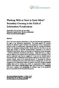
Fig. 1. Values are means 6 SE for leptin mRNA levels in retroperitoneal and epididymal fat of acutely exercised and control animals. Relative levels of leptin mRNA were standardized against 28S rRNA and are expressed in arbitrary units. Acute exercise reduced leptin mRNA in retroperitoneal (P , 0.05) but not epididymal fat. RESULTS
Experiment 1 The average run time to exhaustion was 85.5 6 1.5 min, and acutely exercised animals had low soleus muscle and liver glycogen levels (Table 1). Compared with control animals, leptin mRNA levels in retroperitoneal fat were significantly lower (Fig. 1; P , 0.05) in the exercise group. On the other hand, leptin mRNA in epididymal fat was similar between exercise and control groups. For control animals, leptin mRNA was higher in retroperitoneal than in epididymal fat (Fig. 1; P , 0.01). Retroperitoneal and epididymal fat pad weights for the exercise and control groups were comparable. For retroperitoneal fat, the exercise group fat pad weight averaged 1.24 6 0.08 g, whereas that for the control group averaged 1.41 6 0.08 g. For epididymal fat, the exercise group fat pad weight averaged 2.88 6 0.14 g, whereas that for control averaged 3.19 6 0.13 g. Total body weight, measured before treadmill running, was also similar in the exercise and control groups, averaging 384 6 6 and 381 6 5 g, respectively. Serum glucose and insulin were lower (P , 0.01) in the acutely exercised animals. Serum corticosterone was significantly elevated (P , 0.05) in the exercise group. Serum leptin levels tended to be higher in the Table 2. Serum glucose and hormone levels in experiment 1
Glucose, mg/dl Insulin, ng/ml Corticosterone, ng/ml Leptin, ng/ml
Exercise
Control
127.1 6 8.8* 0.56 6 0.12† 457.3 6 118.6* 3.01 6 0.33
157.52 6 5.2 1.51 6 0.21 130.6 6 47.0 2.67 6 0.41
Values are means 6 SE. * Exercise vs. control group, P , 0.05; † exercise vs. control, P , 0.001.
Fig. 2. Values are means 6 SE for leptin mRNA levels in retroperitoneal (A) and epididymal (B) fat of acutely exercised and control animals receiving either vehicle or vehicle 1 b3-receptor antagonist (SR-59230A). Relative levels of leptin mRNA were standardized against 28S rRNA and are expressed in arbitrary units. Pairwise comparison of least squares means revealed that leptin mRNA levels for exercise were lower than for control (* P , 0.05), but leptin mRNA levels for exercise and control groups receiving SR-59230A (b3antagonist) were not statistically different (P 5 0.42). There was a main effect of treatment (b3-antagonist; P , 0.05) to elevate leptin expression in epididymal fat.
1681
EXERCISE AND LEPTIN
Table 3. Effect of exercise and b3-blockade on serum corticosterone, FFA, glucose, insulin, and leptin in experiment 2
Corticosterone, ng/ml FFA, mmol/l Glucose, mg/dl Insulin, ng/ml Leptin, ng/ml
Control
Control 1 b3-Antagonist
Exercise
Exercise 1 b3-Antagonist
82.9 6 15.3 0.456 6 0.040 140 6 4 1.31 6 0.14 2.86 6 0.28
70.7 6 27.1 0.568 6 0.046‡ 137 6 3 1.58 6 0.15 3.33 6 0.42
380.4 6 47.0† 0.517 6 0.064 141 6 6 1.08 6 0.18* 4.55 6 0.74*
476.4 6 82.4† 0.728 6 0.117‡ 121 6 8 0.77 6 0.14* 4.80 6 1.10*
Values are means 6 SE. FFA, free fatty acids. * Exercise main effect, P , 0.05; † exercise main effect, P , 0.0001; ‡ treatment main effect (b3-antagonist), P , 0.05.
effect, P , 0.05). Pairwise comparison of least squares means revealed that leptin mRNA levels for the exercise group were lower than for control (P , 0.05), but that leptin mRNA levels for control 1 b3-antagonist and exercise 1 b3-antagonist were not statistically different (P 5 0.42). In epididymal fat, there were no significant differences in leptin mRNA levels between exercise and control groups (Fig. 2B); however, animals receiving SR-59230A exhibited higher leptin mRNA (treatment main effect, P , 0.05). Acute exercise was not associated with an increase in serum FFA, but FFA levels were higher in animals receiving SR-59230A (treatment main effect, P , 0.05). Serum leptin levels were higher, and insulin levels were lower, in the exercise groups (exercise and exercise 1 b3-antagonist; exercise main effect for both, P , 0.05). Pairwise comparison of least squares means for insulin indicated that exercise 1 b3-antagonist animals had lower levels than control 1 b3-antagonist animals (P , 0.01), but insulin levels for control and exercise alone were not statistically different (P 5 0.27). Serum glucose was the same across groups, whereas corticosterone levels were elevated in the exercise groups (exercise main effect, P , 0.001). Serum values for FFA, leptin, insulin, glucose, and corticosterone can be found in Table 3. Data for body composition are listed in Table 4. There were no differences in percent body fat, water, and ash among the four groups of animals. Similarly, retroperitoneal and epididymal fat pad weights were the same across the control, exercise, control 1 b3-antagonist, and exercise 1 b3-antagonist groups. DISCUSSION
Our laboratory has previously shown that voluntary wheel running in rats decreases both adipose tissue mRNA and circulating levels of leptin (35). The decrease in leptin was related to reductions in fat mass and cell size associated with exercise training. This observation was in agreement with the findings of
Friedman et al. (8), who showed that 8–12 wk of treadmill exercise training reduced fat mass and leptin mRNA in lean and obese SHHF/Mcc-facp rats. What was unclear from these studies was whether exercise, independent of changes in fat mass and cell size, could reduce white adipose tissue leptin mRNA. Catecholamines, b-agonists, and agents that increase cellular levels of cAMP all acutely reduce leptin mRNA (32, 34). Because exercise significantly increases the activity of the sympathoadrenal system, we hypothesized that acute exercise would result in reduced white adipose tissue leptin mRNA immediately postexercise. Findings from experiment 1 demonstrate that a single bout of treadmill exercise in male Sprague-Dawley rats is sufficient to reduce leptin mRNA in retroperitoneal but not epididymal fat. Reduced leptin mRNA in retroperitoneal fat after acute exercise was independent of a decline in fat mass. Noradrenaline given to mice by subcutaneous injection reduces leptin expression in white adipose tissue by .50% (34), whereas specific b3-adrenergic agonists also suppress leptin mRNA levels (3, 19). During exercise, catecholamine activation of adrenoreceptors on adipose tissue leads to lipid mobilization. Activation of lipolysis in this manner could initiate a signaling cascade that suppresses leptin mRNA. The purpose of the second experiment was to test the hypothesis that b3-adrenergic-receptor stimulation during acute exercise is responsible for the downregulation of leptin mRNA. First, we confirmed our initial observation that acute exercise reduces leptin mRNA in retroperitoneal, but not epididymal, white adipose tissue. However, leptin mRNA levels in retroperitoneal fat after administration of the b3-antagonist were similar in exercised and control animals, suggesting that b3-receptor activation is involved in the downregulation of leptin mRNA. A complete block of the exercise effect was not observed, as leptin mRNA levels for exercise 1 b3-antagonist were not statistically different from those of exercise alone (P 5 0.20). Reduction in leptin mRNA during
Table 4. Body composition of the 4 treatment groups in experiment 2
Control Control 1 b3-antagonist Exercise Exercise 1 b3-antagonist
Fat, %
Retro Weight, g
Epi Weight, g
Water, %
Ash, %
6.9 6 0.4 6.2 6 0.5 6.0 6 0.2 6.9 6 0.6
1.88 6 0.09 1.99 6 0.13 1.75 6 0.28 1.68 6 0.15
4.28 6 0.13 3.78 6 0.09 4.09 6 0.28 3.84 6 0.29
66.6 6 0.3 66.5 6 1.4 66.8 6 0.3 66.6 6 0.8
3.4 6 0.2 3.7 6 0.3 3.6 6 0.3 3.6 6 0.2
Values are mean 6 SE. Retro, retroperitoneal fat pad; Epi, epididymal fat pad.
1682
EXERCISE AND LEPTIN
acute exercise likely involves a combination of mechanisms. Previous studies have shown that insulin levels are related to leptin expression (24, 31, 37) and that rats subjected to streptozotocin-induced diabetes experience a complete suppression of leptin mRNA (21). Exercise and b3-blocked animals tended to have the lowest insulin level, and this likely explains the partial reduction in leptin mRNA, even under conditions of b3-receptor antagonism. We have shown that activation of b3-receptors during exercise is involved in the downregulation of leptin mRNA. Accordingly, blood flow, which is important for catecholamine (b-receptor agonist) delivery, may mediate this response. It has been shown that, at rest, rat retroperitoneal fat receives twice the blood flow as epididymal fat (6). If this regional difference in blood flow were to be maintained during exercise, greater potential for catecholamine delivery to retroperitoneal fat would exist. This could explain why leptin mRNA was reduced in retroperitoneal, but not epididymal, fat after acute exercise. To our knowledge, no study has directly compared metabolic and lipolytic capabilities of rat retroperitoneal and epididymal fat, but regional differences in b-adrenoreceptor type, density, and function (1) and hormone-sensitive lipase mass and function (18, 33) could exist. Thus blood flow and metabolic potential, as well as direct sympathetic activation of fat pads, may predict how leptin mRNA levels are influenced by acute and/or chronic exercise. Reduction in white adipose tissue leptin mRNA after acute exercise was also found in a study by Zheng et al. (38). They reported a 30% reduction in epididymal and periuterine fat after 60–120 min of treadmill running in male and female rats, respectively. Under our exercise conditions, we found leptin mRNA to be reduced in retroperitoneal but not epididymal fat. Zheng et al. did not report on leptin mRNA in retroperitoneal fat, and 6 of their 10 acutely exercised animals were female, each running at 30 m/min up an 8% grade for a total of 120 min. Thus gender and the ability to perform longduration high-intensity exercise may be factors that contribute to the leptin mRNA response during acute exercise. In humans, acute exercise appears to have no effect on serum leptin levels (14, 27) or adipose tissue production of leptin (28). In experiment 2, we found that acute exercise, independent of b3-receptor antagonism, was associated with high serum leptin. Kirchgessner et al. (17) recently reported that tumor necrosis factor (TNF)a-treated mice exhibited high, whereas TNF-a-deficient mice had low, serum leptin levels. Furthermore, they reported that TNF-a treatment of 3T3-L1 adipocytes results in rapid accumulation of leptin in cell culture media. Their work suggests the existence of regulatable, preformed pools of leptin within adipocytes and that TNF-a can stimulate leptin release. Our data are consistent with the idea that, in rats, exercise also stimulates leptin release from a storage site within adipocytes but that this is dependent on the duration of exercise. When exercise time was fixed at 55 min, serum leptin was increased. When rats exercised for
.80 min, no significant increase in serum leptin was observed, suggesting that the stored pool of leptin had been depleted. Precise identification of an exercise time course for serum leptin changes in the rat is beyond the scope of the present study. Additional experiments will be required to confirm that moderate-intensity treadmill running for ,1 h is sufficient to increase serum leptin levels in rats. Interestingly, the increase in serum leptin corresponds to previous experiments that have found exercise to induce an anorectic effect in rats (30); therefore, leptin may be acting as a satiety factor immediately postexercise. It is interesting that the b3-receptor antagonist increased FFA and that there was a trend for the exercise 1 b3-antagonist animals to have the highest levels. Blockade of the b3-receptor may have increased the responsiveness of the b1- and b2-receptors for adrenoreceptor-mediated lipolysis, resulting in the higher FFA levels. Normally, elevated FFA during exercise tends to spare muscle glycogen use (29), but there was little evidence of this in exercise 1 b3-antagonist animals. Thus an alternative explanation may be that SR-59230A blocks fatty acid uptake or oxidation in vivo, thereby giving rise to higher serum FFA. Unfortunately, the present experiments were not designed to fully characterize b3-receptor-antagonism effects on serum FFA turnover and/or oxidation. In summary, we have shown that acute exercise in rats reduces leptin mRNA in retroperitoneal but not epididymal fat. A partial block of the reduction in leptin mRNA after acute exercise was accomplished through the administration of a b3-receptor antagonist (SR59230A). This provides direct in vivo evidence that the b3-receptor in adipose tissue is involved in the regulation of leptin expression. Because b3-receptor antagonism did not completely block exercise-induced downregulation of leptin mRNA, other factors such as low serum insulin contribute to this response. This study was supported, in part, by Department of Defense contract no. DAAH04–93-G-0298. Address for reprint requests and other correspondence: J. J. Zachwieja, Pennington Biomedical Research Center, Louisiana State Univ., 6400 Perkins Road, Baton Rouge, LA 70808 (E-mail: zachwijj@ mhs.pbrc.edu). Received 8 February 1999; accepted in final form 19 July 1999. REFERENCES 1. Arner, P., L. Hellstrom, H. Wahrenberg, and M. Bronnegard. Beta-adrenoreceptor expression in human fat cells from different regions. J. Clin. Invest. 86: 1595–1600, 1990. 2. Campfield, A. L., F. J. Smith, Y. Guisez, R. Devos, and P. Burn. Recombinant mouse OB protein: evidence for a peripheral signal linking adiposity and central networks. Science 269: 546–549, 1995. 3. Collins, S., and R. S. Surwit. Pharmacologic manipulation of ob expression in a dietary model of obesity. J. Biol. Chem. 271: 9437–9440, 1996. 4. Considine, R. V., E. L. Considine, C. J. Williams, M. R. Nyce, S. A. Magosin, T. L. Bauer, E. L. Rosato, J. Colberg, and J. F. Caro. Evidence against either a premature stop codon or the absence of obese gene mRNA in human obesity. J. Clin. Invest. 95: 2986–2988, 1995. 5. Considine, R. V., S. K. Madhur, M. L. Heiman, A. Kriauciunas, T. W. Stephens, M. R. Nyce, J. P. Ohannesian, C. C. Marco, L. J. McKee, T. L. Bauer, and J. F. Caro. Serum
EXERCISE AND LEPTIN
6.
7.
8.
9.
10.
11. 12.
13. 14.
15.
16. 17.
18. 19. 20.
21.
immunoreative-leptin concentrations in normal-weight and obese humans. N. Engl. J. Med. 334: 292–295, 1996. Crandall, D. L., B. M. Goldstein, F. Huggins, and P. Cervoni. Adipocyte blood flow: influence of age, anatomic location, and dietary manipulation. Am. J. Physiol. 247 (Regulatory Integrative Comp. Physiol. 16): R46–R51, 1984. Frederich, R. C., A. Hamann, S. Anderson, B. Lollmann, B. B. Lowell, and J. S. Flier. Leptin levels reflect body lipid content in mice: evidence for diet-induced resistance to leptin action. Nature Med. 1: 1311–1314, 1995. Friedman, J. E., C. M. Ferrara, K. S. Aulak, M. Hatzoglou, S. A McCune, S. Park, and W. M. Sherman. Exercise training down-regulates ob gene expression in the genetically obese SHHF/Mcc-facp rat. Horm. Metab. Res. 29: 214–219, 1997. Galitzky, J., D. Langin, P. Verwaerde, J.-L. Montastruc, M. Lafontan, and M. Berlan. Lipolytic effects of conventional exercise b3-adrenoceptor agonists and of CGP 12,177 in rat and human fat cells: preliminary pharmacological evidence for a putative b4-adrenoceptor. Br. J. Pharmacol. 122: 1244–1250, 1997. Gettys, T. W., P. J. Harkness, and P. M. Watson. The b3-adrenergic receptor inhibits insulin-stimulated leptin secretion from isolated rat adipocytes. Endocrinology 137: 4054–4057, 1996. Harris, R. B. S. The impact of high- or low-fat cafeteria foods on nutrient intake and growth of rats consuming a diet containing 30% energy as fat. Int. J. Obes. 17: 307–315, 1993. Harris, R. B. S., and R. J. Martin. Metabolic response to a specific lipid-depleting factor in parabiotic rats. Am. J. Physiol. 250 (Regulatory Integrative Comp. Physiol. 19): R276–R286, 1986. Harris, R. B. S., T. G. Ramsay, S. R. Smith, and R. C. Bruch. Early and late stimulation of ob mRNA expression in meal-fed and overfed rats. J. Clin. Invest. 97: 2020–2026, 1996. Hickey, M. S., J. A. Houmard, R. V. Considine, G. L. Tyndall, J. B. Midgette, K. E. Gavigan, M. L. Weidner, M. R. McCammon, R. G. Israel, and J. F. Caro. Gender-dependent effects of exercise training on serum leptin levels in humans. Am. J. Physiol. 272 (Endocrinol. Metab. 35): E562–E566, 1997. Igel, M., H. Kainulainen, A. Brauers, W. Becker, L. Herberg, and H. G. Joost. Long-term and rapid regulation of ob mRNA levels in adipose tissue from normal (Sprague-Dawley rats) and obese (db/db mice, fa/fa rats) rodents. Diabetologia 39: 758– 765, 1996. Ji, L. L., D. L. F. Lennon, R. G. Kochan, F. J. Nagle, and H. A. Lardy. Enzymatic adaptation to physical training under b-blockade in the rat. J. Clin. Invest. 78: 771–778, 1986. Kirchgessner, T. G., K. T. Uysal, S. M. Wiesbrock, M. W. Marin, and G. S. Hotamisligil. Tumor necrosis factor-a contributes to obesity-related hyperleptinemia by regulating leptin release from adipocytes. J. Clin. Invest. 100: 2777–2782, 1997. Lafontan, M., M. Berlan, J. Galitzky, and J. L. Montastruc. Alpha-2 adrenoceptors in lipolysis: a2 antagonists and lipidmobilizing strategies. Am. J. Clin. Nutr. 55: 219S–227S, 1992. Li, H., M. Matheny, and P. J. Scarpace. b3-Adrenergicmediated suppression of leptin gene expression. Am. J. Physiol. 272 (Endocrinol. Metab. 35): E1031–E1036, 1997. Lust, W. D., J. V. Passonneau, and S. K. Crites. The measurement of glycogen in tissues by amylo-a-1,4-a-1,6-glucosidase after the destruction of preexisting glucose. Anal. Biochem. 68: 328–331, 1975. MacDougald, O. A., C. S. Hwang, H. Fan, and M. D. Lane. Regulated expression of the obese gene product (leptin) in white adipose tissue and 3T3-L1 adipocytes. Proc. Natl. Acad. Sci. USA 92: 9034–9037, 1995.
1683
22. Manara, L., D. Badone, M. Barone, G. Boccardi, R. Cecche, T. Croci, A. Giudice, U. Guzzi, M. Landi, and G. Le Fur. Functional identification of rat atypical b-adrenoreceptors by the first b3-selective antogonists, aryloxypropanolaminotetralins. Br. J. Pharmacol. 117: 435–442, 1996. 23. Mantzoros, C. S., D. Qu, R. C. Frederich, V. S. Susulic, B. B. Lowell, E. Maratos-Flier, and J. S. Flier. Activation of b3 adrenergic receptors suppresses leptin expression and mediates a leptin-independent inhibition of food intake in mice. Diabetes 45: 909–914, 1996. 24. Mizuno, T. M., H. Bergen. T. Funabashi, S. P. Kleopoulos, Y. G. Zhong, W. A. Bauman, and C. V. Mobbs. Obese gene expression: reduction by fasting and stimulation by insulin and glucose in lean mice, and persistent elevation in acquired (dietinduced) and genetic (yellow agouti) obesity. Proc. Natl. Acad. Sci. USA 93: 3434–3438, 1996. 25. Passonneau, J. V., P. D. Gatfield, D. W. Schulz, and O. H. Lowry. An enzymatic method for measurement of glycogen. Anal. Biochem. 19: 315–326, 1967. 26. Pelleymounter, M. A., M. J. Cullen, M. B. Baker, R. Hecht, D. Winters, T. Boodne, and F. Collins. Effects of the obese gene product on body weight regulation in ob/ob mice. Science 269: 540–543, 1995. 27. Perusse, L., G. Collier, J. Gagnon, A. S. Leon, D. C. Rao, J. S. Skinner, J. H. Wilmore, A. Nadeau, P. Z. Zimmet, and C. Bouchard. Acute and chronic effects of exercise on leptin levels in humans. J. Appl. Physiol. 83: 5–10, 1997. 28. Racette, S. B., S. W. Coppack, M. Landt, and S. Klein. Leptin production during moderate-intensity aerobic exercise. J. Clin. Endocrinol. Metab. 82: 2275–2277, 1997. 29. Rennie, M. J., W. M. Winder, and J. O. Holloszy. A sparing effect of increased free fatty acids on muscle glycogen content in the exercising rat. Biochem. J. 156: 647–655, 1976. 30. Rivest, S., and D. Richard. Involvement of corticotropinreleasing factor in the anorexia induced by exercise. Brain Res. Bull. 25: 169–172, 1990. 31. Saladin, R., P. De Vos, M. Guerre-Millo, A. Leturque, J. Girard, B. Stails, and J. Auwerx. Transient increase in obese gene expression after food intake or insulin administration. Nature 377: 527–529, 1995. 32. Slieker, L. F., K. W. Sloop, P. L. Surface, A. Kriauciunas, F. LaQuier, J. Manetta, J. Bue-Valleshey, and T. W. Stephens. Regulation of expression of ob mRNA and protein by glucocorticoids and cAMP. J. Biol. Chem. 271: 5301–5304, 1996. 33. Sztalryd, C., and F. B. Kraemer. Differences in hormonesensitive lipase expression in white adipose tissue from various anatomic locations of the rat. Metabolism 43: 241–247, 1994. 34. Trayhurn, P., J. S. Duncan, and D. V. Rayner. Acute coldinduced suppression of ob (obese) gene expression in white adipose tissue of mice: mediation by the sympathetic system. Biochem. J. 311: 729–733, 1995. 35. Zachwieja, J. J., S. L. Hendry, S. R. Smith, and R. B. S. Harris. Voluntary wheel running decreases adipose tissue mass and expression of leptin mRNA in Osborne-Mendel rats. Diabetes 46: 1159–1166, 1997. 36. Zhang, Y., R. Proenca, M. Maffer, M. Barone, L. Leopold, and J. M. Friedman. Positional cloning of the mouse obese gene and its human homologue. Nature 272: 425–432, 1994. 37. Zheng, D., J. P. Jones, S. J. Usala, and G. L. Dohm. Differential expression of ob mRNA in rat adipose tissues in response to insulin. Biochem. Biophys. Res. Commun. 218: 434–437, 1996. 38. Zheng, D., M. H. Wooter, Q. Zhou, and G. L. Dohm. The effect of exercise on ob gene expression. Biochem. Biophys. Res. Commun. 225: 747–750, 1996.