Prpe, G., Reboul, J. P., Brun, P. & Zylber, J. (1995). Acta Cryst. C51,. 729-732. Sheldrick, G. M. (1976). SHELX76. Program for Crystal Structure. Determination.
G. PEPE, J. ZYLBER, A. TUBUL AND E BRUN Marson, C. M., Grabowska, U., Walsgrove, T., Eggleston D. F. & Baurs, P. W. (1994). J. Org. Chem. 59, 284-290. Prpe, G., Reboul, J. P., Brun, P. & Zylber, J. (1995). Acta Cryst. C51, 729-732. Sheldrick, G. M. (1976). SHELX76. Program for Crystal Structure Determination. Univ. of Cambridge, England. Toja, E., Gorini, C., Zirotti, C., Barzaghi, F. & Galliani G. (1987). Eur. Patent 229 566. Woo, E. P. & Mullins, M. J. (1991). US Patent 4 943 640; Chem. Abstr. (1991), 114, 23798c.
1917
k
o
oO> o
I
Acta Cryst. (1995). C51, 1917-1919
6-[1-(4-Ethoxyphenyl)ethyl]-5-methoxy- 1,3benzodioxole
(I)
The structure determination reveals that the fused dioxole and phenyl rings and the unfused phenyl ring are both planar and almost perpendicular to each other. The relative orientation between these two ring systems is likely to be an important factor that enables the derivative to bind tubulin. The only other report of a crystal structure of a 6-benzyl-l,3-benzodioxole derivative ap-
PAUL ALA AND DANIEL S. C. YANG
~
Department of Biochemistry, McMaster University, Hamilton, Ontario, Canada L8N 3Z5 (Received 14 December 1993; accepted 13 March 1995)
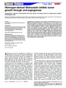
C(18)
~) O(4)
C(13)f,~
Abstract The title compound, C18H2004, is structurally similar to podophyllotoxin. It contains a fused dioxole and phenyl ring system and an unfused phenyl ring which are almost perpendicular to each other. However, this compound lacks the cyclohexyl and lactone rings which are present in podophyllotoxin. In addition, the unfused phenyl ring in podophyllotoxin contains three methoxy groups in the para and meta positions, whereas the title compound contains only an ethoxy group in the para position.
C(14)
~ C(15) C(16) / C( 1 0 ) ~ ~
C(6) C ( 7 ) ~ C(2) C(9)'~"~~'-'~
0(2)
C(I)
C(4)
Comment The title compound, (I), belongs to a series of 6benzyl-l,3-benzodioxole derivatives that are structurally similar to podophyllotoxin and have podophyllotoxinlike antimitotic activity (Batra, Jurd & Hamel, 1985). These derivatives have been used to study structurefunction relationships of podophyllotoxin. For example, tubulin polymerization is inhibited if the 6-benzyl-l,3benzodioxole derivative contains an intact dioxole ring, a methoxy group at the para position of the unfused phenyl ring and a methoxy or ethoxy group at the 5position of the fused rings (Batra et al., 1985). The title compound contains all these key structural features except that the methoxy group at the para position of the unfused phenyl ring is replaced by an ethoxy group, which reduces its potency as an inhibitor of tubulin polymerization by only twofold (Batra et al., 1985). © 1995 International Union of Crystallography Printed in Great Britain - all rights reserved
C
(
8
)
~
Fig. 1. ORTEPII (Johnson, 1976) plot of the molecular structure of the benzyl-benzodioxole derivative (ellipsoids represent 50% probability).
-~
v
• \
I
Fig. 2. PLUTO (Motherwell & Clegg, 1978) stereoplot of the packing of the benzyl-benzodioxole derivative. Acta Crystallographica Section C ISSN 0108-2701 01995
1918
C18H2004
p e a r e d r e c e n t l y ( S i c h e r i , D e r r y , G u p t a & Y a n g , 1992). A structural analysis of other benzyl-benzodioxole derivat i v e s m a y h e l p us to b e t t e r u n d e r s t a n d t h e i m p o r t a n c e o f the relative orientations between the two ring systems and identify additional features that make podophyll o t o x i n a p o t e n t m i t o t i c inhibitor.
Experimental The title c o m p o u n d was obtained in crystalline form from the National Cancer Institute (NSC 353648). This c o m p o u n d has one chiral centre but crystallized in an achiral space group. Therefore, both enantiomers crystallized together.
0(4) c(1) c(2) c(3) c(4) c(5) c(6) c(7) c(8) c(9) c(10) c(i I) c(12) c(13) c(14) c(15) c(16) c(17) c(18)
0.3273 (1) 0.1536 (2) 0.1394 (2) 0.1062 (2) 0.0736 (2) 0.0748 (2) 0.1414 (2) 0.1080 (2) 0.0110 (2) 0.1066 (2) 0.0488 (2) 0.1634 (2) 0.1674 (2) 0.2210 (2) 0.2716 (2) 0.2687 (2) 0.2154 (2) 0.3325 (2) 0.3970 (2)
0.2573 (3) 0.8982 (6) 0.6703 (5) 0.6454 (5) 0.4981 (6) 0.3724 (5) 0.5468 (5) 0.3943 (5) 0.1844 (6) 0.2547 (5) 0.2742 (6) 0.2555 (5) 0.3262 (6) 0.3295 (5) 0.2596 (5) 0.1851 (5) 0.1824 (5) 0.3268 (6) 0.3162 (7)
-0.2084 (1) 0.0858 (3) 0.0108 (2) 0.0654 (2) 0.0706 (2) 0.0156 (2) -0.0423 (2) -0.0407 (2) 0.0718 (2) -0.0998 (2) -o.1563 (2) -0.1335 (2) -0.2012 (2) -0.2288 (2) -0.1880 (2) -0.1198 (2) -0.0940 (2) -0.2782 (2) -0.2870 (3)
4.2 (1) 5.7 (2) 3.9 (2) 3.8 (2) 3.8 (2) 3.4 (2) 3.9 (2) 3.4 (2) 5.5 (2) 3.6 (2) 5.0 (2) 3.4 (2) 4.1 (2) 3.9 (2) 3.2 (2) 3.6 (2) 3.5 (2) 4.8 (2) 6.1 (2)
crystal data C18H2004
Cu Ko~ radiation A = 1.54178 ,~ Cell parameters from 25 reflections 0 = 25.05-32.7 ° # = 0.683 m m T = 296 K Rectangular 0.14 × 0.06 × 0.04 m m Transparent
Mr = 300.35 Monoclinic
C2/c a = 22.662 ( 5 ) / ~ b = 7.671 (1)/~, c = 18.443 ( 1 ) / ~ /3 = 99.60 (1) ° V = 3161.2 A 3 Z=8 Dx = 1.262 M g m -3
Data collection Rigaku AFC-6R diffractometer w-20 scans Absorption correction: scan (North, Phillips & Mathews, 1968) Tmin - 0.87, Tmax = 1.00 2644 m e a s u r e d reflections 2567 independent reflections 1416 observed reflections [1 > 3~r(/)]
Rint = 0.043 0max = 60.05 ° h = 0 ---~ 25 k = 0---~ 8 1 = - 2 0 ~ 20 3 standard reflections monitored every 150 reflections intensity decay: none
Refinement Refinement on F R = 0.050 wR = 0.061 S = 1.41 1416 reflections 200 parameters H-atom parameters not refined w = 1/tr2(F) (A/cr)ma~ = 0.03
T a b l e 1.
/~pmax = 0.14 e ,~,-3 mpmin = - 0 . 1 3 e ,~-3 Extinction correction: MITHRIL (Gilmore, 1984) Extinction coefficient: 0.14012 x 10 -5 Atomic scattering factors from International Tables
for X-ray Crystallography (1974, Vol. IV)
Fractional atomic coordinates and equivalent isotropic displacement parameters (,~2) Beq = (87r2/3)~i~jUijaTaf ai.aj.
O(1) 0(2) 0(3)
x 0.1101 (I) 0.1668 (2) 0.0442 (1)
y 0.7891 (4) 0.8318 (4) 0.2172 (3)
z
0.1109(2) 0.0189 (2) 0.0148 (2)
Beq 5.6(2) 6.3 (2) 4.4 (1)
T a b l e 2.
Selected geometric parameters (/~,, o)
o(1)--c(i) o(1)---c(3) o(2)---c(i) o(2)-----c(2) o(3)----c(5) o(3)--c(8) o(4)--c(14) o(4)----c(17) c(2)---c(3) c(2)--c(6) c(3)--c(4) c(4)----c(5) c(1)----o(1)----c(3) c(1)----o(2)--c(2) c(5)----o(3)----c(8) c(14)----o(4)----c(17) o(1)----c(1)--o(2) o(2)----c(2)---c(3) o(2)----c(2)----c(6) c(3)---c(2)--c(6) o(1)----c(3)---c(2) o(1)--c(3)----c(4) c(2)---c(3)----c(4) c(3)----c(4)--c(5) o(3)----c(5)----c(4) o(3)--c(5)----c(7) c(4)----c(5)--c(7) c(2)----c(6)--c(7) c(5)---c(7)--c(6)
1.428 (5) 1.379 (4) 1.412 (5) 1.382 (5) 1.376 (4) 1.414 (5) 1.376 (4) 1.417 (5) 1.368 (5) 1.368 (5) 1.362 (5) 1.4o2 (5) 104.5 (3) 105.1 (3) 118.3 (3) 117.6 (3) 109.3 (3) 109.8 (4) 129.1 (4) 121.1 (4) 110.4 (4) 127.0 (4) 122.7 (4) 116.4(3) 122.2 (3) 115.5 (3) 122.3 (4) 119.0 (3) i 18.6 (4)
c(5)---c(7) c(6)----c(7) c(7)----c(9) c(9)---c(10) c(9)----c(11) c(11)--c(12) c(11)--c(16) c(12)----c(13) c(13)----c(14) c(14)----c(15) c(15)---c(16) c(17)----c(18) c(5)---c(7)----c(9) c(6)----c(7)----c(9) c(7)---c(9)---c(10) c(7)----c(9)----c(11) c(10)----c(9)---c(11) c(9)---c(11)----c(12) c(9)---c(11)---c(16) c(12)----c(11)----c(16) c(11)---c(12)----c(13) c(12)----c(13)----c(14) o(4)--c(14)---c(13) o(4)----c( 14)----c(15) c(13)----c(14)---c(15) c(14)----c(15)--c(16) C(l 1)----c(16)----c(15) o(4)----c(17)---c(18)
1.392 (5) 1.396 (5) 1.525 (5) 1.540 (5) 1.519 (5) 1.377 (5) 1.395 (5) 1.394 (5) 1.371 (5) 1.393 (5) 1.371 (5) 1.499 (6) 120.0 (4) 121.4 (3) 109.2 (3) 111.8 (3) 114.0 (3) 124.1 (4) 118.9 (3) 117.0 (3) 122.1 (4) 119.5(3) 125.1 (3) 115.1 (3) 119.8 (3) 119.7 (3) 121.9(3) 108.1 (4)
M e a s u r e m e n t s were m a d e on a AFC-6R diffractometer with graphite-monochromated radiation and a 12 k W rotating anode generator. The data were corrected for Lorentz and polarization effects. The structure was solved by direct methods using MITHRIL (Gilmore, 1984). H atoms were generated using o p t i m u m bonding g e o m e t r y and included in Ft. H-atom displacement parameters and positions were not refined. NonH atoms were refined anisotropically. Anomalous-dispersion effects were included in Fc (Ibers & Hamilton, 1964). All calculations were performed using TEXSAN (Molecular Structure Corporation, 1985). This research was supported by the National Cancer Institute of Canada (DSCY). Lists of structure factors, anisotropic displacement parameters, Hatom coordinates and torsion angles have been deposited with the IUCr (Reference: BK1028). Copies may be obtained through The Managing Editor, International Union of Crystallography, 5 Abbey Square, Chester CHI 2HU, England.
PAUL ALA AND DANIEL S. C. YANG References Batra, J. K., Jurd, L. & Hamel, E. (1985). Mol. Pharmacol. 27, 94102. Gilmore, C. J. (1984). J. Appl. Cryst. 17, 42-46. Ibers, J. A. & Hamilton, W. C. (1964). Acta Cryst. 17, 781-782. Johnson, C. K. (1976). ORTEPII. Report ORNL-5138. Oak Ridge National Laboratory, Tennessee, USA. Molecular Structure Corporation (1985). TEXSAN. TEXRAY Structure Analysis Package. MSC, 3200 Research Forest Drive, The Woodlands, TX 77381, USA. Motherwell, W. D. S. & Clegg, W. (1978). PLUTO. Program for Plotting Molecular and Crystal Structures. Univ. of Cambridge, England. North, A. C. T., Phillips, D. C. & Mathews, F. S. (1968). Acta Cryst. A24, 351-359. Sicheri, F. V., Derry, W. B., Gupta, R. S. & Yang, D. S. C. (1992). Acta Cryst. C48, 1687-1689.
Acta Cryst. (1995). C51, 1919-1921
2,6,6-Trimethyl-2-oxo-l,3-dioxa-6-azonia-2phosphocyclooctane Iodide G. KUMARAVELAND RICHARDD. GANDOUR*
.Department of Chemistry, Virginia Polytechnic Institute and State University, Blacksburg, VA 24061-0212, USA FRANK R. FRONCZEK
Department of Chemistry, Louisiana State University, Baton Rouge, LA 70803-1804, USA (Received 2 November 1993; accepted 10 March 1995)
Abstract The eight-membered ring in the title compound, C7H17NOaP+.I - , has a boat-chair conformation, with the local mirror plane passing through the cyclic O atom and methylene C atom adjacent to the N atom. The ~ bond is pseudo-axial and the P---CH3 bond is pseudo-equatorial. The P - - N distance is 3.821 (2)A. Comment We are interested in the design and synthesis of conformationally restricted reaction-intermediate-analogue inhibitors of carnitine acyltransferases (Gandour et al., 1992). Our inhibitor design suggests that an eightmembered ring would be an excellent match for the putative tetrahedral intermediate in the reactions catalyzed by carnitine acyltransferases. We have produced the title compound, (I), in order to perform the conformational analysis of the eight-membered phosphorus-containing ring. ©1995 International Union of Crystallography Printed in Great Britain - all rights reserved
1919
Oe,N+S" Me/ ~ . . .
. O/ \ Me l-
(I) Eight-membered phosphorus heterocycles with heteroatoms located in positions 1, 2, 3 and 6 provide interesting models for studying transannular interactions. For example, 2-methyl-2-oxo-6-phenyl1,3 - dioxa- 6- aza- 2- phosphacynan (Kalinin, Andrianov & Struchkov, 1975) exists in the solid state in the chair-chair or crown conformation and a close contact between the P and N atoms [3.22 A] indicates a transannular P...N interaction. Another compound, 2-thio2,6-dimethyl- 1,3-dioxa-6-aza-2-phosphacynan (Dutasta, 1980), also exists in the solid state in the crown conformation (Dutasta, Robert & Wiesenfeld, 1980) with a transannular P . . . N distance shorter than the sum of the van der Waals radii (Piccinni-Leonardi et al., 1986). Unlike the neutral phosphorus compounds mentioned above, the title compound exists in the solid state in a boat-chair conformation, with the local mirror plane passing through the cyclic O atom and methylene C atom adjacent to the N atom. The M bond is pseudoaxial and the P---CHs bond is pseudo-equatorial. GoA S
_
~.R
0~0,
0-
/\
(II) In this conformation, the P . . . N distance [3.821 (2)/~] mimics the distance (d = 3.84 ~,) between the tetrahedral C atom and the quaternary N atom of the proposed (Gandour et al., 1992) intermediate, (II), which was calculated by PCMODEL (Gajewski & Gilbert, 1992), in the acyl transfer to carnitine. In addition, the N - -
Fig. 1. Plot (ORTEP; Johnson, 1965) of the heterocyclic cation. Displacement ellipsoids are drawn at the 40% probability level, with H atoms shown as spheres of arbitrary radii.
Acta Crystallographica Section C ISSN 0108-2701 0 1 9 9 5