Physiol. Res. 53: 403-408, 2004
Pinealectomy Increases and Exogenous Melatonin Decreases Leptin Production in Rat Anterior Pituitary Cells: an Immunohistochemical Study I. KUS1, M. SARSILMAZ1, N. COLAKOGLU2, A. KUKNER2, O. A. OZEN1, B. YILMAZ3, H. KELESTIMUR3 Departments of Anatomy1, Histology and Embryology2 and Physiology3, Medical School, Firat University, Elazig, Turkey Received May 16, 2003 Accepted October 15, 2003
Summary Melatonin, the main hormone of the pineal gland, informs the body about the environmental light and darkness regimen, which in turn contributes to the photoperiodic adaptation of several physiological functions. Leptin, the hormone secreted mainly by adipocytes and some other tissues including the pituitary, informs the brain about the mass of adipose tissue, which plays an important role in energy homeostasis. Melatonin has been shown to decrease circulating leptin levels. It is currently not known whether melatonin has an effect on leptin synthesis in the pituitary. The aim of this study was to immunohistochemically examine the effects of pinealectomy and administration of melatonin on leptin production in the rat anterior pituitary. The pituitary samples obtained from 18 male Wistar rats including sham-pinealectomized, pinealectomized and melatonin-injected pinealectomized groups were immunohistochemically evaluated. Immunostaining of leptin was moderate (3+) in sham-pinealectomized rats, heavy (5+) in pinealectomized rats and low (1+) in melatonin-treated pinealectomized rats, respectively. The present results indicate that pinealectomy induces leptin secretion in anterior pituitary cells, and this increase of leptin synthesis can be prevented by administration of melatonin. Thus, melatonin seems to have both physiological and pharmacological effects on leptin production in the anterior pituitary of male rats.
Key words Leptin • Melatonin • Pinealectomy • Anterior Pituitary • Rat
Introduction Melatonin, the main hormone of the pineal gland, informs the body about the environmental light and darkness regimen, which provides the photoperiodical adaptation of some physiological functions. Leptin, the hormone secreted by adipocytes,
informs the brain about the mass of adipose tissue, which plays an important role in energy homeostasis. Therefore, melatonin and leptin may be regarded as “darkness” and “fatness” hormones, respectively. Actions of melatonin include antioxidative (Reiter 2000) and neuroprotective effects (Kilic et al. 1999, Baydas et al. 2002), stimulation of immune system (Guerrero and Reiter 1992),
PHYSIOLOGICAL RESEARCH © 2004 Institute of Physiology, Academy of Sciences of the Czech Republic, Prague, Czech Republic E-mail:
[email protected]
ISSN 0862-8408 Fax +420 241 062 164 http://www.biomed.cas.cz/physiolres
404
Vol. 53
Kus et al.
modulation of smooth muscle contraction (Ayar et al. 2001). The pineal gland may also exert a modulating influence on the pituitary (Pallotti et al. 2002). It inhibits the release of luteinizing hormone (LH) from the neonatal rat anterior pituitary gland (Vaněček 1999) induced by the gonadotropin-releasing hormone (GnRH) and may have antigonadal effects in the adult rats (Kus et al. 2000, Yilmaz et al. 2000). Exogenous melatonin can affect the spontaneous release of LH and prolactin (PRL) in humans (Ninomiya et al. 2001). Melatonin stimulates the accumulation of thyroid stimulating hormone (TSH) in the rat pars tuberalis (PT) – TSH cells via secretory granule formation, and it has been suggested that melatonin regulates TSH release from PT-TSH cells (Sakamoto et al. 2000). Leptin is secreted mainly by adipocytes and has important effects on the regulation of food intake and energy expenditure. Leptin exerts its effects by interacting with leptin receptors in the brain and many other tissues. Leptin provides information about the state of fat stores to the brain, and the neuroendocrine systems adapt their function to the current state of energy homeostasis and fat stores (Casanueva and Dieguez 1999). Although leptin is produced mainly by the white adipose tissue, a growing number of tissues including the anterior pituitary gland have been shown to produce low amounts of leptin (Lloyd et al. 2001). It may act as the critical link between adipose tissue and the reproductive system, indicating whether adequate energy reserves are present for normal reproductive function (Moschos et al. 2002). Leptin stimulates pituitary cells to synthesize and secrete both LH and the follicle-stimulating hormone (FSH) bringing about the onset of puberty (Tezuka et al. 2002). Leptin has a direct enhancing effect on the pituitary secretion of growth hormone (GH) induced by growth hormone releasing hormone (GHRH) (Mizuno et al. 1999). In the rat anterior pituitary gland, there are paracrine relationships between leptin-producing cells and cells with the leptin receptor (leptin-R) that may regulate the function of GH cells (Sone et al. 2001). Leptin has an acute stimulatory effect on TSH release in vivo, acting probably in the hypothalamus. However, the direct pituitary effect of leptin is inhibitory and there is also evidence that leptin may act as an autocrine/ paracrine inhibitor of TSH release in the rat pituitary (Ortiga-Carvalho et al. 2002). Melatonin has recently been suggested to have a role in leptin release. Our previous studies (Canpolat et al. 2001, Baydas et al. 2001) and other studies (Rasmussen et al. 1999, Wolden-Hanson et al. 2000) have shown that melatonin suppresses plasma leptin
levels. It has been reported that leptin is produced in human (Jin et al. 1999), rat and murine anterior pituitary cells (Jin et al. 2000). Melatonin receptors are expressed within the pituitary gland (Hazlerigg 2001). We hypothesized that melatonin may have an effect on leptin synthesis in the adenohypophysis. The present study therefore examined the effects of pinealectomy and exogenous melatonin on leptin production in the rat anterior pituitary.
Methods Adult male Wistar rats (weighing 180-200 g, n=18) were used in this study. The animals were maintained under controlled temperature (21±1 oC) and light conditions (light 07:00-19:00 h). Food (standard pellet diet) and tap water were supplied ad libitum. The animals were divided into three groups. Group I (n=6) and group II (n=6) were designated as control (sham-pinealectomized, sham-PNX) and pinealectomized (PNX) rats, respectively. They received 10 % ethanol (0.1 ml s.c.) alone. Rats in group III (n=6) were pinealectomized and received a daily injection of melatonin (3 mg/kg dissolved in 0.1 ml 10 % ethanol s.c.; Sigma) for 2 months commencing on day 7 after surgery. The animals were killed by decapitation at the end of the experiments. The pituitary glands of all rats were removed and fixed in Bouin’s solution. The specimens were embedded in paraffin and serially sectioned (thickness, 5 µm). All the protocols in the present study were approved by the local Ethics Committee of the Medical School. Immunohistochemical procedures Avidin-biotin-peroxidase technique was used for determination of leptin protein expression in this study. Paraffin sections (5 µm) were dewaxed in xylene, treated with 0.1 % hydrogen peroxide in methanol for 10 min to block endogenous peroxidase, blocked with 10 % normal goat serum in PBS for 20 min and incubated overnight at 4 ºC with Leptin Ob (A-20) rabbit polyclonal IgG antibody (Santa Cruz, California). Sections were then incubated with biotinylated goat anti-rabbit IgG for 30 min, followed by avidine-peroxidase for 30 min and treated with 0.5 mg/ml diaminobenzidine with 0.1 % hydrogen peroxide until the brown reaction product was obtained. Finally sections were counterstained with hematoxylin, dehydrated in alcohol, cleared in xylol and mounted. Sections were viewed and photographed under a BH2 Olympus photomicroscope.
2004
Melatonin Affects Leptin Synthesis in the Anterior Pituitary
405
Immunohistochemical leptin staining of the cytoplasm of anterior pituitary cells was evaluated semiquantitatively by two independent histologists in a blind test. The more leptin antigen (leptin protein) present in the cell, the more binding will occur and as a result, darker staining will be seen. The intensity of immunostaining was scored as follows: no staining (0), minimal (1+), low (2+), moderate (3+), strong (4+), heavy (5+).
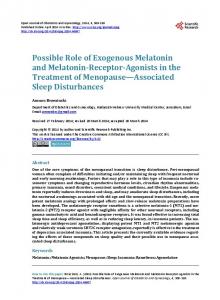
Results Fig. 1. Immunohistochemical staining of leptin in control anterior pituitary gland, showing moderate levels of leptin protein in the cytoplasm of anterior pituitary cells (arrow). Magnification x40.
According to the density of the observed immunohistochemical staining the content of leptin was moderate (3+) in sham-pinealectomized (control) rats (Fig. 1), heavy (5+) in pinealectomized rats (Fig. 2) and minimal (1+) in pinealectomized rats that were treated with melatonin (Fig. 3). Thus leptin production in anterior pituitary cells increased after pinealectomy, and these effects were reversed when melatonin was administered to PNX animals.
Discussion
Fig. 2. Immunohistochemical staining of leptin in anteror pituitary gland of a pinealectomized rat, showing strong leptin staining in the cytoplasm of anterior pituitary cells (arrow). Magnification x40.
Fig. 3. Immunohistochemical staining of leptin in anteror pituitary gland of a pinealectomized rat that was treated afterwards with melatonin, showing low levels of leptin protein in the cytoplasm of anterior pituitary cells (arrow). Magnification x40.
The results of the present study have confirmed leptin production in the rat anterior pituitary, as previously reported by Jin et al. (1999, 2000). Furthermore, our study clearly demonstrates that exogenous melatonin decreases and pinealectomy increases leptin production in the rat anterior pituitary. Increased leptin secretion following pinealectomy implicates that melatonin modulation of leptin in the anterior pituitary is also a physiological effect. This may be important in the control of various physiological functions. Firstly, there seems to be a functional antagonistic interaction between melatonin and leptin in the timing of puberty. Melatonin is suggested to delay puberty (Kennaway and Rowe 1997), whereas leptin has been reported to have a permissive role in puberty onset (Gueorguiev et al. 2001). The failure of the pineal gland to produce sufficient melatonin causes precocious puberty (Commentz and Helmke 1995), whereas insufficient leptin release may result in pubertal delay (Wauters et al. 2000). Thus, sexual maturation seems to be signaled by a decrease of melatonin levels and an increase in leptin levels. It has been suggested that there is an inverse interaction between plasma melatonin levels and sexual maturation (Reiter 1998). Thus, reduction in melatonin release may facilitate transition into puberty.
406
Kus et al.
Melatonin may exert its puberty-delaying effect by means of suppressing leptin production in the anterior pituitary. Lack of leptin-reducing effect of melatonin near puberty may be responsible for the puberty-accelerating effect of leptin. This concept needs confirmination because the cell types in which leptin production was affected were not separated in this study. In the rat anterior pituitary, LH and FSH secreting cells have been shown to express 4±1 % and 3±0.5 % of leptin, respectively (Jin et al. 2000). In the present study, melatonin and pinealectomy may have affected leptin production in these cells. Leptin is synthesized and stored in the human pituitary gland and is suggested to modulate secretion of other pituitary hormones, although its contribution to changes in plasma leptin levels is currently unknown. Pituitary leptin has therefore been suggested to be a novel paracrine regulator of pituitary function (Korbonits et al. 2001). Colocalization studies of leptin and anterior pituitary cells have shown that 70 % of ACTH cells are positive for leptin, 21 % of GH cells, 29 % of LH cells, 33 % of FSH cells, 32 % of TSH cells, 64 % folliculostellate cells, whereas very few PRL cells (3 %) were positive (Popovic et al. 2001). Leptin is also expressed in TSH cells of the rat anterior pituitary (Jin et al. 2000). Leptin has been shown to have many functions in the anterior pituitary. It directly influences GH regulation at the pituitary level (Baratta et al. 2002) and leptin also has a stimulatory effect on LH release in the
Vol. 53 pituitary in vivo and in vitro (De Biasi et al. 2001, Borowiec et al. 2002). All the anterior pituitary cell types express the leptin receptor. However, leptin has been localized in specific subtypes of anterior pituitary cells indicating cell type-specific production of leptin in the anterior pituitary (Lloyd et al. 2001). To the best of our knowledge, the effects of melatonin hormone on the leptin production in pituitary cells have not previously been reported. We have shown that melatonin suppresses not only plasma leptin levels (Canpolat et al. 2001, Baydas et al. 2001) but also leptin production in anterior pituitary cells. The mechanism by which melatonin reduces leptin production in the anterior pituitary remains to be determined. Melatonin receptors are mainly expressed in the pars tuberalis and pars distalis. It has been suggested that pars tuberalis mediates the seasonal effects of melatonin on prolactin secretion, whilst the pars distalis may be involved in photoperiodic programming of the developing gonadotropic axis (Hazlerigg 2001). Melatonin may exert its effects by affecting leptin production within these regions. In conclusion, the present findings suggest that melatonin may have an important role in the control of leptin production in the anterior pituitary. To date, this is the first study to report melatonin modulation of leptin in the pituitary gland. Further studies investigating leptinproducing cells in the anterior pituitary affected by exogenous melatonin and pinealectomy are needed.
References AYAR A, KUTLU S, YILMAZ B, KELESTIMUR H: Melatonin inhibits spontaneous and oxytocin-induced contractions of rat myometrium in vitro. Neuroendocrinol Lett 22: 199-207, 2001. BARATTA M, SALERI R, MAINARDI GL, VALLE D, GIUSTINA A, TAMANINI C: Leptin regulates GH gene expression and secretion and nitric oxide production in pig pituitary cells. Endocrinology 143: 551-557, 2002. BAYDAS G, GURSU F, CANPOLAT S, KONAR V, YASAR A, CANATAN H, KELESTIMUR H: Effects of pinealectomy on the circadian release pattern of leptin in male rat. Neuroendocrinol Lett 22: 449-452, 2001. BAYDAS G, GURSU MF, CIKIM G, CANPOLAT S, YASAR A, CANATAN H, KELESTIMUR H: Effects of pinealectomy on the levels and the circadian rhythm of plasma homocysteine in rats. J Pineal Res 33: 151-155, 2002. BOROWIEC M, WASILEWSKA-DZIUBINSKA E, CHMIELOWSKA M, WOLINSKA-WITORT E, BARANOWSKA B: Effects of leptin and neuropeptide Y (NPY) on hormones release in female rats. Neuroendocrinol Lett 23: 149-154, 2002. CANPOLAT S, SANDAL S, YILMAZ B, YASAR A, KUTLU S, BAYDAS G, KELESTIMUR H: Effects of pinealectomy and exogenous melatonin on serum leptin levels in male rat. Eur J Pharmacology 428: 145-148, 2001. CASANUEVA FF, DIEGUEZ C: Neuroendocrine regulation and actions of leptin. Front Neuroendocrinol 20: 317-363, 1999.
2004
Melatonin Affects Leptin Synthesis in the Anterior Pituitary
407
COMMENTZ JC, HELMKE K: Precocious puberty and decreased melatonin secretion due to a hypothalamic hamartoma. Horm Res 44: 271-275, 1995. DE BIASI SN, APFELBAUM LI, APFELBAUM ME: In vitro effect of leptin on LH release by anterior pituitary glands from female rats at the time of spontaneous and steroid-induced LH surge. Eur J Endocrinol 145: 659665, 2001. GUEORGUIEV M, GOTH ML, KORBONITS M: Leptin and puberty: a review. Pituitary 4: 79-86, 2001. GUERRERO JM, REITER RJ: A brief survey of pineal gland-immune system interrelationships. Endocr Res 18: 91113, 1992. HAZLERIGG DG: What is the role of melatonin within the anterior pituitary? J Endocrinol 170: 493-501, 2001. JIN L, BURGUERA BG, COUCE ME, SCHEITHAUER BW, LAMSAN J, EBERHARDT NL, KULIG E, LLOYD RV: Leptin and leptin receptor expression in normal and neoplastic human pituitary: evidence of a regulatory role for leptin on pituitary cell proliferation. J Clin Endocrinol Metab 84: 2903-2911, 1999. JIN L, ZHANG S, BURGUERA BG, COUCE ME, OSAMURA RY, KULIG E, LLOYD RV: Leptin and leptin receptor expression in rat and mouse pituitary cells. Endocrinology 141: 333-339, 2000. KENNAWAY DJ, ROWE SA: Controlled-release melatonin implants delay puberty in rats without altering melatonin rhythmicity. J Pineal Res 22: 107-116, 1997. KILIC E, OZDEMIR YG, BOLAY H, KELESTIMUR H, DALKARA T: Pinealectomy aggravates and melatonin administration attenuates brain damage in focal ischemia. J Cereb Blood Flow Metab 19: 511-516, 1999. KORBONITS M, CHITNIS MM, GUEORGUIEV M, JORDAN S, NORMAN D, KALTSAS G, BURRIN JM, GROSSMAN AB: Leptin in pituitary adenomas a novel paracrine regulatory system. Pituitary 4: 49-55, 2001. KUS I, SARSILMAZ M, OGETURK M, YILMAZ B, KELESTIMUR H, ONER H: Ultrastructural interrelationship between the pineal gland and the testis in the male rat. Arch Androl 45: 119-124, 2000. LLOYD RV, JIN L, TSUMANUMA I, VIDAL S, KOVACS K, HORVATH E, SCHWEITHAUER BW, COUCE ME, BURGUERA B: Leptin and leptin receptor in anterior pituitary function. Pituitary 4: 33-47, 2001. MIZUNO I, OKIMURA Y, TAKAHASHI Y, KAJI H, ABE H, CHIHARA K: Leptin stimulates basal and GHRHinduced GH release from cultured rat anterior pituitary cells in vitro. Kobe J Med Sci 45: 221-227, 1999. MOSCHOS S, CHAN JL, MANTZOROS CS: Leptin and reproduction: a review. Fertil Steril 77: 433-444, 2002. NINOMIYA T, IWATANI N, TOMODA A, MIIKE T: Effects of exogenous melatonin on pituitary hormones in humans. Clin Physiol 21: 292-299, 2001. ORTIGA-CARVALHO TM, OLIVEIRA KJ, SOARES BA, PAZOS-MOURA CC: The role of leptin in the regulation of TSH secretion in the fed state: in vivo and in vitro studies. J Endocrinol 174: 121-125, 2002. PALLOTTI S, NORDIO M, GIULIANO S: Melatonin/circadian rhythm. Is there a feedback between epiphysis and hypophysis? Minerva Endocrinol 27: 73-77, 2002. POPOVIC V, DAMJANOVIC S, DIEGUEZ C, CASANUEVA FF: Leptin and the pituitary. Pituitary 4: 7-14, 2001. RASMUSSEN DD, BOLDT BM, WILKINSON CW, YELLON SM, MATSUMOTO AM: Daily melatonin administration at middle age suppresses male rat visceral fat, plasma leptin, and plasma insulin to youthful levels. Endocrinology 140: 1009-1012, 1999. REITER RJ: Melatonin and human reproduction. Ann Med 30: 103-108, 1998. REITER RJ: Melatonin: lowering the high price of free radicals. News Physiol Sci 15: 246-250, 2000. SAKAMOTO S, NAKAMURA K, INOUE K, SAKAI T: Melatonin stimulates thyroid-stimulating hormone accumulation in the thyrotropes of the rat pars tuberalis. Histochem Cell Biol 114: 213-218, 2000. SONE M, NAGATA H, TAKEKOSHI S, OSAMURA RY: Expression and localization of leptin receptor in the normal rat pituitary gland. Cell Tissue Res 305: 351-356, 2001. TEZUKA M, IRAHARA M, OGURA K, KIYOKAWA M, TAMURA T, MATSUZAKI T, YASUI T, AONOT: Effects of leptin on gonadotropin secretion in juvenile female rat pituitary cells. Eur J Endocrinol 146: 261266, 2002. VANĚČEK J: Inhibitory effect of melatonin on GnRH-induced LH release. Rev Reprod 4: 67-72, 1999. WAUTERS M, CONSIDINE RV, VAN GAAL LF: Human leptin: from an adipocyte hormone to an endocrine mediator. Eur J Endocrinol 143: 293-311, 2000.
408
Kus et al.
Vol. 53
WOLDEN-HANSON T, MITTON DR, MCCANTS RL, YELLON SM, WILKINSON CW, MATSUMOTO AM, RASMUSSEN DD: Daily melatonin administration to middle-aged male rats suppresses body weight, intraabdominal adiposity, and plasma leptin and insulin independent of food intake and total body fat. Endocrinology 141: 487-497, 2000. YILMAZ B, KUTLU S, MOGULKOC R, CANPOLAT S, SANDAL S, TARAKCI B, KELESTIMUR H: Melatonin inhibits testosterone secretion by acting at hypothalamo-pituitary-gonadal axis in the rat. Neuroendocrinol Lett 21: 301-306, 2000. Reprint requests Professor Haluk Kelestimur, Department of Physiology, Medical School, Firat University, 23119 Elazig, Turkey. Fax: +90 424 2333770. E-mail:
[email protected]