Martha Campbell-Thompson,2 and. Desmond A. Schatz1 .... Steffes MW, Sibley S, Jackson M, Thomas W. ... Campbell-Thompson M, Fu A, Kaddis JS, et al.
Diabetes Care Volume 39, July 2016
CLINICAL IMAGES IN DIABETES
1292
Presumptive Type 1 Diabetes With Comorbidities and Rapid Progression Despite Numerous Insulin-Positive Islets
Laura M. Jacobsen,1 Mark A. Atkinson,1,2 Martha Campbell-Thompson,2 and Desmond A. Schatz1
Diabetes Care 2016;39:1292–1294 | DOI: 10.2337/dc16-0737
CASE SUMMARY c c c c c
c
c
c c c
African American female died at age 26 years Diabetes duration 15 years (diagnosed at age 11 years with type 1 diabetes) Overweight; BMI 26.6 kg/m2 Protective class II HLA (A*03:01, 03:02; DRB1*12:01, 13:03; DQA1*01:01, 05:01; DQB1*02:01, 05:01) Type 1 diabetes–associated autoantibodies positive for anti–glutamic acid decarboxylase (GADA; 99 U/mL, radioimmunoassay cutoff 20) and C-peptide (0.48 ng/mL) collected at terminal hospitalization (performed 12 days after brain death); HbA1c not availablednot recorded in chart and not obtained by the organ procurement organization (OPO) Medical comorbidities included hypertension, hyperlipidemia, diabetic retinopathy, gastroparesis, end-stage renal disease on hemodialysis, cardiomyopathy and congestive heart failure with recent myocardial infarction and stent placement (2 months prior), history of gastrointestinal bleeding, anemia, seizures, and bipolar disorder Listed home medications included insulin (dosage, type, and frequency unavailable), atorvastatin, gabapentin, clonazepam, omeprazole, digoxin, aspirin, clopidogrel, sevelamer, metoclopramide, senna, and valproic acid Found unresponsive, asystolic, and apneic and resuscitated several times before and at admission; glucose of 860 mg/dL, pH 7.07, and HCO32 13 mEq/L Head computed tomography showed diffuse cerebral edema and signs of anoxic injury with progression to brain death 40 h after admission Cause of death attributed to anoxia following diabetic ketoacidosis
CASE NARRATIVE
The subject was a 26-year-old African American female diagnosed with type 1 diabetes at the age of 11 years, treated with insulin injections, who died because of an anoxic brain injury likely secondary to diabetic ketoacidosis. The patient was found with agonal respirations and had acidosis along with severe hyperglycemia on arrival to the emergency department. In the 15 years since being diagnosed with diabetes, she had developed significant signs of cardiovascular disease including cardiomyopathy and congestive heart failure with recent myocardial infarction and stent placement, hypertension, and hyperlipidemia. She was positive for anti–glutamic acid decarboxylase autoantibodies (GADA) and persistent C-peptide (0.48 ng/mL) below the normal range for healthy control subjects but detectible using standard assays. Residual C-peptide secretion of this level is generally detectable in only 3–9% of patients diagnosed prior to 18 years of age after 10–19 years duration (1); however, recent studies using ultrasensitive assays have demonstrated prolonged secretion of small amounts of C-peptide even three to four decades after diabetes onset (2,3). The Diabetes Control and Complications Trial (DCCT) showed that those patients with type 1 diabetes with even low levels of persistent stimulated C-peptide
1 Department of Pediatrics, University of Florida, Gainesville, FL 2 Department of Pathology, Immunology and Laboratory Medicine, University of Florida, Gainesville, FL
Corresponding author: Desmond A. Schatz, schatz@ ufl.edu. Received 4 April 2016 and accepted 25 April 2016. © 2016 by the American Diabetes Association. Readers may use this article as long as the work is properly cited, the use is educational and not for profit, and the work is not altered.
care.diabetesjournals.org
developed fewer microvascular complications (retinopathy, nephropathy) and less severe hypoglycemia (4). Despite the presence of C-peptide, this donor already had significant comorbidities, mentioned above, that were likely attributable to her diabetes and suggest a rapid progression of disease. Thus, we expected to observe significant loss of b-cells when presented with this severe clinical picture of type 1 diabetes. Through the Network for Pancreatic Organ Donors with Diabetes (nPOD), transplant-quality pancreas and immunological tissues that are not used for transplantation are recovered for research applications. Clinical history obtained is
Jacobsen and Associates
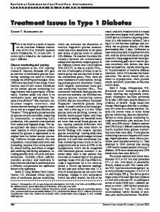
limited by that which is collected by the OPO, and no earlier records were available. Following whole-pancreas processing, representative formalin-fixed samples were stained to evaluate organ pathology (5). The pancreas from this donor showed a majority of islets containing residual b-cells (Fig. 1A, B, and D). A large number of residual b-cells is atypical in type 1 diabetes of 15 years duration (6). Numbers of b-cells per islet varied widely and included islets without b-cells, so-called pseudoatrophic islets. Pseudoatrophic islets are synonymous with type 1 diabetes (7). Increased proportions of a-cell to b-cell islets were frequent (Fig. 1C). Both
Figure 1—Representative islet images from nPOD donor 6196. Serial paraffin sections were stained by double immunohistochemistry for Ki67+Insulin (A, B, and D) or CD3+Glucagon (C and E), Congo Red (F), or hematoxylin and eosin (G and H). Numerous insulin-positive islets were observed with a range of normal sizes in all sections (red, A). Islets from the boxed area in A are shown in higher magnification for b-cells (red, B and D) and a-cells (red, C and E). Rare islets showed scattered CD3+ cells (brown, C and E). Several islets had amyloid, seen as intraislet redstaining amorphous material by Congo Red staining (F). Arteriosclerosis was observed including calcification within arterial walls (G) and expansion of the subintimal layer (H). Scale bars: 700 mm (A), 200 mm (B and C), and 100 mm (D–H).
cell types were reduced in islets with amyloidosis (Fig. 1D–F). Islet amyloidosis is reported in 90% of patients with type 2 diabetes (8). Advanced atherosclerosis was also observed in this patient with calcification within the arterial walls and subintimal proliferation (Fig. 1G and H). Secondary complications of atherosclerosis are common to both type 1 and type 2 diabetes (9). Classic type 1 diabetes clinical progression is thought to eventually reach a state of absolute insulin deficiency, and the rate at which this state is reached is dependent on many factors, most notably glycemic control and age at onset; however, more literature on long-term insulin microsecretors is emerging (2,3,10). We wondered why this patient had such severe comorbidities, presumed rapid disease progression, and early death despite the presence of insulin-positive b-cells, appreciable C-peptide, and HLA associated with protection against type 1 diabetes. Perhaps these observations were solely due to poor glycemic control; alternatively, peripheral insulin resistance (11) and/or b-cell dysfunction (related to impaired blood glucose sensing, signal transduction, insulin processing, or insulin secretion) (12) might have been at play. Flatbush diabetes, also referred to as atypical or ketosis-prone diabetes, is a subgroup of diabetes with clinical features of type 2 diabetes but severe presentations of ketoacidosis followed by periods of recovery where b-cells are functioning, insulin is secreted, and exogenous insulin treatment is not necessary. Different forms have been described, distinguished by the presence or absence of autoantibodies in addition to variation in the amount of C-peptide secretion present (13). It is possible the patient presented herein had Flatbush diabetes, which may explain the presence of numerous insulinpositive islets and detectable C-peptide. As we refine our diagnostic skills and laboratory methods, we will be able to identify these cases sooner and progress to offering specialized treatment. There is a spectrum even within these disease variants that dictate the level of C-peptide production, and this may influence the severity as well as the speed with which complications develop. Whether these complications were precipitated by glucose toxicity, severe insulin resistance, or dysfunctional insulin secretion/action requires further exploration, which will
1293
1294
Severe Diabetes Yet Numerous b-Cells and C-Peptide
hopefully lead to the identification of new therapies for patients with similar disease characteristics. This severe presentation and progression of autoantibody-positive diabetes with incongruous C-peptide and histologic findings provides another unique example of disease variability and heterogeneity. Perhaps not all long-standing diabetes reaches absolute insulin deficiency. It is likely that a subset of African American patients with presumed type 1 diabetes have Flatbush diabetes or even a hybrid form of the disease. The pathogenesis may involve b-cell dysfunction or other factors contributing to cardiovascular disease and severe complications despite the presence of b-cells and measurable C-peptide; however, the specific underlying mechanism(s) have yet to be elucidated. The rapid progression of clinical disease discordant with the histological analysis demonstrates a mixed picture of diabetes pathology, and further research is needed in this area. Acknowledgments. The authors acknowledge the nPOD staff members and OPOs that partner with nPOD to recover organ donors. Additional donor details can be obtained through the JDRF nPOD website (www.jdrfnpod.org). This study used data from the Organ Procurement and Transplantation Network. Donor data sets are available through nPOD DataShare, an online
database for collaborative communication organized around the nPOD specimen repository. Funding. This work was supported by JDRF (grants 25-2013-268, 17-2012-3, and 25-2012516 to M.C.-T. and D.A.S.). Duality of Interest. No potential conflicts of interest relevant to this article were reported. Author Contributions. L.M.J. researched the data and wrote the manuscript, M.A.A. contributed to the discussion and reviewed/edited the manuscript, M.C.-T. researched the data and reviewed/edited the manuscript, and D.A.S. conceived of the study and reviewed/edited the manuscript. D.A.S. is the guarantor of this work and, as such, had full access to all the data in the study and takes responsibility for the integrity of the data and the accuracy of the data analysis.
References 1. Davis AK, DuBose SN, Haller MJ, et al.; T1D Exchange Clinic Network. Prevalence of detectable C-peptide according to age at diagnosis and duration of type 1 diabetes. Diabetes Care 2015; 38:476–481 2. Wang L, Lovejoy NF, Faustman DL. Persistence of prolonged C-peptide production in type 1 diabetes as measured with an ultrasensitive C-peptide assay. Diabetes Care 2012;35:465–470 3. Oram RA, Jones AG, Besser RE, et al. The majority of patients with long-duration type 1 diabetes are insulin microsecretors and have functioning beta cells. Diabetologia 2014;57:187–191 4. Steffes MW, Sibley S, Jackson M, Thomas W. b-Cell function and the development of diabetesrelated complications in the Diabetes Control and Complications Trial. Diabetes Care 2003;26:832–836 5. Campbell-Thompson M, Wasserfall C, Kaddis J, et al. Network for Pancreatic Organ Donors with Diabetes (nPOD): developing a tissue biobank for
Diabetes Care Volume 39, July 2016
type 1 diabetes. Diabetes Metab Res Rev 2012;28: 608–617 6. Campbell-Thompson M, Fu A, Kaddis JS, et al. Insulitis and b-cell mass in the natural history of type 1 diabetes. Diabetes 2016;65:719–731 7. Foulis AK, Stewart JA. The pancreas in recentonset type 1 (insulin-dependent) diabetes mellitus: insulin content of islets, insulitis and associated changes in the exocrine acinar tissue. Diabetologia 1984;26:456–461 8. H¨oppener JW, Ahre´ n B, Lips CJ. Islet amyloid and type 2 diabetes mellitus. N Engl J Med 2000; 343:411–419 9. Lacy ME, Wellenius GA, Carnethon MR, et al. Racial differences in the performance of existing risk prediction models for incident type 2 diabetes: the CARDIA study. Diabetes Care 2016;39:285–291 10. Barker A, Lauria A, Schloot N, et al. Agedependent decline of b-cell function in type 1 diabetes after diagnosis: a multi-centre longitudinal study. Diabetes Obes Metab 2014;16:262–267 11. Bjornstad P, Maahs DM, Duca LM, et al. Estimated insulin sensitivity predicts incident micro- and macrovascular complications in adults with type 1 diabetes over 6 years: the coronary artery calcification in type 1 diabetes study. J Diabetes Complications 2016;30:586–590 12. Krogvold L, Skog O, Sundstr¨om G, et al. Function of isolated pancreatic islets from patients at onset of type 1 diabetes: insulin secretion can be restored after some days in a nondiabetogenic environment in vitro: results from the DiViD study. Diabetes 2015;64:2506–2512 13. Maldonado M, Hampe CS, Gaur LK, et al. Ketosis-prone diabetes: dissection of a heterogeneous syndrome using an immunogenetic and beta-cell functional classification, prospective analysis, and clinical outcomes. J Clin Endocrinol Metab 2003;88:5090–5098