Original Article
© Schattauer 2009
Prospective epidemiological study on the beginning of varicose veins* Bochum Study I–IV U. Schultz-Ehrenburg1; S. Reich-Schupke1; B. Robak-Pawelczyk1; T. Rudolph1; C. Moll1; N. Weindorf2; H. Hirche3; P. Altmeyer1; M. Stücker1 1Department
of Dermatology and Allergology , Ruhr University Bochum; 2Department of Dermatology and Allergology, St. Elisabeth Hospital, Oberhausen; 3Institute of Medical Informatics, Biometry and Epidemiology, University of Duisburg-Essen, Germany
Keywords Longitudinal vein study, varicogenesis, preclinical saphenous refluxes, risk factor of puberty, CEAP
Summary Objective: To conduct a longitudinal vein study in a young study population on when and how varicose veins develop in healthy veins. Population, method: The initial study population consisting of pupils aged 10–12 (BO I, n = 740) underwent clinical and ultrasound follow-up at the ages of 14–16 (BO II, n = 518), 18–20 (BO III, n = 459) and 29–31 (BO IV, n = 136). During BO I-IV all venous findings detected (including preclinical refluxes of the saphenous veins) were recorded. Results: The data were broken down to reveal the incidence and prevalence of venous refluxes (VR), varicose veins (VV) and venous abnormalities (VA) for each part of the study. Furthermore, the data were analyzed longitudinally to identify any correlations between VR and VV in the two saphenous veins. Conclusions: Since none of the study subjects exhibited VV during BO I, the study permits evaluation of the venous situation in the subjects from birth on. The manifestation of a
Correspondence to Prof. Dr. med. Markus Stücker Klinik für Dermatologie im St. Josef Hospital, Universitätsklinik der Ruhr-Universität Bochum Gudrunstr. 56. 44791 Bochum, Germany Tel. + 49/(0)234/879 23 77 Fax + 49/(0)234/879 23 76 E-mail:
[email protected] www.venenzentrum-uniklinik.de
truncal VV is preceded by a VR in the same vein (p = 0.039). VR occurred mainly during puberty (BO I: 2.5%, BO III: 18.5%, BO IV: 25%). A preclinical VR represents a 30% risk (95% CI: 13–53%) of developing a truncal VV within four years, as a consequence, subsequent preventive 2-year follow-up examinations are recommended.
Schlüsselwörter Longitudinale Venenstudie, Varikogenese, präklinische Saphenarefluxe, Risikofaktor Pubertät, CEAP-Klassifikation
Zusammenfassung Ziel dieser longitudinalen Venenstudie ist, an einer jungen Population zu erforschen, wann und wie es von einer unversehrten Vene zur Krampfader kommt. Studienteilnehmer, Methoden: Schulkinder der Altersstufe 10–12 Jahre (BO I, n = 740) wurden folgenden (klinischen und sonographischen) Follow-up-Untersuchungen unterzogen: 14–16 Jahre (BO II, n = 518), 18–20 Jahre, (BO III, n = 459) und 29–31 Jahre (BO IV, n = 136). Dabei wurden alle auftretenden Venenbefunde einschließlich der
Prospektive Epidemiologische Studie über die Entstehung der Krampfadern* – Bochumer Studie I–IV Phlebologie 2009; 38: 17–25 Received: January 12, 2009 accepted: January 14, 2009
*
präklinischen Saphena-Refluxe erfasst. Ergebnisse: Inzidenz und Prävalenz von VR (venöse Refluxe), VV (varicose veins, so genannte große Varizen) und VA (venous abnormalities, so genannte kleine Varizen) werden aufgeschlüsselt. Darüber hinaus werden für beide Saphenastämme die longitudinalen Befundverläufe auf Interdependenzen zwischen VR und VV analysiert. Schlussfolgerungen: Da in BO I noch keine VV vorkamen, erlaubt die Studie eine Analyse ab der Geburt. Der Manifestation einer Stammvarize geht mit statistischer Signifikanz (p = 0,039) ein VR an derselben Vene voraus. VR traten vor allem in der Pubertät auf (BO I: 2,5%, BO III: 18,5%, BO IV: 25,0%). Ein präklinischer VR bedeutet ein 30%iges Risiko (95% CI: 13–53%), binnen vier Jahren eine Stammvarikose zu bekommen, so dass zweijährige prophylaktische Kontrolluntersuchungen indiziert sind.
Mots clés Etude phlébologique longitudinale, varicogenèse, reflux saphénien préclinique, puberté comme facteur de risque, classification CEAP
Étude épidémiologique prospective sur le développement de veines variqueuses* – Étude de Bochum I–IV
Lecture held on the occasion of the awarding of the Ratschow Commemorative Medallion to Professor Dr. med. Ulrich Schultz-Ehrenburg by the Curatorium Angiologiae Internationalis at the 50th Annual Meeting of the German Society of Phlebology held in Bochum, Germany, on October 15–18, 2008. Recipient of the First Prize for the Best Communication at the 4th Annual Meeting of the European Venous Forum held in Lisbon, Portugal, on June 27–29, 2003 and of the Platinum Abstract Award of the American College of Phlebology at the World Congress Chapter Meeting of the Union Internationale de Phlébologie held in San Diego, California, USA, on August 27–31, 2003.
Phlebologie 1/2009
17
18
Schultz-Ehrenburg et al.: Bochum Study I–IV
Résumé Objectif : Étude longitudinale sur l'état veineux d'une population jeune pour tenter de comprendre comment et quand les varices se développent dans une population saine. Population, méthode : Le premier groupe concerne des adolescents de 10 à 12 ans (BO I, n = 740) soumis à un examen clinique et ultrasonique suivi, de 14 à 16 ans (BO II, n = 518), de 18 à 20 ans (BO III, n = 459) et de 29 à 31 ans (BO IV, n = 136). Durant la période entière, les constatations veineuses (incluant le reflux préclinique des veines saphènes) ont été rassemblées. Résultat : Les données ont été analysées pour étudier l'incidence et la prévalence des reflux veineux (VR), des veines variqueuses (VV) et des anomalies veineuses (VA) pour chaque partie de l'étude. De plus, les résultats ont été analysés de manière longitudinale pour identifier toute corrélation entre VR et VV pour les deux troncs saphéniens. Conclusion : Aucun sujet n'a présenté de VV au cours de BO I, ce qui correspond à une situation veineuse normale de naissance. L'apparition d'une VV tronculaire est précédée par une VR dans la même veine (p = 0,039). Les VR surviennent essentiellement durant la puberté (BO I = 2,5% BO III = 18,5% BO IV = 25%). Un VR préclinique représente un risque de 30% (95% CI : 13–53%) de développer une VV tronculaire dans les 4 ans. Ainsi, une prévention d'un suivi de 2 ans est recommandée pour ce genre de situation.
A number of epidemiological cross-section studies have unanimously substantiated the high prevalence of varicose veins and venous disease and the rise in their prevalence with increasing age (1, 3, 5, 8, 9, 10). The conclusions reached by these studies concerning possible risk factors for the development of venous disease are less congruent. A question that has hardly been explored to date is the sequence of events in the development of varicose veins from their very beginnings to the appearance of more advanced stages.
What is lacking are longitudinal epidemiological studies measuring and following the incidence and prevalence of chronic venous disease. The only study of this kind so far, namely the Framingham Study (2), is mainly of historical interest since it did not use standardized methods of data collection for its analysis. Our own study, the Bochum Study instituted in 1982 on the occurrence and longitudinal development of chronic venous disorders in the second and third decade in life, aims to close this gap (13–17). The international CEAP classification standardized the grading of the various signs and features of chronic venous disorders. This system has recently undergone further refinement culminating in the Revised Version of the CEAP Classification for Chronic Venous Disorders (4). According to the CEAP system, telangiectasias and reticular veins are no longer classified as varicose veins (VV) but as venous abnormalities (VA). VA alone do not justify a diagnosis of chronic venous disease or chronic venous insufficiency since telangiectasias and reticular veins can also be found in healthy individuals. This view is in general becoming increasingly accepted and is supported by recent epidemiological studies with a study design reflecting the CEAP classification (3, 8, 10, 12). The presented prospective study, which covers a period of almost 20 years, has explored the incidence and progression of all phlebological findings (including VR, VV and VA) occurring between childhood and adulthood. In particular, it aimed to answer the following questions: ● Which phlebological findings can be detected? When and how often did they occur in this study? ● When and for what reason does a varicosity develop in a healthy vein? ● Are visible varices preceded by refluxes? If so, what is the latency period? ● Is it possible to identify individuals at risk? If so, what consequences can be derived from this?
study phase
BO I
BO II
BO III
BO IV
year
1982/83
1986/87
1990/91
2001/02
age (years)
0010–12
0014–16
0018–20
0029–31
n
0740
0518
0459
0136
women / men
0396 / 344 0292 / 226 0253 / 206 0087 / 49
Phlebologie 1/2009
Tab. 1 Survey of the Bochum Study (BO)
Study population, methods Study population The data were collected during the Bochum Study (BO), a prospective longitudinal epidemiological study in four parts that commenced in 1982/83 (13–17). The initial study population consisted of pupils aged 10–12 attending the first year of college-preparatory high school (Gymnasium) in Bochum, a city with a population of approx. 400 000 in the Ruhr district in Germany. Informed consent was obtained from the pupils, their parents and the directors of the 11 schools participating in the study. The study participants were contacted and examined again at three later points in time (BO II, III and IV) (씰Tab. 1). An increased number of dropouts was recorded at each of the three follow-up examinations. The dropout rate remained relatively low as long as the pupils were still attending school (BO I to BO III). The highest number of dropouts was noted as expected at the follow-up examination performed 11 years after the pupils had left school. The reasons most frequently cited for this disproportionate increase were change of address, lack of time and insufficient further interest in the study. Of the 740 pupils taking part in the study when it started in 1982/83, 136 were still available for follow-up in 2001/02. All of the individual longitudinal analyses are based on this finally remaining follow-up population of 136 subjects (씰Fig. 3, 씰Fig. 4). The four cross-sectional analyses of the individual parts of the study, in contrast, were always based on the total number of study participants in each part (씰Fig. 1)
Examination methods On each of the four examination dates, the study participants were individually examined and interviewed. The following methods were employed: ● questionnaire, ● various body measurements, ● photoplethysmography, ● physical examination, ● CW Doppler (all study series), ● colour-coded duplex scanning (BO IV only).
© Schattauer 2009
Schultz-Ehrenburg et al.: Bochum Study I–IV
© Schattauer 2009
80
74.3
BO I (n = 740) BO II (n = 518) BO III (n = 459) BO IV (n = 136)
60
50.4
%
40
35.3 30.3
25.7 17.7
20 11
0 a)
0
1.6 2
12.9
10.7 0 0.2 1.3 1.5
0 0.8
trunk varices trunk varices GSV SSV
4.1 5.2
5
0
0
tributary varices
incomp. perf. veins
reticular veins
3.7
telangiectasias
25 20.6
20 %
This analysis explores the morphological and ultrasound venous findings. The measurements of venous function over time performed with the aid of photoplethysmography have already been published (17). An additional publication on the importance of possible risk factors is in preparation. The morphological venous assessment – like the ultrasound examination – was performed in each study participants by an experienced phlebologist on our physician team. During BO I to BO III these examinations were carried out on at the respective schools. The investigators were allotted two class periods – a total of 1,5 hours – to examine a group of subjects consisting of about 20 to 25 pupils; instruction by the teachers was cancelled during this time. Venous sonographic examination was performed with a directional CW-Doppler device (Blood Velocimeter BV 380, Kranzbühler & Sohn, Germany). The examination method of duplex sonography was not yet available when the study began in 1982/83. For the Bochum Study IV, the investigators first had to locate the study participants. They subsequently contacted them and asked them to report to our hospital for a follow-up examination. Since the method of color-coded duplex sonography was now available to the study team, both techniques (CW Doppler: Sonodop 7000 CW, Sonotechnik GmbH, Markt Schwaben, Germany, and color-coded duplex scanning: Sonoline Elegra, Siemens AG, Erlangen, Germany) were used during BO IV. The clinical findings for each patient were graded according to the CEAP classification (4). Further subclassification was undertaken on the basis of anatomic criteria. The venous abnormalities (clinical class C1) were subdivided into telangiectasias and reticular veins. The findings of varicose veins (clinical class C2) were subdivided into trunk varices of the great saphenous vein (GSV) and the small saphenous vein (SSV), incompetent perforating veins and tributary varices. In the category of venous refluxes, both superficial refluxes (GSV and SSV) and deep axial refluxes (popliteal vein and ileofemoral transition) were diagnosed. Among the refluxes of the GSV and SSV, refluxes of clinically manifest varicose veins as well as preclinical refluxes (i. e. without detectable varicose dilatation of the vein) were registered; in each case the reflux length was classified by Hach stage (6, 7).
15
13.5 11
10.4
10
6.3
5 0 b)
0
1.6
2
trunk varices GSV
2.4 0
refluxes GSV
1.5 0.2 1.3
trunk varices SSV
0.1
5.9
1.9
refluxes SSV
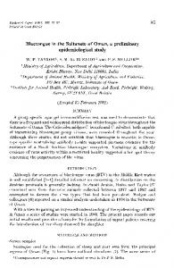
Fig. 1
Bochum Study I–IV a) incidence and prevalence of venous abnormalities and varicose veins b) correlation of varicose veins with venous refluxes
Statistics The results are presented descriptively in the text, tables and graphics. The prevalence of VA, VV and VR was described consecutively from BO I to BO IV and the clinical course evaluated. Exact confidence intervals for the percentage values were taken from the scientific tables in Documenta Geigy (7th edition 1977). To explore the question of whether the manifestation of truncal varicose veins is preceded by a reflux in the same vein, a Sign Test of statistical significance was performed (11). The threshold for significance was defined in advance as p = 0.05 for a two-tailed question. The empirically found p value was given descriptively.
Results Incidence and prevalence of venous abnormalities, varicose veins and venous refluxes (BO I to BO IV) The first step in the final study analysis was to perform statistical cross-section analyses of
the individual study phases. The comparison of these four statistical „snapshots“ provides valuable information on the dynamic development of all the phlebological findings elicited during the 19-year observational period of the study, i. e. during the second and third decades of life (씰Fig. 1). During BO I, when the subjects were aged 10–12, none of the study participants had varicose veins of any kind. A few preclinical refluxes, all without visible varicose dilatation, were found, however, in the great saphenous vein (GSV) in 2.4% of the study participants, and in the small saphenous vein (SSV) in 0.1%. In the category of venous abnormalities, telangiectasias were totally absent; however, reticular veins of a very discrete nature were already found in 10.7% of the study participants (씰Fig. 1, 씰Fig. 2a). VV were encountered for the first time when the study participants were aged 14–16 (BO II) and covered the entire spectrum of VV types (씰Fig. 1, 씰Fig. 2c). However, these varices were very discrete and affected only a small percentage of the study population (truncal varices of the GSV in 1.6%, truncal varices of the SSV in 0.2%, tributary varices in Phlebologie 1/2009
19
20
Schultz-Ehrenburg et al.: Bochum Study I–IV
0.8% and incompetent perforating veins in 4.1%). The number of refluxes was distinctly higher than the number of manifest truncal varices (refluxes of the GSV in 10.4%, refluxes of the SSV in 1.9%). Likewise, all types of VA were now represented including telangiectasias (reticular veins in 30.3%, telangiectasias in 3.7%) (씰Fig. 1, 씰Fig. 2b). These were also without exception minimal clinical findings. A further increase was recorded when the study participants were 18–20 (BO III). An increase was noted in both VV (truncal varices of the GSV in 2.0%, truncal varices of the SSV in 1.3%, varices of tributary veins in 5.0% and incompetent perforating veins in 5.2%) and VA (reticular veins in 35.3%, telangiectasias in 12.9%). The same observation applied for the VR (refluxes of the GSV in 13.5%, refluxes of the SSV in 6.3%). The
prevalence of these VR continued to be several times higher than that of the visible truncal varices. The clinical picture presented by the VV was still negligible without exception (씰Fig. 1, 씰Fig. 2d, 2e). Severe or moderate findings, i. e. findings of the kind seen regularly in the patients coming to our consultation hours, were completely absent in the study participants. The same is true of the venous abnormalities: here the insignificance of the clinical findings was in sharp contrast to the high prevalence values, which reached two digits for both forms of VA. During BO IV, carried out when the subjects were aged 29–31, the highest prevalence recorded to date in the study was noted for all forms of VV and VA (truncal varices of the GSV in 11%, truncal varices of the SSV in 1.5%, varices of tributary veins in 17.7%, in-
a)
b)
c)
d)
competent perforating veins in 25.7%, reticular veins in 74.3%, telangiectasias in 50.4%). Among the venous refluxes the same trend was evident in the GSV, where refluxes were found in as many as 20.6% of the study population; the prevalence figures for refluxes in the SSV, in contrast, were characterized by stagnation (5.9%) (씰Fig. 1). The clinical findings were predominantly harmless. During BO IV, however, findings in CEAP clinical grades higher than C0 and C1 were seen for the first time: 11 subjects (8.1%) displayed pathology in Grade C3, four (2.9%) displayed pathology in Grade C4a (3 × pigmentation, 1 × eczema) and one study subject (0.7%) exhibited a lesion in Grade C4b (mild atrophie blanche) (씰Tab. 2).
Individual longitudinal development of varices and refluxes On the basis of the core study population (n = 136) which remained in the study from BO I to BO IV, it is possible to estimate the longitudinal course in the individual study participants. This is of great interest in particular with respect to a possible correlation between the consecutive development of VR
e)
Fig. 2
Some cases as examples a) reticular veins, Bochum Study I, 11-year-old boy; b) telangiectasia, Bochum Study II, 15-year-old girl; c) trunk and tributary VV of the GSV, Bochum Study II, 15-year-old boy; d) the same boy four years later during the Bochum Study III; e) initial truncal VV of the SSV, Bochum Study III, 19-year-old boy
Phlebologie 1/2009
© Schattauer 2009
Schultz-Ehrenburg et al.: Bochum Study I–IV
Tab. 2 CEAP classification of findings in the Bochum Study IV (multiple answers permitted)
clinical classes
total (n = 136)
men (n = 49)
women (n = 87)
p value
%
n
%
n
%
n
men/women
C0
no signs of venous disease
17.6
024
24.5
12
13.8
12
n. s.
C1
telangiectasias, reticular veins
77.2
105
67.4
33
83.8
72
< 0.01
C2
varicose veins*
28.7
039
36.7
18
24.1
21
n. s.
C3
oedema
08.1
011
04.1
02
10.3
09
n. s.
C4
a) pigmentation, eczema
02.9
004
04.1
02
02.3
02
n. s.
b) lipodermatosclerosis, white atrophy
00.7
001
00.0
00
01.1
01
n. s.
C5
healed venous ulcer
00.0
000
–
C6
active venous ulcer
00.0
000
–
* all types (truncal, tributary and perforating veins); n. s.: not significant
and VV. In this context the individual longitudinal developments taking place in both saphenous veins are of particular interest. Basically, it is possible to make statements about the dynamics underlying the development of varices and refluxes over time only for the truncal varices but not for tributary varices and incompetent perforating veins. The reason for this is that only the venous trunks of GSV and SSV were examined with Doppler ultrasound to detect refluxes in any case, i. e. regardless of whether there were visible VV or not. Since the variance in tributary varices and incompetent perforating veins is too large, systematic prospective reflux diagnostics in advance would not have been practicable for these varix types.
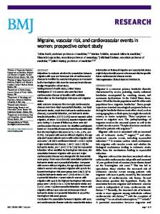
Great saphenous vein First, the findings in the GSV for which the larger figures were recorded will be described (씰Fig. 3a). At the provisional end of the study a total of 18 refluxes was recorded in the study population of BO IV (n = 136). Of these 18 cases, 13 had also developed visible truncal varices in the course of the study . The statistical test shows with sufficient certainty (p = 0.039) that the manifestation of varicosity is preceded by a reflux in the same vein. The individual follow-up findings can be broken down as follows: In the 10–12 age group (BO I), six study participants displayed a reflux of the GSV; none of them exhibited a visible trunk varicosity of this vein. The follow-up of these six subjects yielded the findings described below. One of them displayed a trunk varicosity in © Schattauer 2009
this vein four years later (BO II), a second eight years later (BO III), and two others during the follow-up 19 years later (BO IV). Even at the last follow-up examination, the latter two subjects displayed only a reflux of the GSV without any detectable varicose transformation. The incidence of refluxes and varices of the GSV detected in the study population up to this age (10–12 years) is thus 4.4% (95% CI: 1.5–9%) and 0% (95% CI: 0–2.5%), respectively. In the 14–16 age group (BO II) four new refluxes were recorded in the GSV. In one subject this was accompanied by a manifest trunk varicosity. The second and third subjects each displayed a trunk varicosity of this vein at the follow-up examination performed four years later (BO III) and the fourth at the follow-up examination after 15 years (BO IV). During BO II the investigators noted the only (apparently) paradoxical development in the study: a visible trunk varicosity for which a reflux of the GSV was not recorded until BO IV. This case will be discussed separately. The 4-year incidence of refluxes and varices of the GSV detected in the study population during this period is thus 3% (95% CI: 1–7%) and 2% (95% CI: 0.5–6%), respectively. In the 18–20 age group (BO III) four new refluxes emerged. In one subject, the reflux was already accompanied by a visible trunk varicosity. In another subject, a visible trunk varicosity was not seen until the follow-up 11 years later (BO IV), while the last two subjects still displayed only a reflux without a manifest varicosity even as late as BO IV. The corresponding 4-year incidence of refluxes and varices of the GSV detected in the study
population during this period is 3% in both cases (95% CI: 1–7%). During the last part of the study (BO IV), which was performed when the study participants were aged 29–31, four additional refluxes were again detected. One of these belonged to the „paradoxical“ varix-reflux sequence already described in BO II. Two of the new refluxes were already associated with a trunk varicosity which had also developed during the preceding 11-year interval. The latter subject displayed a reflux without a visible varicosity. The corresponding 11-year incidence of refluxes and varicosities of the GSV detected in the study population during this period is 3% (95% CI: 1–7%) and 4.4% (95% CI: 1.5–9%), respectively.
Small saphenous vein In the SSV only five refluxes and three clinically manifest truncal varices were detected up to the end of the study (BO I-IV) (씰Fig. 3b). This corresponds to an incidence of 4% (95% CI: 1–8%) for refluxes of the SSV and 2% (95% CI: 0.5–6%) for varices of the SSV during the observation period of 19 years. The first reflux of the SSV was noted during BO I, the second during BO II. As late as BO IV, both of these subjects still demonstrated only a reflux without a manifest varicosity. During BO III two additional refluxes were recorded; both were accompanied by a visible trunk varicosity. An additional new reflux, which was also accompanied by a visible trunk varicosity, was detected during BO IV.
Phlebologie 1/2009
21
22
Schultz-Ehrenburg et al.: Bochum Study I–IV
Length of saphenous refluxes On the basis of the core study population remaining until BO IV, the individual progression of reflux length in the second and third decades of life can be analyzed (씰Fig. 4). For all refluxes of the GSV and SSV involving the junction zone – the type of reflux encountered almost exclusively during our study – reflux length was determined, in addition to the qualitative reflux diagnosis, and classified by Hach stage (6, 7). The development of the 18 refluxes of the GSV over time is described below (씰Fig. 4a). In the 10–12 age group (BO I) the investigators already detected six short refluxes of the GSV (four at stage I and two at stage II according to Hach). In the course of time reflux length reached stage IV in four cases and stage III in one case. However, one reflux remained at stage II during the entire 19-year observation period up to the end of the study. In the 14–16 age group (BO II) four new refluxes were found (one at stage I, two at stage II and one at stage III according to Hach). Three of them displayed progression in the further course of the study (one to stage IV and two to stage III). The fourth reflux remained at stage III up to the end of the study. In the 18–20 age group (BO III) four additional refluxes were diagnosed (two at stage I, one at stage II and one at stage III according to Hach). Both of the new refluxes at stage I were still at this stage in BO IV, while the remaining two – at stage II and III, respectively – had progressed to stage IV.
In the 29–31 age group (BO IV) an additional four refluxes were detected that had developed during the preceding 11-year interval. One of these was already at stage IV. In contrast, the remaining three were only at stage I; their further development remains to be seen. The five truncal refluxes found in the SSV developed as follows (씰Fig. 4b): The only reflux of the SSV that was already present in BO I was at stage II and attained the maximal reflux length for the SSV (stage III) in BO III. The next reflux of the SSV, the only new one detected during BO II, was also at stage II and also reached stage III during BO III. The next two refluxes that appeared during BO III were both detected at stage I and exhibited no progression up to the end of the study. The fifth (and to date last) reflux recorded during BO IV was at stage II. Its further development remains to be seen.
Discussion One of the most important questions in this context is certainly whether varices are preceded by refluxes, whether these refluxes exist from birth on or develop later in life, and how much time elapses between the detection of a VR without detectable VV and the manifestation of a VV in this vein. Information on this subject would be useful for devising preventive measures or perhaps even preventive interventions.
29–31 years/BO IV
29–31 years/BO IV
18–20 years/BO III
18–20 years/BO III
14–16 years/BO II
14–16 years/BO II
10–12 years/BO I
10–12 years/BO I
a) GSV
venous refluxes
varicose veins
b) SSV
From preclinical saphenous reflux to manifest truncal varicosity The single steps in varicogenesis taking place in young people can be broken down rather clearly by evaluating the individual longitudinal courses of all the study participants exhibiting venous pathology in one or both saphenous veins. This is especially true of the GSV, where the body of data was sufficiently large to permit a detailed analysis. Refluxes were demonstrated in the GSV in 18 cases and a trunk varicosity developed in the same vein in 13 cases. The following sequence of events was observed: ● In five cases only a VR without manifest VV was demonstrated. ● In eight cases the VR occurred one or more study intervals in advance of the VV. ● In four cases the VR was detected together with the VV. ● In one case a paradoxical sequence was found in connection with a VV in BO II; here the VV seemed to appear earlier than the VR, which was not detected in this vein until BO IV. In eight of these twelve truncal VV cases (if we disregard the last case which does not lend itself to evaluation), the reflux occurred demonstrably first and varix formation came afterwards. The consecutive or simultaneous manifestation of VR and VV in the GSV, and the associated time intervals, can be seen directly in 씰Figure 3a.
venous refluxes
varicose veins
Fig. 3 Correlation of venous refluxes with varicose veins during the Bochum Study (core study population of BO IV, n = 136) in the a) great saphenous vein; b) small saphenous vein Phlebologie 1/2009
© Schattauer 2009
Schultz-Ehrenburg et al.: Bochum Study I–IV
Since there were two cases in BO IV, however, with a reflux of the GSV that had already been detected in BO I and had not undergone varicose transformation even after 19 years, the latency period between the appearance of a VR and the manifestation of a VV can presumably be 20 years or longer in single cases. On the other hand, there were two cases in which the VR and the VV appeared „simultaneously“ in BO II and BO III, i. e. they were detected at the same follow-up examination after a four-year interval. These cases (and two similar cases in BO IV) do not by any means rule out the possibility that this particular VV was also preceded by a VR in the preceding follow-up interval. A time period of four years (or 11 years between BO III and BO IV) is simply too long to allow us to draw such a conclusion. Instead, these cases support the conclusion that the progression from a preclinical reflux to a clinical varicosity can take place within a relatively short period of time. The time that may elapse between the occurrence of a VR in a previously healthy vein and the varicose dilatation of this vein obviously ranges from a relatively short time period to many years. With respect to the one case in which the visible trunk varicosity of the GSV apparently developed before the venous reflux, this „paradoxical“ finding is probably attributable to technical causes. Owing to the time limitation of a double class period (90 min), the investigators used a relatively schematic examination sequence during the CWDoppler examinations performed during BO I to BO III: in general they first looked for refluxes at the junction and subsequently classified the length of these refluxes by Hach stage. With this approach, all VR involving the junction were naturally detected by the examiners whereas a segmental VR in a more distal part of the GSV was more likely to be overlooked. This reflux was probably indeed overlooked in previous study phases and first discovered during BO IV, during which the examinations were performed at our hospital without such a time constraint. Because of the way in which we discovered this constellation, this case cannot be used for assessing the order of VR/ VV development since varices can naturally not exist without (at least segmental) refluxes. This particular case is rather evidence suggesting that the less common type of segmental non-junctional refluxes occurs in this © Schattauer 2009
IV
III
II
I
0
a)
Fig. 4 Individual courses of reflux length of the VR (core study population of BO IV, n = 136) in the a) great saphenous vein b) small saphenous vein
b)
age group, too, even if only in exceptional cases. Certainly, one of the most exciting observations made in our longitudinal evaluations is that many VR develop into VV early on. Out of the total of 23 VR which were found from BO I to BO IV in the two saphenous veins (18 in the GSV and 5 in the SSV as described below), there were seven which proceeded to a VV fast, i.e. with a latency period between 0 and 4 years. This proportion (7 out of 23 VR) corresponds to a percentage of 30% (95% CI: 13–53%). The percentage of cases with a fast progression may even be higher than 30%, since there were three more simultaneous VR/VV constellations detected in BO IV. However, the long time interval of 11 years between BO III and BO IV does not allow us to classify these courses in more detail. Our data on the sequence in which varices and refluxes develop in the SSV (씰Fig. 3b) are based on very small case numbers. Up to the end of the study there were three instances of manifest VV and five instances of VR of this vein. The two findings of reflux alone
were made during BO I and BO II; after 19 or 15 years, respectively, neither of these had developed into a VV. In all three cases in which a VV became manifest, the VR occurred during the same interval in which the VV manifested itself. On the whole these observations are congruent with the spectrum of possible developments obtained from our GSV evaluation. However, the two steps of varicogenesis cannot be broken down nearly as well for the SSV as for the GSV subgroup since the case numbers are simply too small.
Progression of reflux length The examinations performed to study the development of reflux length can provide additional insights on varicogenesis (씰Fig. 4). Trendelenburg already postulated descending reflux propagation from the junction to the distal lower leg for both saphenous veins. This progression was later subdivided by Hach – depending on the reflux length attained – into four (GSV) and three (SSV) Phlebologie 1/2009
23
24
Schultz-Ehrenburg et al.: Bochum Study I–IV
stages, respectively (6, 7, 18). It has not been documented precisely so far whether refluxes in the GSV and SSV actually develop in such a consecutive descending manner. Our study provides evidence for this mode of reflux prolongation. All the refluxes of the GSV and SSV – the newly developed as well as the ones already present in BO I – were initially at a more or less short stage. Their lengths increased in the course of the study. This increase took place at different speeds; in some cases, the length remained constant for a longer period of time. In no case did a reflux, once it had developed, recede entirely or partially (씰Fig. 4). Downwards lengthening from the junction was the predominant – and almost exclusive – mode of development seen in our study. As already mentioned above, there was some evidence, that non-junctional segmental refluxes can also occur as a rare exception in these young age groups.
Limitations We are aware that the Bochum Study does not include all socioeconomic groups because of our decision to choose pupils at college preparatory high schools as our study population. The decisive reason was that the resulting study population would stay together in relatively unchanged form for nine years in the second decade of life during which period most of the expected vein changes would start. There is no other group in Germany offering the same opportunity of investigating the very beginnings of varicose vein formation not only numerically, but also on an individual longitudinal basis.
Impact of puberty on varicogenesis and outlook for the fourth decade of life In view of the study findings obtained during the parts of the study (BO I-III) when the subjects were still at school, our study population proved to be properly chosen for data collection, since there was a close correlation between the appearance and increase in prevalence of these findings and the period of puberty (씰Figs. 1–4). At the start of the study in BO I (10–12 age group), there were no varicose veins of any Phlebologie 1/2009
kind. In the further course of the study, however, VV occurred and it was possible to systematically follow the increase in their prevalence and progression. In BO II (14–16 age group), only a small number of varicose veins were detected, but these already included for the first time all the different types of varicose veins, i. e. truncal varices of the GSV, truncal varices of the SSV, tributary varices and incompetent perforating veins. The prevalence of all of these findings increased steadily during the subsequent periods. The first varicose veins thus developed during puberty; this is true of all subtypes. The development of the saphenous VR differs slightly from the development of the truncal VV (씰Fig. 1b, 씰Fig. 3). A small number of VR were already encountered at the beginning of the study. The first VR thus occurred somewhat before the first truncal VV, namely during or shortly before puberty. In any case, the vast majority of the VR seen in our study population were not present at birth. This observation rules out a congenital valve defect as possible cause of venous decompensation leading to varicose vein formation later in life – analogous to a congenital heart failure leading to dilatation and insufficiency of the myocardium over time. The prevalence of VR increased in each part of the study and was always higher than the prevalence of truncal varices (씰Fig. 1b). This was true of both saphenous veins. A comparison of the development of the prevalence of VR and VV, respectively, provides additional information and makes it possible, in particular in the GSV subgroup, to look ahead to the near future. The prevalence of VR in the GSV was 13.5% among the study participants aged 18–20 in BO III. The varices did not attain a similar prevalence (namely 11%) until 11 years later in BO IV (29–31 age group). Since the figures for VR were much higher (20.6%) in BO IV as well, we can expect to see additional truncal VV appearing successively in the upcoming fourth decade of life. A similar trend was observed in the incidence and prevalence figures for VA (reticular veins and telangiectasias) as well as for VV; however, the percentages are much higher in this CEAP class (씰Fig. 1a). The first telangiectasias occur during puberty, the first reticular veins perhaps somewhat earlier. Reticular veins were in any case the earliest morphologically detectable venous finding. They were already present in two-digit percentages during BO I (10.7%); however, their clinical appear-
ance was quite unassuming at this stage. The prevalence of both C1 findings rose sharply in the course of the study. Among the study participants aged 29–31 in BO IV, about 75% had reticular veins and 50% telangiectasias, while only 18% were free of any signs (씰Tab. 2). As in all comparable studies performed to date, findings in CEAP class C1 accounted for the largest share of the detectable venous changes recorded in our study. These data will be commented on only briefly here, since findings in grade C1 were not the focus of our study.
Conclusions The study results presented here provide good substantiation of our postulated concept in which varicogenesis takes place in three (more or less) successive stages from the healthy vein over the preclinical reflux to the manifest varicosity. Owing to the total or almost total absence of venous findings at the beginning of the study, the data analysis allows conclusions to be drawn not only about the 20-year study period itself but also about the preceding decade and in this manner about the first three decades of life. Most VR seen in the study developed during puberty; after puberty, distinctly fewer VR were noted (BO I: 10–12 years, 2.5%; BO III: 18–20 years, 18.5%; BO IV: 29–32 years, 25%). The vast majority of the VR were thus demonstrably not present from birth on. Preclinical VR are therefore as a rule not attributable to a congenital dysplasia or agenesia of the venous valves. Instead, they develop in clinically healthy veins in the course of a person's life. On the basis of our study results, it can be stated with sufficient certainty that manifest truncal varices are preceded in many if not all cases by venous refluxes (p < 0.039). The speed at which this varicose transformation takes place ranges from a relatively short period (i. e. a non-determined period of less than four years) to 20 or more years. In roughly one third of these cases, the transformation takes place during a period of time between zero and four years following the appearance of the reflux. The first occurrence and rapid rise in prevalence of all clinical and preclinical venous findings during the second decade of life indicate that, at a young age (i. e. during the first to third decades), growth and the process of maturing during puberty are the decisive risk fac© Schattauer 2009
Schultz-Ehrenburg et al.: Bochum Study I–IV
tors and not age itself (in the sense of merely an increase in years) as is the case later in life. Genetic factors play an important role here. This applies already to the manifestation of VR (and not only of VV and VA) – an observation that emerged clearly during our longitudinal study. VR occurred earlier in individuals with a familial predisposition and displayed a higher prevalence and longer mean length of reflux in all parts of the study than in the control group with a family history negative for venous disease (data analysis of the Bochum Study on possible risk factors for varicogenesis, publication in preparation). The preclinical saphenous refluxes occurring as early as the second and third decades of life represent a unique precursor sign allowing us to identify individual patients with a high risk of developing venous disease. According to our results, the demonstration of such a VR constitutes a 30% risk (95% CI: 13–53%) that an individual will develop a VV in the same vein within the next four years. Preventive follow-up examinations are therefore indicated and should initially be performed every two years.
Acknowledgements The authors would like to thank Dr. Ulrich Matthes, Ms. Ute Stössl, Ms. Petra Kuhnhenn, Ms. Gudrun Moritz, Ms. Ursula Madri and Ms. Karin Hülshoff for the work they performed for the Bochum Study. Without their energy and commitment it would not have been possible to collect and record this data over a 20-year period. The study was supported by the German Society of Phlebology.
References 1. Allan PL, Bradbury AW, Evans CJ, Lee AJ, Vaughan Ruckley C, Fowkes FGR. Patterns of reflux and severity of varicose veins in the general population – Edinburgh Vein Study. Eur J Vasc Endovasc 2000; 20: 470–477. 2. Brand FN, Dannenberg AL, Abbott RD, et al. The epidemiology of varicose veins: the Framingham study. Am J Prev Med 1988; 4: 96–101. 3. Carpentier PH, Maricq HR, Biro C, Poncot-Makinen CO, Franco A. Prevalence, risk factors, and clinical patterns of chronic venous disorders of lower limbs: a population-based study in France. J Vasc Surg 2004; 40: 650–659. 4. Eklof B, Rutherford RB, Bergan JJ, Carpentier PH, Gloviczki P, Kistner RL, et al. Revision of the CEAP classification for chronic venous disorders: consensus statement. J Vasc Surg 2004; 40: 1248–1252. 5. Evans CJ, Fowkes FG, Ruckley CV, Lee AJ. Prevalence of varicose veins and chronic venous insufficiency in men and women in the general population: Edinburgh Vein Study. J Epidemiol Community Health 1999; 53: 149–153. 6. Hach W, Girth E, Lechner W. Einteilung der Stammvarikose in 4 Stadien. Phlebol Proktol 1977; 6: 116–123. 7. Hach W, Hach-Wunderle V. Die Rezirkulationskreise der primären Varikose. Berlin: Springer 1994. 8. Jawien A, Grzela T, Ochwat A. Prevalence of chronic venous insufficiency in men and women in Poland: multicenter cross-sectional study in 40 095 patients. Phlebology 2003; 18: 110–121. 9. Lionis C, Erevnidou K, Antonakis N, Argyriadou S, Vlachonikolis I, Katsamouris A. Chronic venous insufficiency. A common Health problem in general practice in Greece. Int Angiol 2002; 21: 86–92. 10. Rabe E, Pannier-Fischer, Bromen K, Schuldt K, Stang A, Poncar C, Wittenhorst M, Bock E, Weber S, Jöckel KH. Bonner Venenstudie der Deutschen Gesellschaft für Phlebologie. Epidemiologische Untersuchung zur Frage der Häufigkeit und Ausprägung von chronischen Venenkrankheiten in der städtischen und ländlichen Wohnbevölkerung. Phlebologie 2003; 32: 1–14.
11. Rosner B. Fundamental of Biostatistics. 5th ed. 2000, Duxbury, Pacific Grove, CA 53950 USA. 12. Ruckley CV, Evans CJ, Allan PL, Lee AJ, Fowkes FG. Telangiectasia in the Edinburgh Vein Study: epidemiology and association with trunk varices and symptoms. Eur J Vasc Endovasc Surg 2008; 36: 719–724. 13. Schultz-Ehrenburg U, Weindorf N, von Uslar D, Hirche H. Prospektive epidemiologische Studie über die Entstehungsweise der Krampfadern bei Kindern und Jugendlichen (Bochumer Studie I und II). Phlebol Proktol 1989; 18: 3–11. 14. Schultz-Ehrenburg U, Weindorf N, Matthes U, Hirche H. New epidemiological findings with regard to initial stages of varicose veins (Bochum study I-III). In: Raymond-Martimbeau P, Prescott R, Zumo M. (Eds.), Phlébologie 92, Proceedings of the XIth World Congress of the Union Internationale de Phlébologie. Paris: John Libbey Eurotext 1992: 234–236 15. Schultz-Ehrenburg U, Weindorf N, Matthes U, Hirche H. Étude épidémiologique sur la pathogénèse des varices. Étude de Bochum I-III. Phlébologie 1992; 45: 497–500. 16. Schultz-Ehrenburg U, Stücker M, Reich S, Altmeyer P, Weindorf N. 20-Year Prospective Epidemiological Study on the Development of Varicose Veins from Childhood to Adulthood (Bochum Study I-IV). In: Scuderi, A. (Ed), 15th World Congress of the Union Internationale de Phlébologie, Rio, Brazil, October 2–7, 2005. Medimond International Proceedings 2005: 1–4. 17. Stücker M, Reich S, Robak-Pawelczyk B, Moll C, Rudolph T, Altmeyer PJ, Weindorf NG, Hirche H, Gambichler T, Schultz-Ehrenburg U. Changes in venous refilling time from childhood to adulthood in subjects with apparently normal veins. J Vasc Surg 2005; 41: 296–302. 18. Trendelenburg F. Über die Unterbindung der Vena saphena magna bei Unterschenkelvarizen. Beitr klin Chir 1891; 7: 195.
Kuratoren und wissenschaftlicher Beirat CURATORIUM
Ratschow-Gedächtnismedaille Die Ratschow-Gedächtnismedaille wird vom Curatorium Angiologiae Internationalis an besonders verdiente wissenschaftliche Persönlichkeiten aus dem Gefäßbereich und den sie tangierenden Disziplinen verliehen. Dem Curatorium gehören ausschließlich Wissenschaftler aus dem internationalen Gefäßbereich an. Die Ratschow-Gedächtnismedaille wurde 1969 gestiftet. Mäzen dieser Stiftung ist die © Schattauer 2009
Bauerfeind AG Geschäftsbereich Phlebologie, Zeulenroda.
Präsidenten Prof. Prof. h. c. mult. Dr. Dr. h. c. mult. N. Klüken (1969–1999) Prof. Dr. Dr. h. c. K. U. Tiedjen (1999–2006) Prof. Dr. E. Rabe (seit 2006)
E. Betz (Tübingen), J. M. Coget (Lille), M. Comel (Pisa), U. Dembowski (Wiesbaden), M. Földi (Hinterzarten), R. Fontaine (Strassburg), W. H. Hauss (Münster), K. Ishikawa (Tokio), M. Jacquet (Paris), H. Jellinek (Budapest), M. Jünger (Greifswald), J. Kuiper (Nijmwegen), N. Klüken (Krefeld), P. Langeron (Lille), M. Müller (Santiago de Chile), R. C. Mayall (Rio de Janeiro), J. Merlen (Lille), E. Mian (Pisa), Y. Mishima (Tokio), C. M. Papendieck (Buenos Aires), F. Pratesi (Florenz), E. Rabe (Bonn), T. J. Ryan (Oxford), G. W. Schmid-Schönbein (San Diego), K. U. Tiedjen (Bochum), L. Widmer (Basel), M. Zabel (Recklinghausen). Phlebologie 1/2009
25