Australian Journal of Basic and Applied Sciences, 1(4): 776-784, 2007 ISSN 1991-8178 © 2007, INSInet Publication
Scanning Electron Microscopical Studies on Schistosoma mansoni cercariae Exposed to Ultraviolet Irradiation 1
Saad M. Bin Dajem and
1,2
Osama M. S. Mostafa
1
Biology Department, college of Science, King Khaled University, Abha 61413 PO box 9004, Saudi Arabia. 2 Zoology Department, Faculty of Science, Ain Shams University, Abbassia 11566, Cairo, Egypt. Abstract: In the present work, scanning electron microscopical studies on Schistosoma mansoni cercariae exposed to ultraviolet irradiation (UV-irradiation) were done to answer the question: Is the damage inflicted seen on the adult schistosome worms developed from irradiated cercariae, due to direct effect of UV-irradiation on cercariae or due to the host's immunogenicity induced by UVirradiated cercariae? The present observations showed that there were no changes on the surface topography of S. mansoni cercariae exposed to UV-irradiation. Thus we can suggest that the changes seen in the adult worm developed from UV-irradiated cercariae may be due to the immune responses of the host induced by attenuated cercariae; or it may be due to the mutagenic effects of the UVradiation which appeared in the adult stages. Detailed discussion on the scope of the recent knowledge about the immunological aspects due to irradiated cercariae was presented. Key words: Schistosome, Scanning electron microscopy, Ultraviolet, Irradiation, Cercariae, Vaccine INTRODUCTION Human schistosomiasis, a devastating parasitic worm disease caused mainly by three species of the genus Schistosoma: S. haematobium, S. japonicum, S. mansoni (He et al. 2005). More than 600 million people are at risk with about 200 million actually infected in 74 countries mainly in the tropics and subtropics (Ruelas et al. 2006). Despite the availability of affordable chemotherapy, drug treatment has not influence transmission of the infection. Moreover, there is a concern of drug resistance against chemotherapy of schistosomiasis of choice (e.g. Praziquantel). Therefore, a prophylactic or therapeutic vaccine to eliminate, or substantially reduce, the impact of schistosomiasis remains a highly desirable goal (Bergquist et al., 2002). Large body of vaccine-oriented researches has been carried out using cercariae exposed to various degrees of gamma or ultraviolet radiation and infected different experimental models (Tewari and Biswas 1972; W ebbe et al. 1982; Moloney et al. 1985; McLaren et al. 1990; Osman et al. 1994; Mostafa 1995; Kumar and Ramaswamy 1999; el-Gawish and M oawad 2003; Kariuki et al. 2004; Zhu et al. 2005; Torben and Hailu 2007a,b). Moreover, it was found that vaccination with attenuated infective cercariae is generally more effective at inducing host protection against experimental infection with S.m ansoni than immunization with defined antigens (Ganley-Leal et al., 2005). Scanning electron microscope is an important tool in studying the surface of the parasite when considering the host-parasite relationship (Hockley, 1968). A lot of work has been focused on schistosome normal larval stages. Stereoscan observations on S. mansoni cercariae and miracidia were done by Hockley, 1968, Race et al. 1971 and M iegeville et al. 1979. The ultrastructure of the ventral sucker of S.m ansoni cercariawas studied by Cousin et al. 1995. Dorsey et al. (2002) described the ultrastructure of the cells that comprise the cercaria of S. mansoni at ultrastructure level. However, very few investigations have been done on schistosome larval stages exposed to irradiation and adult worms developed from irradiated cercariae. Harrop and Wilson (1993) studied the morphological differences between normal and gamma irradiated larvae of S. mansoni at the lung stage of development by using scanning electron microscope. Mohamed (1999) studied the surface topography of adult S. mansoni
Corresponding Author: Saad M. Bin Dajem, Biology Department, college of Science, King Khaled University, Abha 61413 P.O. box 9004, Saudi Arabia. Fax: +96672290165 E-mail:
[email protected] 776
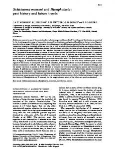
Aust. J. Basic & Appl. Sci., 1(4): 776-784, 2007 originating from ultraviolet-irradiated cercariae. On the other hand, no attention has been paid to the effect of irradiation on the ultrastructure and morphology of irradiated schistosome cercariae, therefore the present work was aimed to study the surface topography of ultraviolet-irradiated S. mansoni cercariae in comparison with non-irradiated cercariae of the same species. M ATERIAL AND M ETHODS Cercariae Production: Ten mice of CD1 strain were exposed to 100±10 cercariae of Egyptian strain of S. mansoni per mouse by the tail immersion method, modified by Oliver and Stirewalt (1952). Mice and cercariae were purchased from the Schistosome Biological Supply Program (SBSP) unit, Theodore Bilharz Research Institute, Giza, Egypt. Six weeks post-exposure, mice were sacrificed and dissected to obtain the liver and intestine. Hepatic and intestinal tissues were cut into pieces, placed in 0.85% saline solution and homogenized. The suspension was poured into a column of sieves arranged in descending order of mesh opening (420 µm, 177 µm, 105 µm and 45 µm). The eggs were collected from the bottom sieve, suspended in dechlorinated tap water and exposed to light to stimulate hatching of miracidia. One hundred Lab-bred Biomphalaria alexandrina (4-5 mm in shell diameter) were individually placed in wells of a 24-well plate, along with freshly hatching five to seven miracidia and 0.5 ml dechlorinated tap water. The snails were left overnight at room temperature to ensure maximum penetration. Then snails were removed from the wells and reared in culture. Snails were placed in plastic trays with a holding capacity of 1.5 liters of water. Each tray contained 25 snails at a time in dechlorinated water and was supplied with lettuce leaves (fresh or boiled and dried) for snail feeding. Trays were loosely covered with a glass plate to reduce evaporation, and maintained at room temperature 27-29 º C. W ater in the trays was changed every two days. At the 5th week post-exposure, snails were examined for cercarial shedding by exposing to strong light and the cercariae released were isolated and used in the present study. UV Irradiation: An S-68 mineral-light lamp, rated to deliver 95% of its output at 254 nM, was used as source of ultraviolet radiation. Thousands of cercariae were exposed to ultraviolet radiation for two and three minutes. Sam ples Preparation and Stereoscan Exam ination: The cercariae were fixed in 4% gluteraldehyde in cacodylate buffer for two hours, washed in the same buffer at pH 7.4, post-fixed in 2% osmic acid, washed in cacodylate buffer, finally dehydrated with ethanol. Specimens were mounted on stubs, coated with carbon and gold, examined with a Joel JEM-1200 EXII electron microscope. Observations: The cercariae of S. mansoni have an oval body or head and a long cylindrical tail which is divided into two furculae at the posterior extremity (Fig.1). The most anterior part of the head is provided with triangular slit surrounded by three spiny tegumental folds (Fig.2). The head of S. mansoni cercariae is covered with numerous spines which are posteriorly directed. The ventral sucker is well developed and provided with numerous, large and sharp spines directed backwardly (Figs. 3& 4). The posterior end of the head is tapered into a spiny collar-like folding over the narrow connection between the head and tail. This area represented the detachment site between the head and tail during penetration (Figs. 5& 8). The tail of S. mansoni has larger and sharper spines than that of the body. They are concentrated on the dorsal and ventral surface, and they are much fewer on the lateral surface. The ventral surface of the tail furculae has few short and pointed spines. The excretory pore is found on the tip of the tail furculae (Figs. 5, 6 & 7). The surface topography of S. mansoni cercariae exposed to 2 and 3 minutes ultraviolet radiation was more or less similar to those of non-irradiated cercariae, since there was no strike differences observed (Figs. 9, 10, 11&12). Discussion: In the present observation on non-irradiated S. mansoni cercariae, the construction between the most anterior part of the head and its middle part was not deep. This observation was agreed with that of Hockley (1968) on his work on S. mansoni cercariae. However, Sobhon et al. (1988) demonstrated that the most anterior part or the head of S. japonicum cercariae is conical and clearly demarcated from the middle section by a deep construction. The body and tail of S. mansoni cercariae were separated by a collar which was
777
Aust. J. Basic & Appl. Sci., 1(4): 776-784, 2007
Fig. 1: Low power scanning electron micrograph of non-irradiated S. mansoni cercaria showing head (H), collar-like structure (arrow), tail (T) and tail furculae (TF).
Fig. 2: Scanning electron micrograph of non-irradiated S. mansoni cercaria showing the most anterior part of the body provided with triangular slit (arrow) surrounded by three spiny folds (SF).
Fig. 3: Scanning electron micrograph of ventral surface of the head of non-irradiated S. mansoni cercaria showing the ventral sucker (VS).
778
Aust. J. Basic & Appl. Sci., 1(4): 776-784, 2007
Fig. 4: Scanning electron micrograph of ventral surface of the head of non-irradiated S. mansoni cercaria showing the ventral sucker (VS) with backwardly directed numerous spines (arrow).
Fig. 5: Scanning electron micrograph of non-irradiated S. mansoni cercaria showing the collar-like structure (C) which connecting the head with the tail (T), the tail is provided with large spines directed posteriorly (arrows).
Fig. 6: Scanning electron micrograph of tail furculae (TF) of non-irradiated S. mansoni cercaria.
779
Aust. J. Basic & Appl. Sci., 1(4): 776-784, 2007
Fig. 7: Scanning electron micrograph of the ventral surface of tail furculae (TF) of non-irradiated S. mansoni cercaria showing short and pointed backwardly directed spines and the excretory pore on the tip of tail furculae (arrow).
Fig. 8: Scanning electron micrograph of the head (H) of non-irradiated S. mansoni cercaria after detachment of the tail showing the collar-like structure(C).
Fig. 9: Scanning electron micrograph of 2 min UV-irradiated S. mansoni cercaria showing the anterior part of the body with three spiny folds (SF).
780
Aust. J. Basic & Appl. Sci., 1(4): 776-784, 2007
Fig. 10: Scanning electron micrograph of 3 min UV-irradiated S. mansoni cercaria showing the anterior part of the body with three spiny folds (SF).
Fig. 11: Scanning electron micrograph of the ventral surface of 3 min UV-irradiated S. mansoni cercaria showing the ventral sucker (VS).
Fig. 12: Scanning electron micrograph of 3 min UV-irradiated S. mansoni cercaria showing the collar-like structure (C) and the proximal part of the tail (T) which provided with large spines directed posteriorly (arrows).
781
Aust. J. Basic & Appl. Sci., 1(4): 776-784, 2007 described by Hockley (1968) as smooth surface collar. In the present investigation the surface of the collar appeared spiny. Sobhon et al. (1988) on their investigations on S. japonicum cercariae described the collar region as a heavily spined tegumental area. The opening of the penetration gland ducts are situated at the anterior tip of the head region in specific pattern in S. japonicum and S. mansoni cercariae (Robson and Erasmus, 1970, Short and Cartrett, 1973 and Sobhon et al., 1988). However, the ducts opening of penetration glands were not observed in the present investigation since the anterior tip of the body is provided with three spiny tegumental folds. It is possible that such tegumental folds covered the ducts opening of penetration glands. The ducts opening of penetration glands were not seen by Hockley (1968) on his work on S. mansoni cercariae. The present observations showed that there were no changes on the surface topography of S. mansoni cercariae exposed to UV-radiation. The radiation-attenuated schistosome vaccine induces a high level of protective immunity in rodents, and > 50% protection to a challenge with normal larvae has been achieved in primates (Yole et al., 1996). Generally, it has been found that immunization with defined antigens is generally less effective at inducing host protection against experimental infection with S. mansoni than vaccination with attenuated infective cercariae (Ganley-Leal et al., 2005). Li et al. (2002) made a preliminary study on S.japonicum cercariae antigens before and after ultraviolet irradiation; they were revealed novel antigens on UV irradiated cercariae. In addition, the protection with irradiated cercariae led to decreased pathology or reduced development of granulomas in the liver and increases the level of the inflammatory tumor necrosis factor- á (TNF-á) that could enhance inflammatory reactions due to the activation effects on macrophages, eosinophils and lymphocytes (Joseph et al., 2004, Torben and Hailu, 2007b). Several possibilities for explanation of the increased immunogenicity of irradiated larvae were summarized by Hewitson et al., 2005. One of them is their delayed and truncated pattern of migration as they move slowly through the skin, skin draining lymph nodes, and are delayed in their arrival in the lungs where migration is truncated. This means there is greater opportunity for interaction of parasite antigen with immune cells at these sites, which may in turn prim of a protective response. In addition, irradiation may alter the antigens present on the surface of the parasite as it may convert a poorly immunogenic surface molecule into one that is highly immunogenic. Irradiation may also result in the greater exposure of certain cryptic antigens that are highly protective but ordinarily only exposed to a limited extent (i.e. by normal larvae). It is also possible that irradiation affects some non-protein component of the parasite in the tegument as normal and irradiated parasites have different lectin-binding properties, indicating differences in their surface carbohydrates. Harrop and W ilson (1993) on their stereoscan study on gamma irradiated larvae of S. mansoni at the lung stage of development reported that irradiation of cercariae impairs neuromuscular function in developing schistosomula, since the irradiated larvae exhibited random constrictions, probably resulting from contraction of circular muscle fibers, at intervals along the length of the body and showed subtle differences in motility. They suggested that these abnormalities account for persistence of attenuated larvae in the skin-draining lymph nodes and lungs, 2 events that are instrumental to the induction of protective immunity in this vaccine model. DNA is often damaged by many environmental agents (natural solar irradiation for example) that lead to the up-regulation of several genes involved in different repair mechanisms. Recently, studies showed that a gene called nucleotide excision repair gene (NER) is involved in mechanism for removing a broad spectrum of different DNA damages. It was revealed that the correlation of these genes with their analogues in other eukaryotes (Silva et al., 2007). It seems that the S.mansoni irradiated cercariae may be has a damage repair genes to some extent as Mohamed (1999) studied the surface ultrastructure of adult S. mansoni worms developed from ultraviolet-irradiated cercariae, she observed a generalized deformities in the different regions of male and female worms originating from irradiate cercariae. These observations suggest that the repairing genes are important repair pathway during the complex life cycle of S.mansoni. Finally, we can suggest that the changes seen in the adult worms developed from UV-irradiated cercariae may be due to the immune responses of the host induced by attenuated cercariae Also; it may be due to the mutagenic effects of the U V-radiation which appeared in the adult stages. Further investigations on the DNA of cercariae exposed to UV-radiation must be done. REFERENCES Bergquist, R., M. Al-Sherbiny, R. Barakat and R. Olds, 2002. Blueprint for schistosomiasis vaccine development. Acta Trop., 82(2): 183-192. Cousin, C., C. Dorsey, V. Kennedy and K. Ofori, 1995. Ultrastructure of the ventral sucker of Schistosoma mansoni cercaria. J. M orphol., 223(2): 215-23.
782
Aust. J. Basic & Appl. Sci., 1(4): 776-784, 2007 Dorsey, C.H., C.E. Cousin, F.A. Lewis and M .A. Stirewalt, 2002. Ultrastructure of the Schistosoma mansoni cercaria. Micron., 33(3): 279-323. El-Gawish, M.A. and M .A. Moawad, 2003. Immunological studies on mice vaccinated with irradiated cercaria and IL-12 against Schistosoma mansoni infection. Egypt. J. Immunol., 10(2): 39-48. Ganley-Leal, L.M., J. Guarner, C.W . Todd, A.A. Da'dara, G.L. Freeman, Jr., A.E. Boyer, D.A. Harn and W .E. Secor, 2005. Comparison of Schistosoma mansoni irradiated cercariae and Sm23 DNA vaccines. Parasite Immunol., 27: 341-9. Harrop, R. and R.A. W ilson, 1993. Irradiation of Schistosoma mansoni cercariae impairs neuromuscular function in developing schistosomula. J. Parasitol., 79(2): 286-289. He, Y.X., B. Salafsky and K. Ramaswamy, 2005. Comparison of skin invasion among three major species of Schistosoma. Trends Parasitol., 21: 201-203. Hewitson, J.P., P.A. Hamblin and A.P. Mountford, 2005. Immunity induced by the radiation-attenuated schistosome vaccine. Parasite. Immunol., 27(7-8): 271-280. Hockley, D.J., 1968. Scanning electron microscopy of Schistosoma mansoni cercariae. J. Parasitol., 54(6): 1241-3. Joseph, S., F.M. Jones, G. Kimani, J.K. Mwatha, T. Kamau, F. Kazibwe, J. Kemijumbi, N.B. Kabatereine, M. Booth, H.C. Kariuki, J.H. Ouma, B.J. Vennervald and D .W . Dunne, 2004. Cytokine production in whole blood cultures from a fishing community in an area of high endemicity for Schistosoma mansoni in Uganda: the differential effect of parasite worm and egg antigens. Infect. Immunol., 72: 728-734. Kariuki, T.M., I.O. Farah, D.S. Yole, J.M. Mwenda, G.J. Van Dam, A.M. Deelder, R.A. W ilson and P.S. Coulson, 2004. Parameters of the attenuated schistosome vaccine evaluated in the olive baboon. Infect. Immun., 72(9): 5526-9. Kumar, P. And K. Ramaswamy, 1999. Vaccination with irradiated cercariae of Schistosoma mansoni preferentially induced the accumulation of interferon-gamma producing T cells in the skin and skin draining lymph nodes of mice. Parasitol. Int., 48(2): 109-19. Li, H., X.G. Chen, P.L. Yang, X.H. Zhou, S.M. Shen, H.J. Peng and K . W u, 2002. Preliminary study of cercaria antigen of Schistosoma japonicum before and after ultraviolet irradiation. [Article in Chinese] Di Yi Jun Yi Da Xue Xue Bao., 22(8): 697-699. McLaren, D.J., V.S. Delgado, J.R. Gordon and M.V. Rogers, 1990. Schistosoma mansoni: analysis of the humoral and cellular basis of resistance in guinea-pigs vaccinated with radiation-attenuated cercariae.Parasitology, 100(1): 35-44. Miegeville, M., M. Marjolet and C. Vermeil, 1979. Scanning electron microscopic observation of Schistosoma mansoni miracidia and cercariae. [Article in French] Bull. Soc. Pathol. Exot. Filiales., 72(1): 51-55. Mohamed, S.H., 1999. Scanning electron microscopical studies on the tegument of adult worms of Schistosoma mansoni originating from ultraviolet-irradiated and non-irradiated cercariae. J. Helminthol., 73: 157-161. Moloney, N.A., Q.D. Bickle and G. W ebbe, 1985. The induction of specific immunity against Schistosoma japonicum by exposure of mice to ultraviolet attenuated cercariae. Parasitology, 90(2): 313-23. M ostafa, O.M.S., 1995. Parasitological and biochemical studies on albino mice infected by Schistosoma mansoni cercariae irradiated by ultraviolet rays. M. Sc. Thesis, Zoology Department, Faculty of Science, Ain Shams University, Cairo, Egypt. Oliver, L. and M.A. Stirewalt, 1952. An efficient method for exposure of mice to cercariae of Schistosoma mansoni. J. Parasitol., 38: 19-23. Osman, A., R. El Ridi, N. Guirguis and D.A. Dean, 1994. Identification of Schistosoma mansoni antigens recognized by T cells of C57BL/6 mice immunized with gamma-irradiated cercariae. J. Parasitol., 80(3): 421-31. Race, G.J., J.H. M artin, D.V. Moore and J.E. Larsh, Jr., 1971. Scanning and transmission electronmicroscopy of Schistosoma mansoni eggs, cercariae, and adults. Am. J. Trop. Med. Hyg., 20(6): 914-24. Robson, R.T. and D.A. Erasmus, 1970. The ultrastructure, based on stereoscan observations, of the oral sucker of the cercaria of Schistosoma mansoni with special reference to penetration. Z Parasitenkd., 35(1): 76-86. Ruelas, D.S., D . Karentz and J.T. Sullivan, 2006. Lethal and sub-lethal effects of UVB on juvenile Biomphalaria glabrata (Mollusca: Pulmonata). J. Invertebr. Pathol., 93: 192-200. Short, R.B. and M.L. Cartrett, 1973. Argentophilic "papillae" of Schistosoma mansoni cercariae. J. Parasitol., 59(6): 1041-1059. 783
Aust. J. Basic & Appl. Sci., 1(4): 776-784, 2007 Silva, C.S., S´.H. Silva, O.S. Pereira-J´unior, F.J. Cabral, J.M. Costa-Cruz and V. Rodrigues 2007. Schistosoma mansoni : Gene expression of the nucleotide excision repair factor 2 (NEF2)during the parasite life cycle, and in adult worms after exposure to different DNA-damaging agents. Acta Trop., 104: 52-62. Sobhon, P., V. Anupunpisit, H.C. Yuan, E.S. Upatham and P. Saitongdee, 1988. Schistosoma japonicum (Chinese): changes of the tegument surface in cercariae, schistosomula and juvenile parasites during development. Int. J. Parasitol., 18(8): 1093-1104. Tewari, H.C. and G. Biswas, 1972. Experimental studies on the immunology of Schistosoma incognitum Chandler 1926 by vaccination with gamma irradiated cercariae and passive transfer. Z Parasitenkd., 38(1): 48-53. Torben, W . and A. Hailu, 2007a. Serum cytokines of the 20 Krad-irradiated S. mansoni cercariae vaccinated, primary and superinfected Cercopethicus aethiops aethiops. Exp. Parasitol., 115(2): 121-126. Torben, W . and A. Hailu, 2007b. The development of hepatic granulomas in 20 Krad irradiated Schistosoma mansoni cercaria vaccinated grivet monkeys (Cercopithecus aethiops aethiops). Exp. Parasitol., 117(4): 376-81. W ebbe, G., R.F. Sturrock, E.R. James and C. James, 1982. Schistosoma haematobium in the baboon (Papio anubis): effect of vaccination with irradiated larvae on the subsequent infection with percutaneously applied cercariae. Trans. R. Soc. Trop. Med. Hyg., 76(3): 354-61. Yole, D.S., G.D. Reid and R.A. W ilson, 1996. Protection against Schistosoma mansoni and associated immune responses induced in the vervet monkey Cercopithecus aethiops by the irradiated cercaria vaccine. Am. J. Trop. Med. Hyg., 54(3): 265-270. Zhu, X., Z.S. Zhang, M.J. Ji, H.W . W u, Y. W ang, X.P. Cai, L. Zhang, S.Y. Hu, L.L. Fu, F. Liu, C. Su and G.L. W u, 2005. Gene transcription profile in mice vaccinated with ultraviolet-attenuated cercariae of Schistosoma japonicum reveals molecules contributing to elevated IFN-gamma levels. Acta. Biochim. Biophys. Sin. (Shanghai)., 37(4): 254-64.
784