Undefined area. Then, separate the brain into left and right to specify Ischemic Stroke Area. As a result of experiment in 5 Stroke patients sample by setting CBV ...
Signal Processing Medical Signal Processing & Medical Imaging Paper 101531
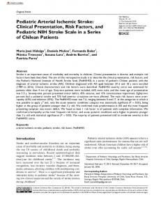
Simulation Program of Specifying Ischemic Stroke Area from CT Perfusion images Based on Digital Image Processing Techniques 2 l l S. Fueanggan , S. Chokchaitam , and S. Muengtaweepongsa I
Department of Electrical and Computer Engineering, Thammasat University, Thailand. 2 Division of Neurology, Thammasat University, Thailand.
Abstract- This research paper presents a new designed program to initially analyze Ischemic Stroke Area from Computed Tomography Perfusion (CTP) based on Digital Image Processing Techniques. The MATLAB program is applied to develop the designed software to analyze the Ischemic Stroke Area. The new designed software can specify Ischemic Stroke Area by assigning Threshold level of CTP from CBV (Cerebral Blood Volume) , CBF (Cerebral Blood Flow) and MTT (Mean Transit Time) images. Our experimental results will be shown in N-Match (normal tissue areas),D-Match (dead tissue areas ), Mismatch (blood cot tissue areas) and Undefined area. Then, separate the brain into left and right to specify Ischemic Stroke Area. As a result of experiment in 5 Stroke patients sample by setting CBV Threshold level to 2.5 ml/lOOg (±1.5) , CBF Threshold level to 20 ml/lOOg/min (±1O) and MTT Threshold level to 7.5 sec (±4.5) , the distribution of N-Match, D-Match, Mismatch and Undefined depends on their threshold. So, it is possible to sort elementary information of left and right side of the brain to specify Ischemic Stroke Area in order to compare the results with brain specialists.
I.
C
that studied to find Ischemic Stroke Area and lead to a program that help doctors to analyze by finding Mismatch from CBV and CBF images.[5] The idea of this research is to design a program to analyze Ischemic Stroke Area, comparing image data from CT scanned images of CBV, CBF and MTT which were collected from CT Perfusion, which rely on the relationship of all three scattered data to identify Ischemic Stroke Area based on data gathered from Mismatch of CBV, CBF images and calculate the Threshold of MTT for preliminary data analysis for the doctors which is easy to use and at cheap price, also to reduce the time for doctor to analyze the case and increase the accuracy of analysis for effective treatment. II. PROCEDURE AND RELATED THEORIES A. Medical principle in CT Scan diagnosis
CTP is a blood brain status checking method after the injection of contrast media, which is expected to be clogged by checking average time of blood in brain at the point that needed to be considered. Afterward, apply the information
INTRODUCTION
to create images and show CBV , CBF and MTT in order
erebrovascular Disease : Stroke is a frequently found
to specify Ischemic Stroke Area, so the doctor could have a
disease
treatment on time.
and
becoming
a
public
health
issue
in
Thailand. According to Public Health Statistics (A.D.
2005), Stroke is in the top three of Thai people cause of death
and
like
to
rise.[l]
In
correspond
with
joint
educational research between Ministry of Public Health and WHO (World Health Organization), Stroke comes first in cause of death in female and second in male.[2] It is also the main reason of Disability Adjusted Life Year in both male and female.[3] At present, there is shortage of personnel to provide sufficient service for treatment of brain and nerve. There's also a large number of patients compared to doctors, so the doctor may made fault analysis from color value from CT-Scan images with similar color scale. Therefore, if there are tools that help to analyze Ischemic Stroke Area images from CT-Scan, it will help doctors to get more correct results. Diagnosis:
CT Scan will assist doctor to diagnose
Patient's status by comparing variable images, such as CBV images. CBF images, MTT images, TTP images and to specify Ischemic Stroke Area on the CT Scan images, doctor normally consider CBV images and CBF images then find the location of mismatch.[4] There are research
The 8th Electrical Engineering/ Electronics, Computer, Telecommunications and Information Technology (ECTI) Association of Thailand - Conference 2011
(b)
(c)
Fig. 1. CT scan of brain which is replaced by color after CTP.
Page 1031
Signal Processing Medical Signal Processing & Medical Imaging
From Fig. l(a) CBV image is the amount of blood per CBV
unit of brain tissue. Comparison of images is done by setting Threshold value, if any area has color value lower
t
than prescribed, will be considered as less value area. And if any area has color value than prescribed,
will be
CBV more than TH
considered as much value area.
MISMATCH • • •
+
From Fig. l(b) CBF image is the amount of blood flow
MTT
•
T�.�--------�------�
per unit of brain tissue per minute. In comparison by setting
t +
Threshold value, if any area has color value lower than prescribed, will be considered as less value area. And if any
UNDEFINED
CBV less than TH
area has color value than prescribed, will be considered as much value area as for CBV images. From Fig. I(c) MTT image was defined as the time
® �Th"
..
difference between the arterial inflow and venous outflow.
CRF less than TH
F
CRF
�
CRF more than TH
The MTT image was calculated from CBV (Cerebral Blood
Fig.2. The distribution of data which is divided into six parts gathered from
Volume) per CBF (Cerebral Blood Flow) which will be
configuring Threshold time ofMTT by comparing CBV and CBF images.
shown in equation 1.
The distribution of data in Fig.2 by comparing the Threshold value of CBV, CBF and MTT could be found in
CBV MIT =-CBF
(1)
Table I. TABLE I
If the average time value shown in MTT image is high, it means that the blood volume compared to blood flow is high. That means there is possibility of Ischemic Stroke Area. And if the average time value shown in MTT image is low, it means the blood value compared to blood flow is low, which results in good blood circulation and may not be Ischemic Stroke. In Threshold value assignment, the area with color value is lower than prescribed, will be regarded as high value are, and the area with color value is higher than prescribed, will be regarded as low value area.
INDICATION OF DISTRIBUTION DATA BY COMPARISON OF THRESHOLD VALUE OF CBV AND CBF WITHMTT IMAGES (UNDEFINED) Threshold (D-Match) (Mismatch) (N-Match) Min Min Max Max CBV Min Min Max Max CBF Max+Min Max+Min Min Max MTT 2+3 4+5 1 6 Area
Table 1 indicates the relationship of scattering data gathered from CBV, CBF and MTT images, and those area are
needed
to
be
divided
into
4
groups,
which
are
Mismatch, N-Match, D-Match and Undefined then compare appeared color to locate Mismatch of the image in order to observe severity of Ischemic Stroke Area and allows the
B. Comparison of CB V, CBF and Ischemic Stroke Area
images to specifY
doctor to determine appropriate treatment. But in this
Equation 1 shows the relationship between data from
left side of the CT Scan images to eliminate the color
MTT
comparison, each doctor may see color scale value on the
CBV, CBF and MTT images which extract information in
difference. By the way,
order to calculate the Threshold value by determining the
properties of individual.
relationship MTT(sec)
of
Equation
CBV(mlllOOg) =
1
which
is
by using the rule of three In
CBF(mIl J �Og/min)
arithmetic to compare CBV, CBF and MTT units in order to fmd using Threshold value. For example, if CBV was set CBV
to 2.5, CBF was 20, so the -
this depends on the physical
III.
OPERATION OF THE SYSTEM
A. Applying Digital Image Processing (DIP) theories with medical method Digital Image Processing used in the analysis by taking CBV , CBF and MTT images to filter out unwanted
will be 0.125, allowing the
information from the images, such as numbers, letters etc.,
MTT value discovered from conversion of time unit into
Area process to pull out the part we want to compare. Then
CBF
the same value equal to 60*0.125, so the MTT value for assigning Threshold equals to 7.5 . After that, Threshold value of CBV, CBF and MTT can be tested by bringing MTT value with higher or lower than prescribed into comparison with Threshold value of CBV and CBF. The results will be scattered into six area, as shown in Fig. 2 .
The 8th Electrical Engineering/ Electronics, Computer, Telecommunications and Information Technology (ECTI) Association of Thailand - Conference 2011
and then take CBV , CBF and MTT images into IdentifY take the interested color (RGB) from the CBV , CBF and MTT images to make comparison (Grey Scale) in order to determine the relationship from table 1 to gather D-Match, Mismatch, N-Match and Undefined area then specify the area of the brain into two part to identify Ischemic Stroke Area (Left/Right Brain), such progress is shown in Fig. 3 .
Page 1032
Signal Processing Medical Signal Processing & Medical Imaging
D. Comparison on the left and right area of the brain to identifY Ischemic Stroke Area Identifying the area of left and right side of the brain can be done by dividing Region Of Interested (ROJ) image then count the number of Pixel of N-Match if there is more pixels on the left or right side. The side with more pixels indicates that the side is normal and the side with less N-Match pixels is the side with Ischemic Stroke Area.
Determination of D-Match, Mismatch, N-Match and Undefined
E.
Determination
of D-Match,
Mismatch,
N-Match and
Undefined can be done by counting pixels in D-Match, Mismatch, N-Match and Undefined area then compare each pixel group to all the pixels of the brain. The results come out as a percentage by using equation
(4).
Pixel Segmen Volume Segment (%)
Fig. 3. Block diagram of identifying Ischemic Stroke Area progress.
B.
x
Filter out unwanted information (Crop)
By
After considered CBV, CBF and MTT images, rows that
as D-Match, Mismatch and N-Match
are less than row number
and columns that are less than
more than
all images are other information such as
letters,
numbers
and
Pixel Segment is the pixels in interested group, such Pixel Brain is all the pixels of the brain
and rows that are more than
70
row number
470 430 of
stripes.
This
is
an
60
example
(4)
100
Pixel Brain
Volume Segment is percentage amount of appeared
and of
D-Match, Mismatch, N-Match and Undefined According to Fig.
3,
it designed a program that can
information that is not necessary for Mismatch, so it is
interacts with users using Graphic User Interface (GUI)
possible to filter unwanted parts out of the image by
under MATLAB program which divide execution of the program into
changing such rows and columns into black pixels. C. Pulling out parts which is needed for analysis (IdentifY Area) [6]
Take filtered CBV and CBF images into Threshold progress as shown is the equation
(2).
3
parts: data entry part, display part and image
processing part. The program execution will start from receiving DICOM data files and process images using DIP principle. Finally, the display part will show image of area assigned brain, categorized into color: D-Match will be shown in blue, Mismatch will be shown in green, N-Match will be shown in red and Undefmed will be shown in
FT [i,J]
=
if F[i,J] 0 if F[i,J] 2 T otherwise
{255
yellow. Then compare left and right side of the brain to
=
I
o
(2)
program and Ischemic Stroke analysis from CTP image is
Take filtered MTT image into Threshold progress as shown is the equation
(3).
FT [i,J]
=
F [i, j]
shown in Fig.
4. �-
Gl.Jn 1
if F[i,J] 0 if F[i,J] S T otherwise
{255
=
I
o
by
identify Ischemic Stroke Area. Operation of the designed
(3)
.: f§5U:-.�
is original image.
.
I
.
T � �
0--...._.
FT[i, j] is grey scale (Imitate Binary Image, but add
255 value for cases that do not involve the brain)
R .. ul1
T stands for Threshold value that is specified by the analysis in order to divide the area of the brain: which area has more or less in normal level and the area which is in
__ ""lUI)
N,lbtchl.
unusual level in Table 1. Such results are Imitate Binary Images.
O.... althL.
FT[i, j] which is equals to
255
M.Mal.l:hR.
� � 3.£011
indicates that the area is
NIMn.u:hL.
D..JIIIlldlR. U6J,4
MilmatcllR.
not related to the brain. Unllltl�edL
FT[i, j] which is equals to I indicates that the area of the brain is normal. FT[i, j] which is equals to
0
indicates that the area of the
brain is unusual.
The 8th Electrical Engineering/ Electronics, Computer, Telecommunications and Information Technology (ECTI) Association of Thailand - Conference 2011
l.2IJi4
Un�e(tnedA z ..m�
Fig. 4. The operation of the program when Threshold level of CBV = 3 CBF = 30 and MTT=6.
Page 1033
,
Signal Processing Medical Signal Processing & Medical Imaging
IV. SIMULATION RE SULTS Tested by comparing CTP images data of the CBV , CBF and MTT images of patients who want to be analyzed. Those images are images from the same time an same CT machine. The Threshold value of CBV equals to 2.5 mliiOOg (±I.S) , Threshold value of the CBF equals to 20 mIiIOOg/min(±IO) and Threshold value of the MTT equals to 7.5 sec (±4.S) are used in the experiment. Locate the area of the brain from the left side or the right side. Splitting images of the brain that are interested (ROI) then count the number of Pixel ofN-Match to identify the side ofIschemic Stroke. Such information are shown in Table 2. and Fig.S . TABLE II
THE SCATTERING DATA OF THE LEFT/RIGHT SIDE TO IDENTIFY THE SIDE OF ISCHEMIC STROKE.
CTP
Area
TH Mismatch
N-Match
D-Match
Undefined
2V24F5M
9.3792
36.7555
1.0519
2.8134
3V30F6M
11.8024
31.3832
3.6071
3.2074
2V24F5M
11.2715
35.8394
0.6756
3V30F6M
13.8776
30.1725
2V24F5M
7.1098
3V30F6M
LIR
N-Match
D-Match
Undefined
41.3217
1.0774
1.9259
L
6.9920
35.8304
4.7634
2.4142
L
2.2135
5.2463
41.4475
1.2220
2.0841
L
3.3080
2.6419
6.4912
36.3469
4.6862
2.4757
L
39.8733
0.8157
2.2012
4.5333
42.8492
0.8618
1.7557
L
9.2036
35.3743
2.8530
2.5690
5.6431
38.4761
3.6462
2.2346
L
2V24F5M
11.4382
34.3033
1.9992
2.2592
13.1923
29.8385
2.9431
4.0262
R
3V30F6M
12.1156
26.4322
8.9405
2.5117
14.5468
22.9588
8.1526
4.3417
R
2V24F5M
11.4472
34.3681
2.8875
1.2972
12.6133
29.1738
3.6958
4.5171
R
3V30F6M
10.5138
27.8510
10.1179
1.5173
12.8452
23.6126
8.7258
4.8163
R
Mismatch
5.6751
2
3
4
5
the left or right side of the brain is in Table 2 by a margin of error depends on cutting Threshold scale of image data. V. CONCLUSIONS This research has developed a program for Ischemic (a)
(b)
Fig.5. (a) Area ofD-Match(blue), Mismatch(green), N-Match(red) and
Stroke analysis from CTP images by using DigitalImage Processing principle applied to medical imaging. And the
Undefined(yellow) after configured level of Threshold CBV=3 ,
developed program is identify which side of the brain is
CBF=30 and MTT=6 .
suffering from stroke by setting Threshold value of CBV
(b) Ischemic Stroke Area providing from comparison of the Pixel ofN-Match.
According to Table 1 between CBV , CBF and MTT images, if configure Threshold of CBV in a stable value
and CBF and specifying the percentage volume of D-Match, Mismatch, N-Match and Undefined, with acceptable accuracy compared to area identifying by brain specialists.
and Threshold of CBF into high value, the amount of
RE FERENCE
D-Match will go up and the amount of N-Match will go down and Mismatch will be higher. But if the Threshold of CBF is in low value, the amount of D-Match will be low and the amount of N-Match will be high and Mismatch will be low. Those result is relate to Fig. 2, when changing the Threshold level of CBV ,CBF and MTT into more or less . and the results of the experiment can be seen by the scatterings of the data comparing to Threshold of CBV , CBF and MTT to identify area on
The 8th Electrical Engineering/ Electronics, Computer, Telecommunications and Information Technology (ECTI) Association of Thailand - Conference 2011
r ll Viriyavcjakul 13 Supp13
A "Stroke in Asia
An Epidemiological consideration,' Clin Ncurophannacol 1990:
526-33
l2J Ministry of Public Health ."Burden of disease and injuries in Thailand Priority setting for policy"2002: A l 4 - A 16 r31 Ministry of Public Health 2002: 58
"Burden of disease and injuries in Thailand Priority setting for policy,"
r41 Judy Rosc Jamcs, Olaniyi Osuntokun, Kaoncn Yodcr, Askicl Bruno, Evan D. Morris ," A superviscd mcthod for calculation of pcrfusion/diffusion mismatch volumc in acutc strokc ," IEEE, pp 1295-129�( 2004 r51 S. Fucanggan , S.CllOkchaitam , S. Mucngtawccpongsa "Simulation program of Finding Ischcmic Strokc Arca with CT-SCAN imagc Bascd on Digital hnagc Proccssing Tcchniqucs," biomcd-con2 . Bangkok , Thailand 20 I 0 r61 Ratncsh Jain. Rangachar Kasturi hltcOlational Edition Vo14.1995
Brain G. Schunck . "machinc Vision." McGRAW-HILL
Page 1034