ubiquitous bone-matrix protein, osteopontin (OPN). Because OPN has been shown to be a potentially important protein in bone remodelling, we investigated.
Spatial and temporal distribution of CD44 and osteopontin in fracture callus M. Yamazaki, F. Nakajima, A. Ogasawara, H. Moriya, R. J. Majeska, T. A. Einhorn From Chiba University School of Medicine, Japan and Mount Sinai Medical Centre, New York, USA
he multifunctional adhesion molecule CD44 is a major cell-surface receptor for hyaluronic acid (HUA). Recent data suggest that it may also bind the ubiquitous bone-matrix protein, osteopontin (OPN). Because OPN has been shown to be a potentially important protein in bone remodelling, we investigated the hypothesis that OPN interactions with the CD44 receptor on bone cells participate in the regulation of the healing of fractures. We examined the spatial and temporal patterns of expression of OPN and CD44 in healing fractures of rat femora by in situ hybridisation and immunohistochemistry. We also localised HUA in the fracture callus using biotinylated HUA-binding protein. OPN was expressed in remodelling areas of the hard callus and was found in osteocytes, osteoclasts and osteoprogenitor cells, but not in cuboidal osteoblasts which were otherwise shown to express osteocalcin. The OPN signal in osteocytes was not uniformly distributed, but was restricted to specific regions near sites where OPN mRNA-positive osteoclasts were attached to bone surfaces. In the remodelling callus, intense immunostaining for CD44 was detected in osteocyte lacunae, along canaliculi, and on the basolateral plasma membrane of osteoclasts, but not in the cuboidal osteoblasts. HUA staining was detected in fibrous tissues but little was observed in areas of hard callus where bone remodelling was progressing.
T
M. Yamazaki, MD, PhD, Assistant Professor F. Nakajima, MD, Fellow A. Ogasawara, MD, Fellow H. Moriya, MD, PhD, Professor and Chairman Department of Orthopaedic Surgery, Chiba University School of Medicine, 1-8-1 Inohana, Chuo-ku, Chiba 260-8677, Japan. R. J. Majeska, PhD Department of Orthopaedics, Mount Sinai Medical Centre, Fifth Avenue 100th Street, New York, NY 10029-6574, USA. T. A. Einhorn, MD, Professor and Chairman Department of Orthopaedic Surgery, Boston University School of Medicine, One Boston Medical Centre Place, Boston, Massachusetts 02118-2393, USA. Correspondence should be sent to Dr M. Yamazaki. ©1999 British Editorial Society of Bone and Joint Surgery 0301-620X/99/39398 $2.00 508
Our findings suggest that OPN, rather than HUA, is the major ligand for CD44 on bone cells in the remodelling phase of healing of fractures. They also raise the possibility that such interactions may be involved in the communication of osteocytes with each other and with osteoclasts on bone surfaces. The interactions between CD44 and OPN may have important clinical implications in the repair of skeletal tissues. J Bone Joint Surg [Br] 1999;81-B:508-15. Received 17 August 1998; Accepted after revision 4 November 1998
The healing of fractures is a complex morphogenetic process involving the co-ordinated participation of many 1 types of osteoblastic and osteoclastic cell. The control of such morphogenetic events requires regulatory interactions between different types of cell and between the 2 cells and the extracellular matrix. Recent studies have shown that several cell-surface receptors for extracellular matrix molecules are present on osteoblasts and osteo3-7 clasts and can regulate their activity. The cell-matrix interactions mediated by these receptors may have an important role in normal bone metabolism and in the healing of fractures. Osteopontin (OPN), a secreted glycosylated phosphoprotein, is one of the major non-collagenous proteins of bone extracellular matrix. It binds with high affinity to hydroxyapatite, possibly through its asparatic acid-rich region, and it has been suggested that it participates in physiological 8 tissue mineralisation. In addition, OPN also interacts with the vitronectin receptor (�v�3 integrin) on osteoclasts through its Arg-Gly-Asp (RGD) sequence, implicating it as 7,8 a potentially important participant in bone resorption. Analyses by in situ hybridisation have shown that OPN mRNA is expressed by osteoblasts, osteocytes and hyper9 trophic chondrocytes in normal skeletal tissues of mice 10,11 and rats as well as by osteoclasts in human osteo12 phytes. OPN mRNA is also expressed by cells surround13 ing the osteotomised sites of rat femora. These findings suggest that OPN, a multifunctional protein, may be involved in normal bone turnover as well as in the repair of skeletal tissue. Nevertheless, the physiological significance of OPN in skeletal tissues has not been fully elucidated. THE JOURNAL OF BONE AND JOINT SURGERY
SPATIAL AND TEMPORAL DISTRIBUTION OF CD44 AND OSTEOPONTIN IN FRACTURE CALLUS
Recently, OPN was found to serve as a ligand for the transmembrane glycoprotein CD44. This interaction 14 induced chemotaxis in mouse embryonic fibroblasts. CD44 has been described as a cell-surface receptor for 15 hyaluronic acid (HUA), but it also binds other extracellular matrix components including collagens types I and 16 17 VI and fibronectin. It is widely distributed and is expressed in many cell types including haematopoietic and 18 epithelial cells. In skeletal tissues, CD44 was identified on osteoclasts and osteocytes in adult and growing rat 3,4 tibiae, implicating it as a mediator of cell-matrix interactions in bone. We hypothesised that interactions between OPN and CD44 may be of importance during the healing of fractures. We have therefore analysed the spatial and temporal distributions of OPN mRNA and CD44 protein within fracture callus by using a standardised rat fracture model. Particular attention was paid to the location of bone-resorbing osteoclasts, and the associations between cells expressing the gene for OPN and those on which CD44 is distributed.
Materials and Methods Fracture model and tissue preparation. We used 34 male Sprague-Dawley rats weighing approximately 400 g each. A standard closed mid-diaphyseal fracture was produced in the right femur of each rat by the method of Bonnarens and 19 Einhorn. The animals were killed at 3, 7, 10, 14, 21 and 28 days after operation by an intracardiac injection of 4% paraformaldehyde after anaesthesia with ketamine and xylazine. The fractured femora were fixed with 4% paraformaldehyde and 0.1M phosphate-buffered saline at a pH of 7.0 at 4°C for 24 hours. After a radiograph had been taken, the tissue was decalcified at 4°C with 20% formic acid or with 0.25M EDTA and 0.05M Tris-HCl, at a pH of 7.0 to 7.2, then bisected sagittally in the median plane, and embedded in paraffin. Midsagittal sections 4 µm thick were stained with haematoxylin and eosin and Toluidine Blue at pH 4.1. In situ hybridisation. We prepared digoxigenin-11-UTPlabelled single-strand RNA probes (antisense and sense probes) for rat OPN (259 to 1025, 767bp) and carried out in 9,20 situ hybridisation as previously described. After signal detection, sections were counterstained with Safranin-O. For the identification of osteoblasts, they were also hybridised with an antisense probe for rat osteocalcin (OC) (218 20 to 346, 129bp) also as previously described. The sense strand probes were used to exclude the possibility of nonspecific signals. Immunohistochemistry. We performed immunohisto21 chemical staining as previously described. Sections were reacted with a monoclonal antibody against rat CD44 (clone: MRC OX-50) (Serotec, Kidlington, UK). Signals were detected using diaminobenzidine (DAB). In some instances, the DAB chromophore was enhanced with DABenhancing solution (Vectastain, Burlingame, California) VOL. 81-B, NO. 3, MAY 1999
509
according to the manufacturer’s directions. Counterstaining was performed with Delafield’s haematoxylin. Detection of hyaluronic acid. Labelling of HUA was 4 performed as previously described, using a biotinylated HUA-binding protein (Seikagaku Corporation, Tokyo, Japan) which contains the HUA-binding regions of aggrecan and link protein. The sections were counterstained with Delafield’s haematoxylin. The specificity of HUA detection (negative control) was confirmed by preincubating the probe (biotinylated HUA-binding protein) with an excess of HUA for 24 hours at 4°C. Tartate-resistant-acid phosphatase (TRAP) staining. To identify mature osteoclasts we assayed TRAP activity 12 histochemically as previously described, using naphthol AS-BI as the substrate.
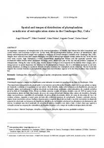
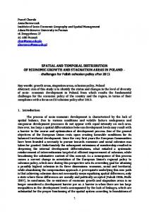
Results Expression of OPN mRNA. On day 3 after fracture, formation of periosteal callus occurred near the site of the fracture, and woven bone was observed in the subperiosteal region (hard callus). Most of the osteoprogenitor cells in the newly-formed woven bone expressed a moderately intense signal for OPN (Figs 1A and 1B), but only a slight OPN signal was seen in the cells of the proliferating periosteum (Fig. 1B). At this stage, an OC signal was also detected in the cells of subperiosteal woven bone (Fig. 1C) and its pattern of expression resembled that of OPN. Near the fracture site, granulation tissue covered the fracture gap (soft callus), but no signal for OPN was seen in it (Fig. 1A). In both the soft and hard callus, there was no signal when the OPN sense probe was used (Fig. 1D). On day 7, the thickness of the hard callus increased as the intramembranous ossification proceeded. TRAP-positive multinuclear cells (mature osteoclasts) were detected on the surface of the trabecular bone in the deep layer of the hard callus, indicating that remodelling of the callus had begun in these tissues at this stage of healing of the fracture. A moderately intense signal for OPN was seen in the hard callus, predominantly in the deep layer (Fig. 2A). OPN mRNA-expressing cells were identified as osteocytes within the bone matrix (Fig. 2C, arrowheads). OC mRNA was also expressed in the hard callus (Fig. 2B), but in contrast to day 3, its pattern of expression was different from that for OPN. OC mRNA was expressed in osteoblasts on the surface of trabecular bone (Fig. 2D). In the soft callus abundant cartilage tissue could be seen adjacent to the hard callus. Both proliferative and early hypertrophic chondrocytes were present at this stage, but none of these chondrocytes expressed OPN mRNA (data not given). At approximately day 10 after fracture, cartilage tissue in the soft callus began to be replaced by trabecular bone and the hard callus expanded towards the fracture gap. On day 14, the hard callus showed a wedge-shaped morphology in the sagittal plane (Fig. 3A). A characteristic pattern of OPN mRNA expression was seen within the callus in that OPN mRNA-
510
M. YAMAZAKI,
F. NAKAJIMA,
A. OGASAWARA,
H. MORIYA,
R. J. MAJESKA,
T. A. EINHORN
Fig. 1 Localisation of OPN and OC mRNAs on day 3 after fracture. Sections were hybridised with OPN (A, B) and OC (C) antisense probes and with the OPN sense probe (D). Figure 1A – Low-power photomicrograph of the OPN signal. The box showing the area of intramembranous ossification is enlarged in Figure 1B. Areas shown in C and D are similar fields to B. Figures 1B and 1C – OPN (B) and OC (C) signals are detected in cells of the newly-formed subperiosteal woven bone, but in Figure 1D no signal is seen (FS, fracture site; PO, periosteum; CB, cortical bone; bars in A = 500 µm, in B, C and D = 100 µm).
Fig. 2 Localisation of OPN and OC mRNAs on day 7 after fracture. Sections were hybridised with OPN (A, C) and OC (B, D) antisense probes. Figures 2A and 2B – Photomicrographs show OPN (A) and OC (B) signals in the hard callus. The boxes indicate the areas of callus remodelling which are enlarged in Figures 2C and 2D respectively. The OPN signal is detected in osteocytes (C, arrowheads) and the OC signal in osteoblasts (D) (PO, periosteum; CB, cortical bone; bars in A and B = 200 µm, in C and D = 50 µm).
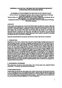
expressing cells were not uniformly distributed, but were restricted to discrete areas (Fig. 3B). Late hypertrophic chondrocytes at the cartilage-bone transitional area also expressed OPN mRNA (Fig. 3D, arrowheads). This pattern of distribution for OPN mRNA clearly differed from that for OC mRNA. The OC signal was detected in osteoblasts of the secondary but not the primary spongiosae (Fig. 3E). Under
high-power magnification, the OPN signal was detected in both osteocytes and osteoclasts (Fig. 3C) and these OPN mRNA-expressing cells were in close contact with each other. OPN-positive osteoclasts (Fig. 3C, arrows) were seen adjacent to bone surfaces in the vicinity of OP-positive osteocytes (Fig. 3C, arrowheads). In contrast, the OC signal was specifically localised in cuboidal osteoblasts (Fig. 3F). THE JOURNAL OF BONE AND JOINT SURGERY
SPATIAL AND TEMPORAL DISTRIBUTION OF CD44 AND OSTEOPONTIN IN FRACTURE CALLUS
511
Fig. 3 Localisation of OPN and OC mRNAs on day 14 after fracture. Sections were hybridised with OPN (A, B, C, D) and OC (E, F) antisense probes. Figure 3A – Low-power photomicrograph of the OPN signal. The area of the box showing soft and hard callus is enlarged in Figure 3B. An area shown in E is a similar field to that in B. Figure 3B – OPN mRNA-expressing cells are not uniformly distributed, but are restricted to discrete areas of the hard callus. The areas in the boxes are enlarged in Figures 3C and 3D, respectively. Figure 3C – The OPN signal is detected in osteocytes (arrowheads) and osteoclasts (arrows). OPN-positive osteoclasts attach to the bone surface or migrate in pits in the vicinity of OP-positive osteocytes. Figure 3D – The OPN signal is detected in late hypertrophic chondrocytes (arrowheads). Figure 3E – OC mRNA-positive cells are uniformly distributed in the secondary spongiosa of the hard callus. The area of the box is enlarged in Figure 3F and the OC signal is detected in cuboidal osteoblasts (FS, fracture site; HC, hypertrophic chondrocytes; bars in A = 500 µm, in B and E = 200 µm, in C, D and F = 50 µm).
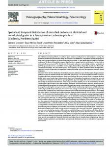
After day 21, osseous bridging occurred over the site of the fracture and remodelling of the hard callus continued. A moderate to weak OPN signal was still seen in osteocytes and osteoclasts in some restricted areas of the hard callus, as had been observed on day 14 (data not given). Immunolocalisation of CD44. On day 3 after fracture, moderately intense immunoreactivity for CD44 was detected in some polygonal cells located in the non-calcified area of the subperiosteal callus (Figs 4A and 4B). Faint staining for CD44 was also seen in osteoprogenitor cells on the surface of woven bone as well as in cells embedded in the bone matrix. Cells in the proliferating periosteum did not react with antibody to CD44 (Fig. 4A). On day 7, CD44 staining was seen in some fibroblastic cells of the soft callus (Fig. 4C), but chondrocytes in the VOL. 81-B, NO. 3, MAY 1999
soft callus, identifiable by metachromatic staining with Toluidine Blue at pH 4.1, were negative for CD44. In the deep layer of hard callus, where remodelling had begun, the surfaces of the osteoclasts were stained with antibody for CD44 (Fig. 4D, arrows), but osteocytes embedded in the bone matrix as well as osteoblasts on the surface of trabecular bone showed only a faint immunoreactivity for CD44 (Fig. 4D). After day 7, remodelling of the callus gradually progressed, and it was possible to detect the typical morphological features of osteoblasts, osteocytes, osteoclasts and chondroclasts. In the hard callus on day 14, the cell surface and canaliculi of the osteocytes showed relatively strong immunoreactivity for CD44 (Fig. 4E, arrows) but this was not seen in the cuboidal osteoblasts (Fig. 4E, arrowheads).
512
M. YAMAZAKI,
F. NAKAJIMA,
A. OGASAWARA,
H. MORIYA,
R. J. MAJESKA,
T. A. EINHORN
Fig. 4 Immunohistochemistry for CD44 on day 3 (A, B), day 7 (C, D), day 14 (E), day 21 (F, G) and day 28 (H) after fracture. Figure 4A – There is CD44 immunostaining in the subperiosteal hard callus. Figure 4B – The area of the box is enlarged and shows CD44 staining in some polygonal-shaped cells in a non-calcified area of the subperiosteal callus. Figures 4C and 4D – There is CD44 staining in some fibroblastic cells in the soft callus (C) and on the surface of multinuclear cells of the hard callus (D, arrows). Figure 4E – In the hard callus the cell bodies and canalicular processes of osteocytes are CD44-positive (arrows), whereas osteoblasts are negative for CD44 (arrowheads). Figure 4F – Intense CD44 staining is detected on the basolateral plasma membrane of osteoclasts in the hard callus (arrows). Figure 4G – At the front of endochondral ossification the CD44 protein is localised on chondroclasts (arrows). Figure 4H – In hard callus CD44 staining is detected in osteocytes (arrows), but not in typical cuboidal osteoblasts (arrowheads) (C and E, DAB-enhanced; PO, periosteum; CB, cortical bone; WB, woven bone; HC, hypertrophic chondrocytes; bars in A = 200 µm, in B to F = 50 µm).
Similar patterns of staining for CD44 were observed in the trabeculae of the hard callus until day 28 after fracture (Fig. 4H). In the mature trabecular bone after day 21, intense staining for CD44 was detected on the apical surface of
osteoclasts (Fig. 4F, arrows), but not on the basal resorbing surface of the cells. Intense CD44 immunostaining was also shown on the surface of chondroclasts located along the ossification front (Fig. 4G, arrows), whereas no staining for THE JOURNAL OF BONE AND JOINT SURGERY
SPATIAL AND TEMPORAL DISTRIBUTION OF CD44 AND OSTEOPONTIN IN FRACTURE CALLUS
513
Fig. 5 Hyaluronic acid (HUA) labelling on day 3 (A, B), day 7 (C, D), day 14 (E, F) and day 21 (G, H) after fracture. Figure 5A – Low-power photomicrograph of HUA labelling. Figure 5B – The area of intramembranous ossification in a box is enlarged and shows relatively strong labelling for HUA in capsule-like fibrous tissue covering the periosteum (A, B, E and G, arrowheads). Subperiosteal non-calcified loose connective tissues are labelled with HUA (arrows). Figures 5C – There is HUA labelling in the hard callus. Figure 5D – The area of the box is enlarged which shows HUA-positive non-calcified loose connective tissues in the superficial layer of the hard callus. Figure 5E – Low-power photomicrograph of HUA labelling. Figure 5F – The area of the box showing endochondral ossification is enlarged and shows intense HUA labelling in the intercellular matrix of hypertrophic chondrocytes. Figure 5G – Low-power photomicrograph of HUA labelling. Figure 5H – The area of hard callus in the box is enlarged and shows a cortical bone-like structure, with HUA labelling within the lacunae of osteocytes (FS, fracture site; PO, periosteum; CB, cortical bone; HC, hypertrophic chondrocytes; bars in A, E and G = 500 µm, in B and C = 200 µm, in D, F and H = 50 µm).
CD44 was seen in hypertrophic chondrocytes (Fig. 4G). Distribution of hyaluronic acid. From day 3 to day 28 after fracture, relatively strong labelling of HUA was detected in capsule-like fibrous tissues covering the periosteum VOL. 81-B, NO. 3, MAY 1999
(Figs 5A, 5B, 5E and 5G, arrowheads). In the hard callus on day 3, moderately intense labelling of HUA was found in the loose connective tissues under the periosteum (Fig. 5B, arrows) which were not yet calcified and were sur-
514
M. YAMAZAKI,
F. NAKAJIMA,
A. OGASAWARA,
rounded by woven bone. Little HUA labelling was seen in the proliferating periosteum and the subperiosteal woven bone (Fig. 5B). In the soft callus at this stage, the intercellular matrix was diffusely labelled with HUA (Fig. 5A). In the superficial layer of hard callus on day 7, the noncalcified loose connective tissues surrounded by bone were HUA-positive (Figs 5C and 5D) as was observed on day 3. In contrast, there was little HUA labelling in the deep layer of the hard callus (Fig. 5C), where TRAP-positive multinuclear cells were present and callus remodelling was underway. On day 14, most of the hard callus contained haematopoietic marrow and there was little HUA labelling in the hard callus (Fig. 5E). In the soft callus at this stage, intense HUA labelling was found in the intercellular matrix surrounding hypertrophic chondrocytes (Fig. 5F). No HUA labelling was seen in the matrix around proliferating chondrocytes. From day 21, a part of the hard callus, especially beneath the periosteum, began to show a cortical bone-like structure (Fig. 5G). In this area, labelling for HUA was first detected within the lacunae of osteocytes (Fig. 5H). In the deep layer of hard callus where predominantly a trabecular bone structure had developed, it was not possible to detect intracellular labelling for HUA at any of the stages of healing of the fracture (Fig. 5G).
Discussion Role of osteopontin during fracture healing. In our study we found that OPN mRNA was preferentially expressed in the mineralisation and remodelling phases of the healing of fractures although with clearly distinct patterns of expression in each. In the mineralisation phase, OPN mRNA expression by osteoprogenitor cells at the early stage of the formation of subperiosteal callus resembled the pattern 9 seen in developing mouse calvariae and mandibulae. In those tissues, OPN mRNA was localised in cells in the mineralising tissues. The expression of OPN by osteoblasts in advance of mineral deposition and its co-localisation with calcified sites in bone and in pathological lesions of 22 23 breast cancer and atherosclerotic plaques have suggested that OPN is a regulator of the mineralisation process. Its precise function remains unclear, however, because both inhibitory and stimulatory effects of OPN on nucleation and growth of mineral crystals have been 24-26 reported. During the remodelling phase in hard callus, OPN was expressed not by osteoblasts, but by cells associated with tissue turnover. We detected OPN mRNA in osteoclasts associated with trabecular bone surfaces and in distinct groups of osteocytes near the OPN-positive osteoclasts. This distribution clearly differed from OC, which was specifically localised in cuboidal osteoblasts. We found a similar pattern in remodelling soft callus, where OPN
H. MORIYA,
R. J. MAJESKA,
T. A. EINHORN
mRNA was localised to mature hypertrophic chondrocytes near multinucleated chondroclasts. Thus, a change in the cell types which expressed OPN mRNA occurred during maturation of callus. This pattern of site- and stage-specific expression suggests that OPN may also play a distinct functional role during the healing of fractures. Previous reports have suggested that OPN is involved in the resorption of non-injured bone. OPN mRNA expression 10 by osteoclasts was shown in trabeculae of the femur and 11 tibia of adult rats, while human osteoclasts were shown to express OPN mRNA and deposit OPN on to resorption 12 surfaces of trabecular bone. OPN expression by osteocytes in rat trabecular bone was also seen after ovariectomy, and was apparently correlated with remodelling 11 activity. The preferential expression of OPN mRNA in fracture callus by groups of osteocytes and by adjacent osteoclasts further supports the idea that osteocytes are 27 active participants in the control of bone metabolism. The distribution pattern also suggests that these groups of osteocytes are acting in a co-ordinated fashion, a likely consequence of their organisation into networks linked by gap 28 junctions. As in bone formation, the function of OPN in bone turnover has yet to be fully established. It has been proposed to anchor osteoclasts to bone based on its association 29 with osteoclasts in situ and its ability to support integrin7 mediated adhesion and osteoclast activation. Our data are consistent with this function for OPN in fracture callus, but raise the possibility that it may have other roles as well. Possible interaction of CD44 with osteopontin and hyaluronic acid during fracture healing. As expected from its widespread distribution, we found CD44 to be expressed by several cell types in fracture callus throughout the healing process, with an immunostaining pattern which often indicates localisation on the cell surface. In remodelling hard callus, a considerable amount of CD44 was localised to osteoclasts and osteocytes including their canalicular processes, but typical cuboidal osteoblasts did not show CD44 expression. These findings are consistent with previous data on CD44 localisation in normal tibiae of adult and newborn 3,4 rats. Of particular interest, however, was the apparent codistribution of CD44 with OPN mRNA in osteoclasts and in nearby groups of osteocytes. This pattern suggests that the same cells may produce both OPN and CD44 and also raises the possibility that OPN may interact with osteoclasts 7 not only through integrins but also through CD44. The report that OPN-CD44 interactions triggered chemotaxis in 14 fibroblasts indicates that this can alter cell-matrix adhesion. We suggest therefore that OPN binding to CD44 may facilitate movement of osteoclasts as they form resorption pits on trabecular surfaces. It is also possible that the OPNCD44 interactions may be involved in the communication between osteocytes and between osteocytes and osteoclasts on bone surfaces. Since OPN is a secreted protein and a major non-collagenous component of bone matrix, the distribution of OPN protein in our rat fracture model needs THE JOURNAL OF BONE AND JOINT SURGERY
SPATIAL AND TEMPORAL DISTRIBUTION OF CD44 AND OSTEOPONTIN IN FRACTURE CALLUS
to be evaluated by immunohistochemistry. A precise localisation of OPN protein, in addition to that of CD44 protein, would give us clues to the further clarification of the OPNCD44 interactions during the healing of fractures. HUA is known to be a ligand for CD44 in a variety of 15 tissues including those of the musculoskeletal system. 4 Noonan et al demonstrated the co-localisation of CD44 and HUA in osteocytes and osteoclasts of the proximal tibiae of weanling rats, suggesting that CD44 is an HUAbinding protein in bone tissues. We found HUA to be distributed abundantly at many sites throughout fracture callus including regions of fibrous connective tissue, the intercellular matrix of hypertrophic cartilage and the lacunae of osteocytes from regions resembling cortical bone. We found little HUA, however, in remodelling hard callus where CD44 was present. These findings suggest that OPN rather than HUA is the predominant ligand for CD44 in this region of callus. The functional consequences resulting from CD44 binding to OPN rather than to HUA have yet to be determined but our data suggest that such a transition may be associated with the onset of bone remodelling. The authors wish to acknowledge Dr Mark Thiede for providing us the cDNAs of osteopontin and osteocalcin. No benefits in any form have been received or will be received from a commercial party related directly or indirectly to the subject of this article.
References 1. McKibbin B. The biology of fracture healing in long bones. J Bone Joint Surg [Br] 1978;60-B:150-62. 2. Thiery JP, Boyer B. The junction between cytokines and cell adhesion. Curr Opin Cell Biol 1992;4:782-92. 3. Nakamura H, Kenmotsu S, Sakai H, Ozawa H. Localization of CD44, the hyaluronate receptor, on the plasma membrane of osteocytes and osteoclasts in rat tibiae. Cell Tissue Res 1995;280:225-33. 4. Noonan KJ, Stevens JW, Tammi R, et al. Spatial distribution of CD44 and hyaluronan in the proximal tibia of the growing rat. J Orthop Res 1996;14:573-81. 5. Hughes DE, Salter DM, Simpson R. CD44 expression in human bone: a novel marker of osteocytic differentiation. J Bone Miner Res 1994;9:39-44. 6. Jamal HH, Aubin JE. CD44 expression in fetal rat bone: in vivo and in vitro analysis. Exp Cell Res 1996;223:467-77. 7. Miyauchi A, Alvarez J, Greenfield EM, et al. Recognition of osteopontin and related peptides by an (alpha v beta 3) integrin stimulates immediate cell signals in osteoclasts. J Biol Chem 1991; 266:20369-74. 8. Oldberg A, Franzen A, Heinegard D. Cloning and sequence analysis of rat bone sialoprotein (osteopontin) cDNA reveals an Arg-Gly-Asp cell-binding sequence. Proc Natl Acad Sci USA 1986;83:8819-23. 9. Nakase T, Takaoka K, Hirakawa K, et al. Alterations in the expression of osteonectin, osteopontin and osteocalcin mRNAs during the development of skeletal tissues in vivo. Bone Miner 1994;26: 109-22.
VOL. 81-B, NO. 3, MAY 1999
515
10. Ikeda T, Nomura S, Yamaguchi A, Suda T, Yoshiki S. In situ hybridization of bone matrix proteins in undecalcified adult rat bone sections. J Histochem Cytochem 1992;40:1079-88. 11. Ikeda T, Yamaguchi A, Yokose S, et al. Changes in biological activity of bone cells in ovariectomized rats revealed by in situ hybridization. J Bone Miner Res 1996;11:780-8. 12. Dodds RA, Connor JR, James IE, et al. Human osteoclasts, not osteoblasts, deposit osteopontin onto resorption surfaces: an in vitro and ex vivo study of remodeling bone. J Bone Miner Res 1995; 10:1666-80. 13. Hirakawa K, Hirota S, Ikeda T, et al. Localization of the mRNA for bone matrix proteins during fracture healing as determined by in situ hybridization. J Bone Miner Res 1994;9:1551-7. 14. Weber GF, Ashkar S, Glimcher MJ, Cantor H. Receptor-ligand interaction between CD44 and osteopontin (Eta-1). Science 1996;271: 509-12. 15. Underhill C. CD44: the hyaluronan receptor. J Cell Sci 1992;103: 293-8. 16. Carter WG, Wayner EA. Characterization of the class III collagen receptor, a phosphorylated, transmembrane glycoprotein expressed in nucleated human cells. J Biol Chem 1988;263:4193-201. 17. Jalkanen S, Jalkanen M. Lymphocyte CD44 binds the COOHterminal heparin-binding domain of fibronectin. J Cell Biol 1992;116: 817-25. 18. Kennel SJ, Lankford TK, Foote LJ, Shinpock SG, Stringer C. CD44 expression on murine tissues. J Cell Sci 1993;104:373-82. 19. Bonnarens F, Einhorn TA. Production of a standard closed fracture in laboratory animal bone. J Orthop Res 1984;2:97-101. 20. Yamazaki M, Majeska RJ, Yoshioka H, Moriya H, Einhorn TA. Spatial and temporal expression of fibril-forming minor collagen genes (types V and XI) during fracture healing. J Orthop Res 1997;15: 757-64. 21. Motegi H, Yamazaki M, Goto S, Mikata A, Moriya H. Proliferating cell nuclear antigen in hypertrophied spinal ligaments: immunohistochemical localization of proliferating cell nuclear antigen in hypertrophied posterior longitudinal ligament of the cervical spine. Spine 1998;23:305-10. 22. Hirota S, Ito A, Nagoshi J, et al. Expression of bone matrix protein messenger ribonucleic acids in human breast cancers: possible involvement of osteopontin in development of calcifying foci. Lab Invest 1995;72:64-9. 23. Giachelli CM, Bae N, Almeida M, et al. Osteopontin is elevated during neointima formation in rat arteries and is a novel component of human atherosclerotic plaques. J Clin Invest 1993;92:1686-96. 24. Hunter GK, Kyle CL, Goldberg HA. Modulation of crystal formation by bone phosphoproteins: structural specificity of the osteopontinmediated inhibition of hydroxyapatite formation. Biochem J 1994;300: 723-8. 25. Sodek J, Chen J, Nagata T, et al. Regulation of osteopontin expression in osteoblasts. Ann NY Acad Sci 1995;760:223-41. 26. Campbell AA, Ebrahimpour A, Perez L, Smesko SA, Nancollas GH. The dual role of polyelectrolytes and proteins as mineralization promoters and inhibitors of calcium oxalate monohydrate. Calcif Tissue Int 1989;45:122-8. 27. Aarden EM, Burger EH, Nijweide PJ. Function of osteocytes in bone. J Cell Biochem 1994;55:287-99. 28. Palumbo C, Palazzini S, Marotti G. Morphological study of intercellular junctions during osteocyte differentiation. Bone 1990;11: 401-6. 29. Reinholt FP, Hultenby K, Oldberg A, Heinegard D. Osteopontin: a possible anchor of osteoclasts to bone. Proc Natl Acad Sci USA 1990; 87:4473-5.