Symptomatic nonunion after fracture of the ... - Semantic Scholar
Recommend Documents
Nov 25, 2009 - (page number not for citation purposes). Cases Journal. Open Access. Case Report. Patellofemoral arthroplasty for symptomatic nonunion after.
cutaneous vertebroplasty at the thoracolumbar junction (T12, L1). The follow-up period was 15â27 months. The occurrence of new symptomatic vertebral ...
MANAGEMENT USING THE ILIZAROV CIRCULAR FIXATOR. V. R. Patel, D. K. Menon, R. D. Pool, R. B. Simonis. From St Peter's Hospital, Chertsey, England.
contraindications for esophagectomy. 2008 Published by European Association for Cardio-Thoracic Surgery. All rights reserved. Keywords: Esophageal ...
(ACL) reconstruction, synovectomy, chondroplasty and debridement. The types of anaesthesia included general, regional and local. Comorbidities were defined ...
living donors. Samhan and Al-Mousawi showed that the incidence of symptomatic lymphocele is more when cadaveric kidney allografts are used. (6) Hence, this ...
Sixteen patients who underwent a revision operation for nonunion of fractures of the distal humerus following previous internal fixation were reviewed at a mean ...
Aug 1, 2015 - cological systemic treatment for fracture healing or nonunion. ... In both cases, partial bone union began to be observed on radiographs.
autologous bone grafting and osteosynthesis with screws ... J Bone Joint Surg [Br] 2003;85-B:953-5. ... THE JOURNAL OF BONE AND JOINT SURGERY. Fig. 1.
Nov 16, 2014 - bones had absence of intra-operative puncate bone bleeding, which was confirmed ..... fixation by a proximal to distal screw. Ann Chir Main ...
Dec 9, 2014 - menopausal state, age, body weight, and comorbidity) and treatment parameters ... subsequent other pelvic bone fractures [16,20]. Femur neck.
made 1714 reconstructions of the anterior cruciate ligament of the knee using bone-patellar tendon-bone technique, and 7 pa- tients had fracture of the patella ...
Mar 8, 2011 - Ninkovic M, Love S, Tom BDM, Alexander GJM,. Compston JE. ... Black DM, Delmas PD, Eastell R, Reid IR, Boonen S,. Cauley JA, et al.
Jan 1, 2016 - Introduction: Periprosthetic fractures of the femur are uncommon, but at times may lead to complications especially in elderly patients.
Key words: arthroplasty, replacement, knee; fractures, bone; patella .... Stress fracture of the patella following duopatellar total knee arthroplasty with patellar.
Male sexual dysfunction after fracture of the pelvis is more common than previously sup- posed with rates as high as 30% reported when the complaint is ...
Apr 29, 2016 - of nonunion or implant failure.1-4 Subtrochanteric fractures show two age-dependent peaks in frequency, with fractures in younger patients.
Scaphoid fracture nonunion - ResearchGate › publication › fulltext › Scaphoid-... › publication › fulltext › Scaphoid-...by MR Bervian · 2015 · Cited by 21 · Related articlesNov 16, 2014 — of scaphoid fractures heal without surgery, most case seri
Dec 16, 2012 - We describe a case of radiation-associated fracture nonunion of the clavicle, which was treated by locking plate fixation and autologous bone ...
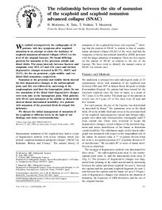
lunate advanced collapse (SLAC) of the wrist, and that the frequency of dorsal intercalated instability (DISI) increases with the longer duration of nonunion.
Nov 19, 2013 - Edward Roddy,1 Martin J Thomas,1 Michelle Marshall,1 Trishna Rathod,1. Helen Myers,1 Hylton B Menz,1,2 Elaine Thomas,1 George Peat1.
Aug 3, 2004 - University School of .... injury in four and a pedestrian vs train accident in one. At .... bler, one retiree, one cook, one mechanic and one un-.
KIC value, however, it remains an open question if KJIC is a useful measure of fracture toughness for such materials. When the results for the UFG state are ...
tors have been associated with recovery after a hip fracture, such as age, sex, ... Data Sources and Search Strategy. In March 2009 a ... Using this format, studies.
Symptomatic nonunion after fracture of the ... - Semantic Scholar
We have treated 14 patients (15 fractures) with nonunion of an intra-articular fracture of the body of the calcaneum. The mean follow-up was six years (2 to 8.5).
Symptomatic nonunion after fracture of the calcaneum DEMOGRAPHICS AND TREATMENT
A. P. Molloy, M. S. Myerson, P. Yoon From Institute for Foot and Ankle Reconstruction, Mercy Medical Center, Baltimore, USA
We have treated 14 patients (15 fractures) with nonunion of an intra-articular fracture of the body of the calcaneum. The mean follow-up was six years (2 to 8.5). A total of 14 fractures (93%) had initially been treated operatively with 12 (86%) having non-anatomical reductions. Four feet (27%) had concomitant osteomyelitis. Of the nonunions, 14 (93%) went on to eventual union after an average of two reconstructive procedures. All underwent bone grafting of the nonunion. The eventual outcome was a subtalar arthrodesis in ten (67%) cases, a triple arthrodesis in four (27%) and a nonunion in one (6%). Three patients had a wound dehiscence; all required a local rotation flap. The mean American Orthopaedic Foot and Ankle Society score at latest follow-up was 69, and the mean Visual analogue scale was 3. Of those who were initially employed, 82% (9 of 11) eventually returned to work. We present an algorithm for the treatment of calcaneal nonunion, and conclude that despite a relatively high rate of complication, this complex surgery has a high union rate and a good functional outcome.
Nonunion is a rarely reported complication of calcaneal fractures. In a series of 157 intra-articular fractures of the calcaneum treated by open reduction and internal fixation (ORIF), Zwipp et al1 had a rate of nonunion of 1.3%. A review of the English literature indicates five case reports, comprising a total of seven patients with calcaneal nonunion. Three accounts (five patients) describe nonunion of the anterior process.2-4 Another describes nonunion of the sustentaculum tali in a middle-aged man with nonunion involving the sustentaculum tali resulting in tarsal tunnel syndrome.5 His symptoms resolved after excision of the ununited fragment. There is only one report of nonunion of an intra-articular fracture of the body of the calcaneum. This case involved a 36-year-old woman with an undisplaced fracture treated initially with early mobilisation. At six months, because of radiological and CT-documented nonunion, she underwent ORIF with a reconstruction plate and union was achieved.6 We present the senior author’s (MSM) experience with a consecutive series of patients treated for nonunion of an intra-articular fracture of the body of the calcaneum.
Patients and Methods Between 1993 and 2004, 18 patients (19 fractures) were treated for calcaneal nonunion and followed up prospectively. Of these, 14 patients
(15 fractures) had follow-up of two years or more since the time of the injury, with a mean of six years (2 to 8.5), and form the basis of this paper. Three patients were unavailable for complete evaluation as they lived too far away, and one was lost to follow-up. Only one had initially been treated at the authors’ institution, the remainder being referred from elsewhere. There were nine men (64%) and five women (36%). Their mean age was 42 years (27 to 64). Six patients (43%) were labourers, primarily roofers and construction workers. Eight patient injuries (57%) were sustained in falls and six (43%) in motor vehicle accidents. At the time of initial injury, seven patients (50%) were smokers. Data regarding demographics, the characteristics of the initial fracture, initial treatment, time to nonunion, and the radiological parameters, including Bohler’s angle, talar declination, heel widening, and the presence of subtalar or calcaneocuboid arthritis, were collected retrospectively at the time of the diagnosis of nonunion (Tables I and II). Subsequently, the following information was collected prospectively: the type of reconstructive surgery to treat the nonunion; the final results; complications; final range of movement of the ankle; return to work; the angle of talar declination; the final visual analogue scale (VAS), and final American Orthopaedic Foot and Ankle Society7 (AOFAS) score. THE JOURNAL OF BONE AND JOINT SURGERY
SYMPTOMATIC NONUNION AFTER FRACTURE OF THE CALCANEUM
1219
Table I. Demographics of initial injury, treatment and complications
16
Non-weightbearing (weeks)
Patient
Sanders grade
Treatment
Type of fixation
Initial complications
1 2 Right Left 3 4 5 6 7 8 9 10 11 12 13 14
3 Not known 2 3 3 3 2 Not known 3 3 Not known 3 TT† TT 4
* ORIF, open reduction and internal fixation † TT, tongue type/joint degression
Table II. Demographics of calcaneal morphology and arthritis Patient 1 2 Right Left 3 4 5 6 7 8 9
Time to nonunion diagnosis (mths) Bohler’s angle (˚) Angle of talar declination (˚) Widening (CT scan) 3 6 6 6 12 12 9 12 6 9
5 30 20 20 0 0 20 0 0 10
22 23 25 25 0 10 10 -20 0 0
Mild Mild None Moderate Moderate Moderate Mild Moderate CT not performed Severe
10 11 12
12 5 6
5 0 10
0 0 20
Severe Severe Moderate
13 14
6 9
0 20
0 10
CT not performed Severe
Operative technique. Careful
pre-operative planning is essential. Thorough clinical evaluation and standard weight-bearing radiographs of the calcaneum, foot and ankle should be carried out and, where possible, compared with the full series of previous pre- and post-operative radiographs. A CT scan with reconstruction views in all planes provides valuable information on the exact nature of the deformity, the ununited fragments and the quality of the bone (Fig. 1). Avascular necrosis and subchondral sclerosis are invariably present, and although this can be demonstrated by plain radiographs, a CT scan is useful to clarify this diagnosis, which is ultimately confirmed intra-operatively. The site of the skin incision is determined by the type of deformity and the reconstruction to be performed. Thus, there is a negative angle of talar declination and painful anterior impingement, a subtalar bone block arthrodesis is
necessary. For these patients, a vertical retrofibular incision is used.8 If, however, an in situ subtalar arthrodesis is to be performed, a standard incision is made centred over the sinus tarsi, regardless of the previous incision which had been used for the ORIF of the fracture.9 Using the original extensile incision to perform an in situ arthrodesis is difficult, unnecessary, and can create the added risk of wound necrosis. We have noted that the subcutaneous dissection performed using either of these incisions does not create an added risk to wound healing if the reconstruction is performed more than six months later. If it is not possible to remove the retained hardware through the chosen approach, it should be approached percutaneously.10 If a bone block subtalar arthrodesis is necessary as part of a triple arthrodesis, then the same vertical retrofibular incision is used as well as a short longitudinal incision over each of the talonavicular and calcaneocuboid joints.
1220
A. P. MOLLOY, M. S. MYERSON, P. YOON
Fig. 1a
Fig. 1b
Fig. 1c
a) Sagittal CT scan showing nonunion of a conservatively-treated calcaneal fracture. b) Lateral radiograph of the same patient demonstrating the nonunion with elevation of the posterior calcaneal tuberosity. c) Post-operative lateral radiograph demonstrating improvement of the talar declination angle, a subtabular arthrodesis and healing of the nonunion.
Whichever arthrodesis is to be performed, the same initial preparation of the subtalar joint is necessary. A lateral approach is made to the joint and the lateral wall of the calcaneum. Because of deformity and increased heel width, a lateral wall exostectomy is usually necessary and is performed following removal of the hardware (Fig. 2). Preparation of the nonunion is then undertaken. Frequently it is necessary to carry out an osteotomy through the old fracture site. This is needed if there is a partial union, which may be fibrous or bony, and is usually on the plantar surface. It can be determined by inserting a non-toothed laminar spreader into the nonunion site, noting hinging rather than longitudinal separation of the main fragments. Thorough debridement of all fibrous tissue and avascular bone is undertaken and continued until punctate bleeding is observed. This is augmented with multiple perforations of the margins of the nonunion with a 2 mm drill bit. Although the bone margins should be debrided in this way,
it may not be possible, particularly distally, at the neck of the calcaneum, which is often densely sclerotic. Here, multiple perforations are relied upon for preparation of the nonunion surface. If a bone block arthrodesis of the subtalar joint is to be performed simultaneously, a laminar spreader is inserted into the subtalar joint and on to the calcaneal tuberosity, which is then depressed towards a more anatomical position (Fig. 3a) This corrects the altered angle of talar declination.11-13 To enable this relative lengthening of the calcaneum, a percutaneous or open lengthening of the tendo Achillis is usually necessary. At this stage, the true size of the defect becomes evident. It is then filled with allograft cancellous chips or a bicortical structural graft, depending on its size and the deformity, as noted above (Fig. 3b to 3d). In order to provide enhanced osteo-induction, a platelet-rich plasma concentrate (The Symphony System; DePuy, Warsaw, Indiana) was used in conjunction THE JOURNAL OF BONE AND JOINT SURGERY
SYMPTOMATIC NONUNION AFTER FRACTURE OF THE CALCANEUM
Fig. 2a
Fig. 2b
1221
Fig. 2c
a) Lateral radiograph showing nonunion of the calcaneum, a decrease in the talar declination angle, and avascular necrosis of the distal calcaneum. b) Lateral radiograph following staged removal of hardware and bone debridement. c) Lateral radiograph following subtalar arthodesis with bone block graft and repair of the nonunion.
Fig. 3c
Fig. 3a
Fig. 3d
Fig. 3b
a) Intra-operative view of a laminar spreader in the subtalar joint and nonunion. b) Intra-operative view of the insertion of the tricortical allograft. c) Intra-operative fluoroscopic lateral view after debridement of the nonunion site, showing the size of the defect once the deformity is corrected with a laminar spreader. d) Intra-operative fluoroscopic view showing the position of the bone graft, the screw fixation, and restoration of both heel height and talar declination.
with the allograft for five of the 15 nonunions when it became available for the later cases. Fixation of the subtalar joint and the nonunion is determined by the size and extent of the defect in the calcaneum and the type of arthrodesis to be performed. The nonunion is usually addressed first, with internal fixation using cannulated screws. Reliable and reproducible fixation cannot be achieved if this fragment is less than 2 cm long anteroposteriorly. Then, a triple arthrodesis should be undertaken. Fixation normally involves the use of 6.5 mm cannulated screws for the subtalar joint and 5.0 mm cannulated screws for the calcaneocuboid and talonavicular joints if a triple arthrodesis is undertaken. If poor-quality bone and comminution are encountered, larger fully threaded screws, multiple smaller divergent partially threaded screws or, especially for a triple arthrodesis, a trapezoidal reconstruction plate, are used. Subluxation or dislocation of the peroneal tendons, associated in some cases with chronic lateral instability of the VOL. 89-B, No. 9, SEPTEMBER 2007
ankle, is often present and will need to be addressed at the same time following the bony reconstruction. This is accomplished by deepening the peroneal groove with a large ovalshaped burr and repair of the retinaculum, supplemented by ligament reconstruction if there is instability. We have found the modification of the Chrisman and Snook procedure14 to be useful if the retinacular and periosteal flap is not sufficient.
Results The fractures included two (13%) open and 13 (87%) closed injuries. Both open injuries were Gustilo15 grade I. It was possible to classify the initial fracture patterns of 12 of the 15 nonunions using the Sanders classification system;16 two of these 12 (17%) were Sanders 2, seven (58%) Sanders 3, one (8%) Sanders 4, and two (17%) tongue-type/joint depression (i.e. complex combinationtype fractures).
1222
A. P. MOLLOY, M. S. MYERSON, P. YOON
Table III. Operations performed Surgery Patient
Number of operations 1
2
3
4
1
2
Debridement, insertion of antibiotic cement
0
0
2 Right
1
0
0
0
Left
3
Debride, osteomyelitis, remove hardware, insertion of antibiotic cement Bone graft nonunion, subtalar fusion Bone graft nonunion, subtalar fusion
Nonunion, subtalar joint revised
0
Bone graft, nonunion, subtalar bone block fusion Bone graft, nonunion, subtalar bone block fusion Debride, osteomyelitis, remove hardware, insertion of antibiotic cement
Abductor flap for wound dehiscence 0
Nonunion subtalar joint revised with triple arthrodesis 0 0
0 Subtalar bone block fusion
Bone graft, nonunion, subtalar bone block fusion Bone graft, nonunion, subtalar bone block fusion Debride, osteomyelitis, remove hardware, insertion of antibiotic cement Bone graft, subtalar and calcaneocuboid fusion Bone graft nonunion, subtalar bone block fusion Cancellous bone graft, subtalar fusion Bone graft, nonunion, subtalar arthrodesis Bone graft, nonunion, subtalar bone block fusion Bone graft, nonunion of subtalar fusion
0
Bone graft subtalar nonunion. Abductor flap for wound dehiscence 0 Debride, osteomyelitis
Bone graft, subtalar fusion 0
3
2
4
1
5
4
6
1
7
4
8
2
9
3
10
1
11
1
12
3
13
1
14
4
Repeat debridement, cement
Abductor flap for wound dehiscence Bone graft nonunion, subtalar bone block fusion
0
0
0
Bone graft and calcaneocuboid Triple arthrodesis fusion 0 0
0
0
0
0
Subtalar arthrodesis
Triple arthrodesis
0
0
0
0
Calcaneal osteotomy
Revise subtalar fusion
Triple arthrodesis with double bone block graft
The initial treatment had consisted of ORIF in 14 fractures (93%) and non-operative treatment in one (7%). Of the patients initially treated operatively, the quality of reduction based on the initial post-operative radiographs was noted. The initial reduction was poor in 12 patients (86%) and good or excellent in two (14%). One of the fractures (7%) treated with ORIF also underwent primary subtalar arthrodesis. Three (20%) were complicated by osteomyelitis and a further three (20%) by wound dehiscence without deep infection (Table I). The mean time to diagnosis of nonunion was 7.9 months (3 to 12). The neck of the calcaneum was involved in 12 nonunions (80%), with the rest involving either the body and/or the tuberosity; 14 (93%) had subtalar and three (20%) calcaneocuboid arthritis. There were CT scans available for review in 13 cases and showed evidence of heel widening in 12 (92%), which was deemed to be mild in three (25%), moderate in five (42%) and severe in four (33%). The mean Bohler’s angle was 9.3˚ (0˚ to 20˚) and the mean angle of talar declination was 8.3˚ (-20˚ to +25˚). All 15 nonunions had subtalar arthritis and three (20%) also had calcaneocuboid arthritis (Table II). The 15 nonunions underwent a mean of two (one to four) reconstructive procedures (Table III). Deep bony
0
infections were managed with staged debridement of nonviable bone, copious irrigation, and insertion of antibioticimpregnated cement beads. This was repeated until clinical and radiological evidence of infection had resolved. Three of 15 cases (20%) were so treated and all went on to have clinical, haematological and radiological resolution of infection. Two of these patients required two procedures and one required four because of wound dehiscence. Although bone graft was used in all patients, ten cases (67%) required either a bone block or cancellous impaction grafting to restore heel height with bone blocks used in nine and cancellous impaction grafting in one. Some form of arthrodesis was required in 14 cases (93%). There were 19 subtalar arthrodeses, four triple arthrodeses, two calcaneocuboid fusions, six debridements of osteomyelitis and three local rotation flaps overall. One foot required a Dwyer-type calcaneal osteotomy to correct heel varus. The mean angle of talar declination for the entire group improved from 8.3˚ (-20˚ to +25˚) to 21˚ (10˚ to 27˚) at final review. This improvement was statistically significant (p = 0.001) using Student’s t-test. The final Bohler’s angle was not measured because 13 patients (93%) underwent subtalar arthrodesis. THE JOURNAL OF BONE AND JOINT SURGERY
SYMPTOMATIC NONUNION AFTER FRACTURE OF THE CALCANEUM
Three nonunions (20%) were complicated by wound dehiscence following the subsequent salvage reconstructive surgery. One of these patients developed osteomyelitis. Two of these three patients had developed wound dehiscence after their initial ORIF and two were smokers. Serial clinical and laboratory examination with inflammatory markers was performed. If these variables had improved after eight weeks, definitive intervention was undertaken. If the situation had not resolved by 10 to 12 weeks a repeat debridement with cement insertion was carried out. All eventually healed with debridement and a local rotation flap. The eventual mean AOFAS score for these patients was 73 (60 to 88) at a mean of 6.5 years (5.5 to 8). There was no clinical, haematological or radiological evidence of residual infection at final follow-up. Sural sensation was impaired or absent in eight cases (53%). Eight ankles (53%) regained a full range of movement and seven (47%) had a range of movement which was reduced by 25% to 50%, which significantly affected the clinical outcome. The mean AOFAS score in patients with limitation of movement was 53 (40 to 65), whereas those without limitation of movement had a mean of 71 (45 to 88). This difference was statistically significant (Student’s t-test, p = 0.016). A total of 14 nonunions (93%) eventually healed. Of the 12 non-infected cases, six (50%) healed after the first procedure and six required at least one further operation. The final outcome was ten (67%) subtalar fusions, four (27%) triple arthrodeses and one (7%) nonunion with a cement spacer in situ. This last patient with persistent nonunion is a 52-year-old physician who sustained an open grade I, Sanders 3 calcaneal fracture in a motor vehicle accident. He underwent initial debridement followed by delayed ORIF, with excellent reduction at two weeks after injury. He subsequently went on to develop nonunion, diagnosed at three months, with concomitant osteomyelitis. He underwent debridement twice, with the insertion of antibiotic cement. However, the cement spacer fitted so well that it acted as an in situ arthrodesis. Currently, at 66 months’ follow-up, he has no signs or symptoms of infection and a healed wound. He has an AOFAS score of 88 and a VAS score of 2, and takes no analgesic or anti-inflammatory medication. He has been working at his initial occupation since eight weeks after operation and does not desire further surgery. Clinically, seven patients (50%) still took some form of analgesic medication at final review, and eight (57%) had sural nerve dysfunction. The final mean AOFAS score for the whole group was 69 (40 to 88) and the mean VAS score was 3 (1 to 8). Nine of 11 patients (82%) for whom data were available were able to return to work at their regular or modified occupation at a mean of 9.75 months (1 to 12) after their last surgical procedure.
Discussion Although notable for complications, including wound breakdown, infection and arthritis of the subtalar joint, calVOL. 89-B, No. 9, SEPTEMBER 2007
1223
caneal fractures do not commonly develop nonunion, and recent reports of external fixation and minimally-invasive treatment have given good results with low rates of complications.17,18 There are several reasons for the low rate of nonunion. As open reduction is usually performed via a lateral approach, only the branches of the peroneal artery should potentially be at risk. The extensile ‘L’-shaped lateral incision has been designed specifically to prevent injury to the osseous blood supply. As the calcaneum is composed mainly of cancellous bone it should heal well. However, 14 nonunions (93%) had been treated operatively initially, suggesting either that surgical intervention may compromise this blood supply, or that the more severe injuries, which have greater disruption of the blood supply, are more likely to be treated surgically. Smoking, diabetes, and open fractures have all been identified as independent risk factors for wound complications after internal fixation of calcaneal fractures,19 and it seems reasonable that they would also be risk factors for nonunion. We found that half of the patients in this series were smokers, and identify smoking as a risk factor. Only two patients had open fractures, although one has persistent nonunion as noted above. There was no evidence either clinically, haematologically, radiologically or intra-operatively of latent chronic osteomyelitis being the cause of nonunion, except in the three cases of overt osteomyelitis. The most significant risk factors for nonunion of a calcaneal fracture appear to be the quality of reduction and the appropriateness of fixation. In 86% of patients, the initial reduction and quality of fixation was poor, and the overall alignment gradually deteriorated. The majority of cases with poor reduction were where a lateral plate was used without supplementary screw fixation. Although there was a correlation between poor reduction and nonunion, this was not predictive in this small selected group. All the nonunions were grossly mobile at time of revision surgery, and had lost any stability provided by the initial treatment (Fig. 4). Although subtalar arthritis was present in each patient, their symptoms were caused by a combination of deformity, nonunion and arthritis. Although arthrodesis may have resolved one component, unless the nonunion and associated deformity are addressed simultaneously, the salvage surgery will not be successful. The first major decision of pre-operative planning is whether an allograft is required.13 This is determined by the angle of talar declination and, to a lesser degree, the relative elevation and rotation of the calcaneal tuberosity. A negative, or grossly abnormal, talar angle will cause anterior impingement and limitation of movement. As we have shown that ankle stiffness statistically significantly reduces the functional outcome, we consider it paramount that this deformity be addressed, rather than an in situ arthrodesis. Although there was a statistically significant improvement in the angle of talar declination, anatomical normality was not achieved in every case. However, the range of movement of the ankle was checked per-operatively, and no
1224
A. P. MOLLOY, M. S. MYERSON, P. YOON
One must be cautious about drawing conclusions from a relatively small number of patients. However, to our knowledge, this is the largest series of patients with calcaneal nonunion reported to date. Moreover, it provides useful information on the risk factors, characteristics, and appropriate treatment of this problem, as well as the likely prognosis. With appropriate treatment, this complex surgery can provide a good functional outcome with a high extension of eventual fracture union. No benefits in any form have been received or will be received from a commercial party related directly or indirectly to the subject of this article.
Fig. 4 Lateral radiograph showing nonunion following fixation using a calcaneal plate.
References 1. Zwipp H, Tscherne H, Thermann H, Weber T. Osteosynthesis of displaced intraarticular fractures of the calcaneus. Clin Orthop 1993;290:76-86. 2. Robbins MI, Wilson MG, Sella EJ. MR imaging of anterosuperior calcaneal process fractures. AJR Am J Roentgenol 1999;172:475-9. 3. Levine J, Kenin A, Spinner M. Non-union of a fracture of the anterior superior process of the calcaneus: a case report. J Bone Joint Surg [Am] 1959;41-A:178-80. 4. Piatt AB. Fractures of the promontory of the calcaneus. Radiology 1956;67:386-91.
patient had clinical or fluoroscopic impingement, whether anatomical normality had been achieved or not. The high rate of re-operation of 60% is to be expected because of the complexity of the surgery, the poor quality of the bone and the extent of the defects. Discussing this as part of informed consent will enable the patient to temper their expectations and allow for the inevitable time required for recovery. There was a high rate of wound dehiscence (20%) and all of these patients required a local rotation flap. However, these three patients had significant risk factors for this complication. No problems were found using a standard approach centred over the sinus tarsi for an in situ subtalar fusion even if an extended lateral approach had previously been used. These patients had all initially sustained a high-energy injury and most were manual labourers. They had been treated with protected weight-bearing for a prolonged period, with a low level of function while awaiting union and the mean time to the diagnosis of nonunion was 7.9 months (3 to 12). A pre-operative AOFAS score was not obtained and would have been impossible for the first two patients, who were recruited prior to the inception of this scoring system. The good overall functional outcome is shown by the mean AOFAS score of 69 (40 to 88), especially considering that the maximum score is 94, not 100, as hindfoot motion has been intentionally removed. The rate of returning to gainful employment (82%) is excellent for such a complex salvage operation.
5. Myerson MS, Berger BI. Nonunion of a fracture of the sustentaculum tali causing a tarsal tunnel syndrome: a case report. Foot Ankle Int 1995;16:740-2. 6. Thomas P, Wilson LF. Non-union of an os calcis fracture. Injury 1993;24:630-2. 7. Kitaoka HB, Alexander IJ, Adelaar RS, et al. Clinical rating system for the ankle-hindfoot, midfoot, hallux, and lesser toes. Foot Ankle Int 1994;15:349-53. 8. Myerson MS. Correction of complex hindfoot deformity after calcaneus fracture. In: Myerson MS, ed. Reconstructive foot and ankle surgery. Philadelphia: Elsevier Saunders, 2005;409-17. 9. Myerson M, Quill GE Jr. Late complications of fractures of the calcaneus. J Bone Joint Surg [Am] 1993;75-A:331-41. 10. Stamatis ED, Myerson MS. Percutaneous hardware removal after open reduction and internal fixation of calcaneus fractures. Orthopedics 2002;25:1025-7. 11. Easley MS, Trnka H-J, Schon LC, Myerson MS. Isolated subtalar arthrodesis. J Bone Joint Surg [Am] 2000;82-A:613-24. 12. Trnka H-J, Easley ME, Lam PW, et al. Subtalar distraction bone block arthrodesis. J Bone Joint Surg [Br] 2001;83-B:849-54. 13. Neufeld SK, Uribe J, Myerson MS. Use of structural allograft to compensate for bone loss in arthrodesis of the foot and ankle. Foot Ankle Clin 2002;7:1-17. 14. Myerson MS. Modification of the Christman-Snook procedure. In: Myerson MS, ed. Reconstructive foot and ankle surgery. Philadelphia: Elsevier Saunders, 2005;341-3. 15. Gustilo RB, Anderson JT. Prevention of infection in the treatment of one thousand and twenty five open fractures of long bones: restrospective and prospective analyses. J Bone Joint Surg [Am] 1976;58-A:453-8. 16. Sanders R, Fortin P, DiPasquale T, Walling A. Operative treatment in 120 displaced intra-articular calcaneal fractures: results using a prognostic computer tomography scan classification. Clin Orthop 1993;290:87-95. 17. Magnan B, Bortolozzi R, Marangon A, et al. External fixation for displaced fractures of the calcaneum. J Bone Joint Surg [Br] 2006;88-B:1474-9. 18. Stulik J, Stehlik J, Rysavy M, Wozniak A. Minimally-invasive treatment of intra-articular fractures of the calcaneum. J Bone Joint Surg [Br] 2006;88-B:163441. 19. Barei DP, Bellabarba C, Sangeorzan BJ, Benirschke SK. Fractures of the calcaneus. Orthop Clin North Am 2002;33:263-85.