AFTER CRITICAL POINT DRYING. J. NORMAN GRIM. From the Biology Department, Northern Arizona University, Flagstaff, Arizona. 86001, and the Zoology ...
ULTRASTRUCTURE OF CILIARY MICROTUBULES AFTER CRITICAL POINT DRYING J . NORMAN GRIM. From the Biology Department, Northern Arizona University, Flagstaff, Arizona 86001, and the Zoology Department, University of California, Davis, California 95616 . Author's present address is at Northern Arizona University INTRODUCTION
MATERIALS AND METHODS
Many studies have been conducted on the fine structure of the "9 + 2" microtubules from cilia and flagella . The techniques used include both thin sectioning of whole cilia or flagella and negative staining of isolated microtubules . In this study, cilia from four diverse species of ciliate protozoa :
Cells from which the cilia or flagella were studied were placed between two clean glass slides and ground by circular rotation . Both slides were then dipped into the clean water surface of a Langmuir trough on which a film of cellular contents including cilia or flagella readily spreads . The surface film was picked up on stainless steel, Formvar-coated, carbon-stabilized grids by touching them to the water surface . They were stained in 0 .2% uranyl acetate and dried from liquid CO2 by the critical point method . Cilia from all organisms were studied without fixation prior to the spreading procedure mentioned above, while E . eurystomus was also studied after fixation in osmium tetroxide or glutaraldehyde . In some cases, the grids were shadowed at an angle of 1 :2 with platinum . All figures except Figs . 7 and 8 were calibrated against a
Euplotes eurystomus, Spirostomum ambiguum, Tetrahymena pyriformes, and Colpoda ; and sperm from the insect Oncopeltus fasciatus, were prepared by spread-
ing on a Langmuir trough (12) followed by drying from liquid CO 2 (2) . The combined procedures (5) will hereafter be referred to as LTCD . In each case, small spherical periodic masses occur attached to one of the doublet peripheral microtubules (11) .
466
B
R
I
FF
NOTES
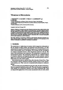
carbon grating of 54,800 lines/inch . Grids were examined with either a Hitachi HU 11-A or J .E .M . 7A electron microscope. RESULTS In each organism in which there was no fixation, ciliary microtubules tend to separate from each other but show no clear substructure such as the protofibrils (16, 3) . The microtubules consistantly possess nodules, possibly the arms which occur on the "A" microtubule (1) . Nodes do not appear to be doublet structures in these preparations . Typical micrographs for each organism are those of Fig . I (E. eurystomus), Fig. 2 (S. ambiguum), Fig. 3 (Colpoda sp .), Fig . 4 (T. pyriformis), and Fig . 5 (0 . fasciatus-sperm) . A high magnification view of an unusually well-dried preparation from T. pyriformis (Fig . 6) shows the doublet microtubule and nodes . Nodes on microtubules which have remained in close association after LTCD appear
Peripheral ciliary microtubules from E . in all photographs . FIGURE 1
FIGURE 9
tubules of
oriented in a straight line (Fig . 4, inset ; Fig . 5) . In each case, the line is at an angle of about 30 ° from a perpendicular to the long microtubule axes . Approximate dimensions of microtubules, their nodes, and spacing of nodes are presented in Table I . An additional feature is the occasional presence of fine strands (Figs . 2, 3 ; arrow) interconnecting fibrils . These often, though not always, are located at points adjacent to the nodes, and may represent the fibrous connection shown by many workers, i .e ., Gibbons (6), and recently Williams and Luft (17) in cross-sections of Tetrahymena cilia . When E . eurystomus is fixed by fuming in osmium tetroxide prior to spreading, the resulting ciliary microtubules (Fig . 7) do not possess nodes . They also have a more rigid appearance than for unfixed material . Fixation of the same organism in glutaraldehyde followed by LTCD routinely reveals
eurystomus . X 60,000.
Bar length equals 0 .1 µ
Ciliary microtubules from S . ambiguum, showing fine strands (arrow) interconnecting microclear view of nodes on a microtubule . X 60,000 .
cilia . Inset,
Ciliary microtubules from Colpoda . Strands are seen interconnecting microtubules (arrow) . Some beam contamination is apparent, and nodes are not so clear as in other preparations . X 60,000 . FIGURE 3
Shadowed, reversal-printed ciliary microtubules from tion view showing linear orientation of nodes . X 60,000 .
FIGURE 4
T . pyriformis. Inset,
low-magnifica-
B R I E F N 0 T E S
467
FIGURE 5
Nodes and microtubules from sperm of 0 . fasciatus . X 41,000 .
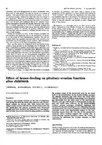
High magnification view of ciliary microtubules from Tetrahymena showing doublet tubules and several nodes . X 125,000 . FIGURE 6
Smooth microtubules from cilia of E. eurystomus. Fixation by Os04 fuming . Several microtubules are in close, side-by-side association . X 60,000 . FIGURE 7
FIGURE 8
Glutaraldehyde-fixed ciliary microtubules from E . ourystomus. X 60,000 .
(15) . Kraneveld (13) air-dried flagella from Trypanosoma evansi and made note of cross-striations
TABLE I
Microtubule and Node Measurements Nodes
Organism
Euolotes Spirostomum Colpoda Tetrahymena Oncopeltus
Microtubule diameter Length Width MIA
MIA
MIA
22 28 20 31 24
22 20 46 33 25
19 20 27 30 25
Center-tocenter my.
92 75-100 110 100 44
with a 50 m i period on the subfibrils ; possibly these, too, were arms . Afzelius (I) first designated them as arms and indicated that they were attached to subfibril A of the doublet . The subfibrils (microtubules) with nodes were first obtained after LTCD preparation by S . L . Wolfe (who kindly contributed Fig . 5) while studying sperm from 0 . fasciatus .
Arms were studied in sectioned material of Pseudotrichonympha by Gibbons and Grimstone (9)
and described as double structures which were 1215 my long by 5 my wide with a center-to-center spacing of 13 mµ . Recently, a number of investigaciliary microtubules (Fig. 8) with poorly defined tions have shown a variety of armlike fibrils radiatnodal regions, and many interconnecting strands . ing from cytoplasmic or nuclear microtubules . These often form a complex interconnecting netDISCUSSION work (see Wilson, 1969) . Gibbons and Rowe (10) It seems reasonable that the nodular periodicities isolated the arms from Tetrahymena, and found 14S on microtubules of cilia and flagella are the "arms" and 30S fractions after sedimentation . The 14S first incompletely described by Manton and Clarke fraction consisted of spherical entities measuring (sperm)
468
B R I E F N O T E S
7-10 mµ high and 18 mg wide . They postulated that these polymerize to form the 30S fraction which was between 40 and 50 mµ long with nodes of 13 mµ center-to-center spacing . It is of particular interest that this period corresponds well with that mentioned above for the flagellate protozoan Pseudotrichonympha . Crane fly spermatids isolated and negatively stained by Behnke and Forer (4) had "regularly spaced projections . . . 600 A apart" on the microtubules and are somewhat similar to the LTCD nodes . In their Fig . 24, however, adjacent nodes appear to be almost touching . Microtubules of the flagellar shaft from developing chicken sperm recently have been reported to have projections with a period of 24 mµ (14) . Whether this was sectioned or isolated-dried material was not mentioned . In the LTCD preparations shown here, the microtubules possess nodes of variable dimensions : 20-46 mµ length (dimension parallel to long microtubule axis), 19-30 mµ width, and a center-to-center distance of 44-110 mµ . These nodes are considerably wider than the arms described by Gibbons and Grimstone, yet they are strikingly similar to the nodular appearance of the isolated 14S fraction . The internode distance is 3%-8 times greater than that for the arms described by earlier workers . Among the possible explanations for the apparent discrepancies in center-to-center spacing of nodes are : (a) Nodes in LTCD preparations are not arms but some other structure . (b) Some arms, occurring at periodic distances, are of a somewhat different nature than the others or are bound to the microtubule differently, and they alone are retained during the preparation . In this regard, Gibbons (8) reported that arms are readily detached during isolation and negative staining of cilia from Tetrahymena pyriformis . This thesis may also be partially supported by the lack of arms or nodes in LTCD preparations of rat sperm tail (19) . (c) Different cilia have arms with different periods . This is clearly the case for the nodes seen after LTCD preparation . A similar variation in centerto-center spacing has not been clearly shown for thin-sectioned material and may be resolved after careful sectioning of cilia from each organism . Comparison of the work of Gibbons and Rowe (10) with the present study suggests a real discrepancy in arm-to-arm distance for Tetrahymena prepared by different methods-13 mµ in the isolated fraction and 110 mu after LTCD . (d) The peripheral
fibrils are stretched during LTCD preparationthough the degree of stretching required for the observed periodicity is very great . The first and fourth explanations are unlikely, but which of these, if any, is the answer awaits subsequent investigation . The second notable discrepancy between this and earlier work is that arms isolated in this way do not appear as double structures . Perhaps, during LTCD the double components collapse upon one another and combine with other cellular proteins to form the rather uniform-looking nodes . This research was supported by a Northern Arizona University Institutional Grant ; University of California, Davis research committee ; and United States Public Health Service Research Grant G. M. 11873-03 from the Institute of General Medical Science . Received for publication 24 October 1969, and in revised form 7 December 1969 .
REFERENCES 1 . AFZELIUS, B . 1959 . J. Biophys . Biochem . Cytol . 5 : 269 . 2 . ANDERSON, T. F . 1950 . C. R ., 1st Congress International de Electron microscopie . 565 . 3 . ANDRÉ, J ., and J . P . THIÉRY . 1963 . J . Microsc . 2 : 71 . 4 . BEHNKE, O ., and A . FORER . 1967 . J. Cell Sci . 2 : 169. 5 . GALL, J . G . 1963 . Science (Washington) . 139 :120 . 6 . GIBBONS, I . R. 1961 . Nature (London) . 190 :1128 . 7 . GIBBONS, I . R ., 1963 . Proc . Nat . Acad. Sci. 50 :1002 . 8 . GIBBONS, I . R . 1965. Arch . Biol . (Liege) . 76 :317 . 9 . GIBBONS, I . R., and A . V. GRIMSTONE . 1960 . J. Biophys . Biochem . Cytol. 7 :697 . 10 . GIBBONS, I . R., and A. J . ROWE. 1965 . Science. (Washington) . 149 :424 . 11 . GRIM, J . N . 1967 . Ph .D . Dissertation, University of California, Davis, California. 12 . KLEINSCHMIDT, A . Z. Naturforsch . 14b :770 . 13 . KRANEVELD, F . C ., A . L . HOUWINK, and J . W . KEIDEL. 1951 . Proc . Acad. Sci. Amst. 54 :393 . 14 . MCINTOSH, J . R. 1968 . J. Cell Biol . 39 :89a. (Abstr . ) 15 . MANTON, I ., and B. CLARKE. 1952 . J . Exp . Bot . 3 :265. 16 . PEASE, D . C . 1963 . J. Cell Biol. 18 :313 . 17 . WILLIAMS, N . E ., and J . H . LUFT . 1968 . J. Ultrastruct . Res. 25 :271 . 18 . WILSON, H . J . 1969 . J. Cell Biol. 40:854. 19 . WOLFE, S . L . 1965 . J. Cell Biol . 25 :408 .
B R I E F
N
0 T E
S
469