MBoC | ARTICLE
Peptide aptamers define distinct EB1- and EB3binding motifs and interfere with microtubule dynamics Karolina Les´niewskaa, Emma Warbrickb, and Hiroyuki Ohkuraa a
Wellcome Trust Centre for Cell Biology, School of Biological Sciences, University of Edinburgh, Edinburgh EH9 3JR, United Kingdom; bDivision of Molecular Medicine, College of Life Sciences, University of Dundee, Dundee DD1 5EH, United Kingdom
ABSTRACT EB1 is a conserved protein that plays a central role in regulating microtubule dynamics and organization. It binds directly to microtubule plus ends and recruits other plus end–localizing proteins. Most EB1-binding proteins contain a Ser–any residue–Ile-Pro (SxIP) motif. Here we describe the isolation of peptide aptamers with optimized versions of this motif by screening for interaction with the Drosophila EB1 protein. The use of small peptide aptamers to competitively inhibit protein interaction and function is becoming increasingly recognized as a powerful technique. We show that SxIP aptamers can bind microtubule plus ends in cells and functionally act to displace interacting proteins by competitive binding. Their expression in developing flies can interfere with microtubules, altering their dynamics. We also identify aptamers binding to human EB1 and EB3, which have sequence requirements similar to but distinct from each other and from Drosophila EB1. This suggests that EB1 paralogues within one species may interact with overlapping but distinct sets of proteins in cells.
Monitoring Editor Francis A. Barr University of Oxford Received: Aug 30, 2013 Revised: Jan 14, 2014 Accepted: Jan 21, 2014
INTRODUCTION Microtubules are a major constituent of the cytoskeleton in all eukaryotic cells. They are essential for cell morphogenesis and motility and form the spindle to segregate chromosomes during mitosis. Microtubules are polar filaments with two differentially regulated ends—a plus end and a minus end. Whereas minus ends are often anchored to subcellular structures, plus ends constantly switch between phases of growth and shrinkage and also interact with subcellular structures (Howard and Hyman, 2003). Therefore precise spatial and temporal regulation of microtubule plus ends is crucial for microtubule organization and function. A growing number of proteins are known to localize to the polymerizing microtubule plus ends and are collectively called This article was published online ahead of print in MBoC in Press (http://www .molbiolcell.org/cgi/doi/10.1091/mbc.E13-08-0504) on January 29, 2014. Address correspondence to: Hiroyuki Ohkura (
[email protected]). Abbreviations used: GFP, green fluorescent protein; ITC, isothermal titration calorimetry; MBP, maltose-binding protein; SxIP, Ser–any residue–Ile-Pro. © 2014 Les´niewska et al. This article is distributed by The American Society for Cell Biology under license from the author(s). Two months after publication it is available to the public under an Attribution–Noncommercial–Share Alike 3.0 Unported Creative Commons License (http://creativecommons.org/licenses/by-nc-sa/3.0). “ASCB®,” “The American Society for Cell Biology®,” and “Molecular Biology of the Cell®” are registered trademarks of The American Society of Cell Biology.
Volume 25 April 1, 2014
microtubule plus end–tracking proteins (also known as +TIPs; Akhmanova and Steinmetz, 2008). These proteins may regulate dynamics of microtubule ends, anchor them to subcellular structures, or be transported as a cargo. Among them, EB1 is considered to play a central role. It is one of a few proteins that directly bind growing microtubule plus ends and is responsible for recruiting most microtubule-tracking proteins through a direct interaction (Busch and Brunner 2004; Dzhindzhev et al., 2005; Akhmanova and Steinmetz, 2008). EB1 is a highly conserved eukaryotic protein (Slep et al., 2005). Although yeasts have only one gene, higher eukaryotes have multiple homologues in their genomes—for example, EB1, EB2, and EB3 in humans (Tirnauer and Bierer, 2000). EB1 and its homologues share an overall structure with a calponin homology (CH) domain in the N-terminal region, a central linker region, the EB1 homology domain (EBH), and a disordered tail containing EEY/F in the C-terminal region (Slep, 2010; Figure 1A). The CH domain is essential for association with microtubule plus ends (Zimniak et al. 2009). The EBH domain and the C-terminal EEY/F motif are the binding sites for EB1-interacting proteins (Honnappa et al., 2006, 2009). These two binding sites mediate two distinct modes of interaction through two types of EB1-interacting motif (Figure 1A). One is
1025
control Apt 37 SRIP SLIP SNIP SMLP SGIP SYIP SAIP SGIP SHIP TLIP SWIP SRLP SYIP SCIP SDLP TRLP SWIP SSIP CRIP SRLP TLIP SWIP SRVP TRIA SMLP SLFP SLLP SKMP SLLP TRLP SMLP CHIP SRVP TWIP SKVP SYLP SLLP CHIP SRIA GKIP CYIP SRVP SNLP GRIP SLLP SLLP TRIA SDIP SGIA SVIP SGIA SHLP GGIP TMLP SVIP SLVP
E
SRAA 1
EBH EEY
prey
MTs SxIP CAP -Gly
Perfect RTRGRSRIPRWVGRRG-
Jumbled RTRGRSRIPWRGVRGR-
G
1026 | K. Les´niewska et al.
EB1
2
0
bits
x5SxIPx7
0.5
Perfect RTRGRSRIPRWVGRRG0.4 4 4
RTRGRSRAARWVGRRG0.3 3
3
0.2
0.1 2
1
0 0 bits
EB1
bits
CH bits
B
Perfect Control SRAA Jumbled
D
37:RCVSRSKIPKLCLSWYL 171:LQSRRSRIPRWVGCRQ380:RSRTRSRIPRWVGFVQ312:RRAGKSRIPVAVRQSSC 188:RWVGVSRIPRWVGWES314:GRCRVSRIPRWVGGIKPerfect:RTRGRSRIPRWVGRRG356:RKRAPSRIPVLKRWPA457:PGKYVSKIPVWRGGRM319:LKLKRSRIPVPTKVRGD 8:IKRGRSKIPRWIGDQH441:TKVQPSRIPRWCRQSE165:ITTRPSLIPRWVGRGG310:SMVGRSRIPVWRGRRG322:VAASRSRIPRPVRKCS167:RTNFVSRIPRWRG429:KRSSVSRIPRWQKRYP448:RLCPKSGIPRWTGRKG377:MRREGSRIPRWRGW390:KWAGRSRIPVRVGRR383:SAAPGSRIPRWVGGGM286:TVRGRSRIPVWVGWAV374:WAGAVSRIPRWVGGRT470:PQSARSRIPRWRGGRI459:RERGGSRIPRWKGG326:YVKARSRIPVWQGARR333:LVYRVSRIPRYVGSGR142:KYKWRSRIPKWTGHVG433:KPRAPSRIPRRVSGRC155:GAVKNSRIPRYIGWRG406:PSLQRSRIPRPTVRRA320:SVKRASRIPVRLKGNR386:RGFRHSRIPRWRGERP219:CRGQRSRIPRWRPAQR291:TRPRRSRIPVRKVW172:RRRVVSKIPRWCGAPT395:SALEGSRIPRWVGWGPC 350:HPRSRSLIPRYVGNMH347:KARKPSRIPRPNSDRH407:PAMGVSRIPRWVRRTG263:WVKWKSRIPVRRGRQA446:TRTGRSMIPRWHGGAG376:RWRRRSGIPRWVGLGG187:LVQRRSGIPRWTGVGK439:EYRGVSRIPVWKGRGT378:HSDGRSFIPRWVGR154:KNCGKSRIPVLQRRVS355:RTRRVSRIPRFVGWPW4:SLASVSRIPVYVARRQC 243:ARMLPSRIPRYKRGGG268:TMRPRSRIPRYVGTWE251:RPVLQSRIPRWAGTSHControl: –
A C 4 4
3 3
1 -5 -4 -3 -2 -1 S x I P +1 +2 +3 +4 +5 +6 +7
XX XX
x
0
strong strong
2
1 -5 -4 -3 -2 -1 S x I P +1 +2 +3 +4 +5 +6 +7
bait
0.8
strong
0.6
0.4
0.2
0
F strong
2
1
0
X XXX
2
1.5
1
0.5
0
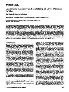
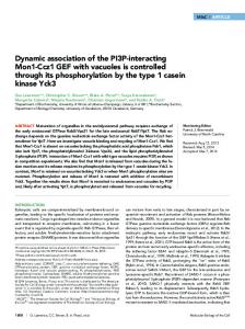
Figure 1: Aptamers revealed amino acid sequences surrounding SxIP that promote interaction with Drosophila EB1. (A) Diagram of the EB1 domain structure. The EBH domain of EB1 interacts with SxIP motifs of many microtubule plus end–binding proteins. (B) Isolation of Drosophila EB1 aptamers by yeast two-hybrid screening. Yeast (Y190) containing the EB1 bait plasmid was cotransformed with a linearized prey plasmid and DNA encoding xxxxxSxIPxxxxxxx flanked by sequences corresponding to a prey plasmid for gap repair. (C) Amino acids surrounding SxIP motif overrepresented
Molecular Biology of the Cell
the CAP-Gly domain, which is present in the dynactin subunit p150Glued and the CLIP-170 family of proteins (Steinmetz and Akhmanova, 2008) and binds the C-terminal EEY/F motif of EB1 (Honnappa et al., 2006). The second motif is the Ser–any residue– Ile-Pro (SxIP) motif, which has been identified in a large number of diverse EB1-interacting proteins (Honnappa et al., 2009; Jiang et al., 2012). This motif binds the EBH domain of EB1 (Honnappa et al., 2009). The SxIP motif was originally characterized as capable of mediating protein interactions with EB1, and an SxIP-containing, 30–amino acid peptide derived from MACF2 has been cocrystallized with EB1 (Honnappa et al., 2009). Although many proteins contain sequences potentially matching the SxIP motifs, only some of these actually bind EB1 and are recruited to microtubule plus ends. Residues comprising and surrounding the SxIP motif of MACF2 were systematically replaced one by one and tested for EB1 binding (Buey et al., 2012), highlighting the contribution of surrounding sequences to EB1 interaction. EB1 influences the microtubule plus end behavior not only by recruiting other proteins, but also by a direct effect on microtubule dynamics (Akhmanova and Steinmetz, 2008). It has been shown that EB1 has important roles in cell division and microtubule organization and dynamics in various cell types. In cultured Drosophila cells, knockdown of EB1 dramatically reduced the dynamicity of microtubules and increased microtubule pausing in interphase. It also resulted in abnormal organization and positioning of the spindle and reduced astral microtubules in mitosis (Rogers et al., 2002). In cultured mouse fibroblasts, EB1 is important for primary cilia assembly (Schroder et al., 2007). At the whole-animal level, mutations in Drosophila EB1 compromise neuromuscular function, especially function of the chordotonal sensory organs (Elliott et al., 2005). The roles of mouse EB1 and EB3 in myogenesis were studied using a cell line in which differentiation can be induced. It was found that knockdown of EB1 or EB3 prevents elongation and fusion of myoblasts into myotubes (Straube and Merdes, 2007; Zhang et al., 2009). Overexpression of EB3 can restore cell elongation but not cell fusion (Zhang et al., 2009). This suggests that EB1 and EB3 have overlapping but distinct functions during myogenesis in mouse. EB1 homologues are likely to play crucial roles in cell morphogenesis during differentiation in many cell types. It is a challenge to investigate roles of proteins essential for mitosis, such as EB1, in differentiated cells, as their removal disrupts cell divisions that form precursors of the differentiated cells. In this study, we use peptide aptamers to further understand the interaction of EB1 with other proteins. Peptide aptamers are small proteins containing a peptide region typically 10–25 amino acids long, which are capable of binding target molecules with high affin-
ity and specificity (Colas et al., 1996; Seigneuric et al., 2011). Highlevel expression of an aptamer can competitively disrupt a specific protein–protein interaction and thus inhibit a specific protein function (Cohen et al., 1998). Peptide aptamers can define residues involved in protein–protein interactions and so can provide the basis for small-molecule design (Fabbrizio et al., 1999; Butz et al., 2000; Baines and Colas, 2006). Aptamers can be used in designing smallmolecule screens in the search for compounds that can bind to the target protein and thus displace the aptamer. Aptamers have advantages over RNA interference (RNAi) or genetic methods for drug target discovery or validation, as they can affect specific interactions rather than lower global levels of a target (Crawford et al., 2003). An ability to potentially inhibit specific protein interactions at desired times and in desired cell types in a whole organism (Kolonin and Finley, 1998, 2000; Yeh et al., 2013) makes them powerful tools to dissect protein functions and molecular pathways in cells and living organisms. Here we describe the isolation of a large number of aptamers that bind Drosophila EB1, human EB1, and human EB3. We use them to identify SxIP-containing peptides that show an increased affinity to EB1 and to identify residues within the motif that determine EB1 homologue binding specificity. We also explore various methods to isolate high-affinity EB1 aptamers that can successfully compete with natural EB1-interacting proteins to displace them from microtubule ends and alter microtubule dynamics.
RESULTS Isolation of EB1 aptamers using yeast two-hybrid screening Peptide aptamers are useful tools for identifying interaction motifs and manipulating protein–protein interactions in vitro and in vivo (Fabbrizio et al., 1999; Wickramasinghe et al., 2010; Yeh et al., 2013; Supplemental Figure S1). We sought to systematically identify EB1 aptamers, peptides that bind to EB1. We used yeast twohybrid screening to screen a large number of semirandom peptides for interaction with full-length Drosophila EB1 (Figure 1B). The peptide prey library used for screening was designed to express peptides with core motif SxIP preceded by five and followed by seven random residues, as this region was shown to be sufficient for interaction with EB1 (Honnappa et al., 2009; Buey et al., 2012). Oligonucleotides encoding these SxIP-containing peptides were synthesized with flanking sequences corresponding to the ends of a linearized prey vector to facilitate library construction by gap repair in yeast. Five million transformants were screened, of which ∼500 showed activation of both reporter genes. Because a large majority of clones in the library contained SxIP (Supplemental Figure S2) but did not interact with EB1, SxIP alone is not sufficient for the interaction.
among EB1 aptamers, and EB1 aptamers with strong interaction. The total height of each stack represents the “information content” in bits at each position and is divided by the frequency of each residue (Bailey et al., 2006). Colored residues indicate that they are more frequently found in aptamers (p < 0.01) than in nonselected peptides and random peptides expected from codon usages. (D) Strength of two-hybrid interactions of the 51 strongest aptamers or aptamer Perfect (marked in red) with EB1. The expression of the reporter gene LacZ was measured by quantitative assay for β-galactosidase activity and normalized for cell density (A420/A600). The empty bait plasmid was used as control. Bars, SEM (n = 3). Full sequence of Aptamer 37, RCVSRSKIPKLCLSWYLIRAREIYES. (E) Two controls (SRAA, Jumbled) generated by mutating aptamer Perfect. Strength of two-hybrid interactions were compared between aptamers Perfect, SRAA, and Jumbled. Bars, SEM (n = 3). (F) Residues within the SxIP motif overrepresented among 56 interactors showing any interaction with EB1 and among the 15 strongest EB1 interactors selected from a library based on the aptamer Perfect sequence in which SRIP was replaced with four random residues (XXXX library). Colored residues indicate that they are more frequently found in aptamers (p < 0.01) than in nonselected peptides and random peptides expected from codon usages. (G) Strength of two-hybrid interactions between EB1 and aptamers from a screen of peptide sequences in which SRIP of aptamer Perfect was replaced with four random residues. It is measured by a quantitative assay for β-galactosidase activity and normalized for cell density (A420/A600). Volume 25 April 1, 2014
Peptide aptamers to EB1 and EB3 | 1027
EB1 aptamers reveal SxIP-flanking sequences that promote EB1 binding To determine which sequences were capable of interacting with EB1, we sequenced the plasmid insert from 45 positive transformants from the two-hybrid screen. As a control, we also sequenced 39 random transformants not selected for activation of reporter genes, which reflect the composition of a library used for screening. A large majority of the sequences from these control nonselected transformants encoded peptides with SxIP and an unbiased mixture of residues at other positions (Supplemental Figure S2). Sequencing of the 45 EB1 aptamers revealed that they have sequences significantly different from those of control nonselected peptides and also from theoretical random peptides (Figure 1C and Supplemental Figure S3). Detailed analysis showed that some residues are significantly overrepresented at certain positions. Of note, we saw preferences of arginine or lysine at the x position of the SxIP motif, arginine or valine at the +1 position (the next residue after SxIP), tryptophan at the +2 position, and glycine at the +4 position. This gives a consensus of (R/K)Tx(R/F)(R/V)S(R/K)IP(R/V)WVGRxG, which represents the most preferred residue at each position (x representing no significant preference).
Selection of strong interactors Our goal was to isolate strong aptamers that tightly associate with EB1 and could potentially compete with endogenous proteins for EB1 interaction. We also hoped that analysis of the sequence of aptamers that showed a particularly high affinity for EB1 would inform us about the residues that were essential for this strong interaction. It is known that activation of two hybrid reporter genes can be roughly correlated with the affinity of a protein–protein interaction (Estojak et al., 1995). To identify the aptamers that potentially had the highest affinity among those identified in our screen, we measured the expression of a reporter, LacZ, in 281 transformants from the screen in a quantitative assay for β-galactosidase activity. To identify aptamers with very strong two-hybrid interactions, we retested 51 transformants with the highest LacZ activation together in triplicate (Figure 1D). Aptamer 37 consistently gave the strongest two-hybrid interaction with EB1. Sequence analysis of these strong aptamers showed that the preferred residues among strong EB1 aptamers were similar to those among EB1 aptamers with any strength of interaction, but these residues appeared more frequently among strong aptamers (Figure 1C).
Creation of designer aptamers To identify the highest-affinity EB1-binding aptamers, we decided to test the hypothesis that we could use semirational design to devise an aptamer sequence based on the consensus information available from the original screen. We generated a potential aptamer (“aptamer Perfect”) that consists of the most frequently presented amino acid at each position, RTRGRSRIPRWVGRRG. The interaction of aptamer Perfect was compared with that of the strongest aptamers found in the screens, using a quantitative two-hybrid assay. Aptamer Perfect gave strong reporter expression, which was comparable to the 10 strongest aptamers, although it was not the strongest (marked in red in Figure 1D). Because the preferred amino acids were chosen from aptamers, most of which have weak interactions, this demonstrated a novel, simple way to design very strong aptamers by examining the consensus without measuring and comparing the interaction of many aptamers. To test the possibility that the strong interaction found for aptamer Perfect may simply reflect overall amino acid composition rather than a sequence specific interaction, we generated two 1028 | K. Les´niewska et al.
control aptamers. In the first control (SRAA), the SRIP motif was mutated to SRAA (Figure 1E), and in the second (Jumbled) the order of the residues downstream of SxIP was changed without changing the overall composition (Figure 1E). Quantitative twohybrid assays showed that these control mutant aptamers do not interact with EB1 (Figure 1E). This demonstrates that the twohybrid interactions between this aptamer and EB1 are sequencespecific interactions.
Importance of the SxIP motif for EB1 binding We then examined the involvement of the conserved residues within the SxIP motif in the EB1 interaction to test the possibility that other residues within this motif might result in a higher affinity. We performed a yeast two-hybrid screen for EB1 interaction using a library based on the aptamer Perfect sequence in which SxIP was replaced with four random residues (Supplemental Figure S4). In total, 57 positive transformants were isolated from ∼1 million screened. Sequence analysis showed a strong and significant preference for serine at the first position, isoleucine or leucine at the third position, and proline at the fourth position (Figure 1F). Of interest, we also found a low frequency of other amino acids among weak interactors, but in such cases this unusual amino acid was limited to only one of the positions (e.g., CRIP, SRVP or SRIA, but not CRVP; Figure 1, F and G). This suggests that some degree of flexibility is allowed within this motif, but only if the surrounding sequence is optimal for interaction with EB1. Although arginine is strongly preferred at the second position for aptamers selected from SxIP-containing peptides, no such strong preference is observed when the SxIP residues are randomized but the flanking regions are kept the same. This suggests that if the surrounding sequence is optimal for interaction with EB1, more flexibility is allowed for the X position. These results underline the importance of SxIP motifs. Change of these conserved residues within the motif can be tolerated to a limited degree but could not further increase the strength of the interaction.
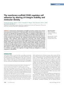
Tandem repeats of aptamers Endogenous EB1-interacting proteins often contain multiple SxIP motifs, and it was previously shown that tandem repeats of SxIPcontaining regions increased the robustness of the localization at microtubule plus ends (Honnappa et al. 2009). We therefore generated constructs expressing tandem repeats of aptamer 37 or aptamer Perfect. DNA encoding tandem repeats of these aptamers with flanking sequences for gap repair were synthesized commercially and tested in a yeast two-hybrid assay. The four-residue spacer SGSG was included between each repeat. Several transformants of each construct were checked for the integrity of the plasmid. From four repeats of aptamer 37, all transformants tested contained the plasmid as designed. In contrast, from seven repeats of aptamer Perfect, the number of the repeats varied between transformants, but within each transformant the number appeared stable. Although we scrambled codon usage as much as possible, it seems that during gap repair, recombination reduced the number of tandem repeats. We tested two, four, and seven repeats of aptamer Perfect and four repeats of aptamer 37 for two-hybrid interaction with EB1 and also tested two repeats of aptamer 37 that we later generated. A quantitative assay for reporter expression showed that whereas only two repeats of aptamer Perfect indeed increased the interaction, all other repeats of aptamers reduced the interaction, and four or seven repeats nearly abolished the interaction (Figure 2A). Molecular Biology of the Cell
A
Sequencing showed that the preference of amino acids at each position in double-constrained aptamers was not significantly different from that seen in single-constrained aptamers (p > 0.08; Figure 2C and Supplemental Figure S5). Quantitative assays of reporter gene expression (LacZ) indicated that the double-constrained aptamers we isolated did not have a stronger interaction than the strongest single-constrained aptamers (Figure 2B). However, it is still possible that double-constrained aptamers embedded in a scaffold protein may be more active in cells because of increased stability.
tandem multimers
1.2 1 0.8 0.6 0.4
B
2x apt 37 4x apt 37
aptamer 37
7x Perfect
4x Perfect
2x Perfect
control
0
Perfect
0.2
double constrained
1 0.8 0.6 0.4
C
T16
T14
T13
T11
T6
Apt. 37
0
control
0.2
4 3 2
1 0
-5 -4 -3 -2 -1 S x I P +1 +2 +3 +4 +5 +6+7
Figure 2: Tandem repeats of aptamers and double-constrained aptamers. (A) Strength of two-hybrid interactions of tandem repeats of aptamers Perfect and 37 with EB1. It is measured by a quantitative assay for β-galactosidase activity and normalized for cell density (A420/A600). Bars, SEM (n = 3). (B) Strength of two-hybrid interactions of double-constrained aptamers embedded in thioredoxin with EB1. Bars, SEM (n = 3; except aptamer 37, n = 1). (C) Residues overrepresented among double-constrained EB1 aptamers.
Isolation of double-constrained aptamers Aptamers embedded in a scaffold protein (“double-constrained” aptamers) are often used, as they potentially have a higher affinity for their target, and more resistance to proteolysis, than single-constrained (or free) aptamers (Geyer and Brent, 2000; Supplemental Figure S1). To isolate double-constrained aptamers, we used random SxIP-containing peptides of 16 residues expressed as a constrained loop in the active site of thioredoxin (Supplemental Figure S4). These were screened for EB1 binding by the yeast twohybrid system, and 18 positive transformants (prefixed with T in figures) were identified from 1 million screened. This was a significantly lower frequency than that seen with singly constrained aptamers (∼100/million), which was not unexpected, as both primary and secondary structures are significant in constrained aptamers. Volume 25 April 1, 2014
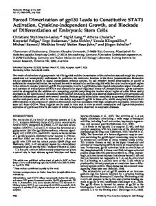
Peptide aptamers tightly bind to EB1 in vitro To estimate the affinity of aptamers to EB1 in vitro, we chemically synthesized peptides corresponding to aptamers and tested their interactions with recombinant maltose-binding protein (MBP) and MBP-EB1 by isothermal titration calorimetry (ITC). ITC measures heat released or absorbed when a protein–peptide interaction occurs, from which interaction parameters, including the dissociation constant (Kd), can be calculated. Peptides corresponding to the two aptamers with strong two-hybrid interactions (Perfect and 37) and two aptamers with weaker interactions (392 and 177) were synthesized. The peptide corresponding to aptamer 37 was not sufficiently soluble for ITC. The solubility of other synthetic peptides was also limited but sufficient to estimate rough Kd values. Recombinant MBP-EB1 and MBP were produced in Escherichia coli and purified. In an isothermal calorimeter, MBP-EB1, MBP, and buffer alone were titrated with aptamer Perfect peptide. The specific heat above the buffer alone was plotted against the molar ratio of the aptamer peptide over MBP-EB1 or MBP (Figure 3, A and B). MBP and the peptide did not show significant interaction. We estimated that the Kd between EB1 and each peptide is roughly 570 nM, 2.6 μM, and 480 nM, and for aptamers Perfect, 177, and 392, respectively (Figure 3, B–D). The Kd measurements by ITC in vitro did not perfectly correlate with the affinity estimates based on yeast two-hybrid data. The Kd between human EB1 and adenomatous polyposis coli protein was reported to be ∼5 μM (Honnappa et al. 2005), whereas more systematic analysis showed that the Kd between human EB1 and various native EB1-interacting proteins range between 1.5 and 140 μM (Buey et al., 2012). Therefore we isolated peptides that appear to have higher affinity for Drosophila EB1 than endogenous interacting proteins for human EB1. To gain insight into the high affinity of the aptamers, we carried out structural modeling of Drosophila EB1 complexed with aptamer Perfect or a fragment of endogenous EB1 interactor Sentin (Supplemental Figure S6). This revealed that aptamer Perfect is likely to interact with EB1 more strongly than the fragment of Sentin. The most crucial residues seem to be the ones at the positions +2 and +3 (WV) of aptamer Perfect. This fits nicely with our results that these two residues are significantly overrepresented at these positions among EB1 aptamers (Figure 1C).
EB1 aptamers can outcompete endogenous proteins for microtubule plus end localization in cells We identified EB1 aptamers with a high affinity of binding in vitro. To test whether these aptamers can bind to EB1 in living cells, we transiently expressed the aptamers as green fluorescent protein (GFP)-fusion proteins under the Actin5C promoter in the Drosophila S2 embryonic cell line. We tested several EB1 aptamers that gave strong two-hybrid interactions in yeast. The fixed cells were immunostained with antibodies against GFP and EB1 or α-tubulin. EB1 localized to microtubule plus ends in a comet-like shape as described (Mimori-Kiyosue et al., 2000). All GFP-aptamer fusion proteins we tested colocalized with EB1 comets at the microtubule plus Peptide aptamers to EB1 and EB3 | 1029
A
0
µcal/sec
B aptamer Perfect (RTRGRSRIPRWVGRRG)
aptamer Perfect MBP-EB1
- 0.1 - 0.2 - 0.3 - 0.4 0 - 0.1 0 - 0.1
C
MBP
Kd=571±74 nM
buffer 0
10
min
20
aptamer 177 (KERHTSRIPRWQSGNS)
30
D aptamer 392
Kd=2.64±0.17 µM
(SGRRRSRIPRWRCKDA)
Kd=479±39 nM
Figure 3: ITC shows a high affinity of aptamers to Drosophila EB1. (A) Raw ITC data showing that aptamer Perfect interacts with MBP-fused Drosophila EB1, but not MBP, in vitro. Heat released by titrations of 100 μM aptamer Perfect into 5 μM solution of MBP-EB1, MBP, and buffer alone. Each peak corresponds to one injection. An initial smaller injection was followed by 10 injections. For MBP-EB1, the heat became smaller for each injection, as the binding site became saturated. For buffer and MBP, it stayed constant, as heat was released only from dilution of the peptide without specific binding. (B–D) Integrated heat peaks subtracted by the heat of dilution and plotted against the molar ratio of the peptide for aptamer Perfect (B), 177 (C), or 392 (D) to MBP-EB1. The line represents the fit to the single-site binding model by the Origin program.
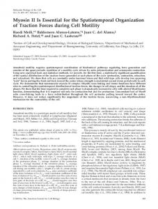
ends, and signals of aptamers 37, Perfect, T14, and T16 at microtubule ends were particularly strong (Figure 4A). Therefore these aptamers can specifically recognize EB1 in the presence of other proteins in cells. To test whether aptamers can compete with endogenous EB1-interacting proteins containing SxIP motifs, we determined the localization of one such protein, Sentin. Sentin contains an SxIP motif in the C-terminal region and is considered to be a major cargo of EB1 in S2 cells (Li et al., 2011). S2 cells were transfected with a plasmid expressing a GFP-aptamer fusion protein as described and immunostained with antibodies against GFP, EB1, and Sentin (Figure 4A). In control cells not expressing aptamers, Sentin colocalized with EB1 at microtubule plus ends as expected. In contrast, in cells expressing aptamers, the Sentin signal was greatly reduced or absent from microtubule plus ends. To quantify this reduction, we measured the intensity of the Sentin signal colocalized with EB1 comets at microtubule plus ends in GFP-aptamer–expressing cells and nonexpressing cells on the same slide (Figure 4, A–D). We found that the Sentin signal was greatly reduced in GFP-aptamer–expressing cells relative to nonexpressing cells. To test whether the effect of the aptamers is due to a sequencespecific interaction with EB1, we tested GFP alone, a mutant aptamer Perfect with SRIP mutated to SRAA (SRAA), and a mutant aptamer Perfect with the residues downstream of SxIP partially jumbled 1030 | K. Les´niewska et al.
(Jumbled). The quantification of Sentin signals overlapping with EB1 comets showed that expression of GFP alone or of the SRAA mutant had no significant effect on Sentin localization (Figure 4C). However, the Jumbled mutant had a small but significant effect on Sentin localization (Figure 4, B and C). This suggests that the overall amino acid composition may contribute, to a small degree, to the interaction with EB1. The binding sites of the two known EB1binding motifs, SxIP motif and CAP-Gly, partially overlap on the C-terminal region of EB1 (Slep, 2010). We investigated whether CLIP-190, which is recruited to microtubules by CAP-Gly but also has a putative SxIP motif, can be outcompeted from the microtubule plus ends by peptide aptamers. Cells transfected with each aptamer and untransfected cells on the same slide were visually examined. CLIP190 colocalized to EB1 at microtubule plus ends in cells transfected with aptamer 37, T14, or T16 to the same degree as in the untransfected cells (Figure 4, D and F). CLIP-190 colocalization with EB1 was weaker in cells expressing aptamer Perfect (Figure 4, E and F). CLIP-190 is predicted to bind to a site of EB1 distinct from but close to the SxIP-binding region (Honnappa et al., 2006), suggesting that this aptamer caused steric hindrance. In conclusion, the aptamers we isolated can specifically bind EB1 in cells and displace endogenous EB1-interacting proteins from microtubule plus ends.
Expression of EB1 aptamers in developing flies alters microtubule dynamics To test whether expression of EB1 aptamers in developing organisms can interfere with vital processes, we made transgenic flies expressing aptamers. Expression of EB1 aptamers fused to GFP controlled by a GAL4-inducible promoter (UASp) was driven by ubiquitously expressed GAL4 under the actin5C promoter. We found that fewer adult flies expressing aptamers were obtained in comparison with the control (Figure 5A). This suggests that aptamers interfere with microtubule function in developing flies. To examine whether microtubule dynamics is altered, we used hemocytes from third-instar larvae because isolated cells can be adhered on a coverslip, allowing good resolution of microtubules. We first confirmed that the GFP-fused aptamers were expressed and localized as comets in hemocytes (Supplemental Movie S1). To test whether expression of the aptamers interferes with microtubule dynamics, we used EB1-GFP driven by the mild ubiquitin promoter to mark growing microtubule plus ends (Shimada et al., 2006; Parton et al., 2011). We compared the microtubule growth rate in hemocytes expressing GFP-fused aptamer and EB1-GFP with that in control hemocytes expressing EB1-GFP alone. We found that the microtubule growth was significantly slower in aptamer-expressing hemocytes than in control hemocytes (p < 0.01; Figure 5B and Supplemental Movies S2 and S3). This fits well with Molecular Biology of the Cell
EB1
Sentin
Jumbled
EB1
Sentin
Merge
Aptamer T14
EB1
CLIP-190
Merge
Apt Perfect
EB1
CLIP-190
Merge
untransfected
untransfected
E transfected
Merge
untransfected
untransfected
transfected
Sentin signal on MT plus ends 1
F
CLIP190 signal on MT plus ends
Apt37
Perfect
T16
T16
T14
Perfect
2x Apt37
Apt37
Jumbled
SRAA
GFP
1
T14
C
GFP
B
D transfected
2x Apt37
transfected
A
Figure 4: Aptamers can be recruited to microtubule plus ends and displace the endogenous EB1 interactor Sentin in Drosophila cells. (A) Aptamer 37 dimer colocalizes with EB1 at microtubule ends and competes out Sentin from microtubule plus ends. (B) A mutant aptamer, Jumbled, does not colocalize with EB1 at microtubule plus ends or compete out Sentin from microtubule plus ends effectively. S2 cells were transfected with an expression plasmid expressing aptamer 37 dimer or aptamer Jumbled fused to GFP and immunostained for GFP, EB1, and α-tubulin. A typical transfected and an untransfected cell on the same slide are shown for comparison. Bar, 10 μm. Yellow boxes indicate areas magnified in the images below. (C) The specific signal intensity of Sentin at microtubule plus ends. Sentin intensities in aptamer-expressing cells relative to those in untransfected cells on the same slide are shown with SEM (n = 30). (D) Aptamer T14 colocalizes with EB1 at microtubule plus ends but competes out CLIP-190 from microtubule plus ends. (E) Aptamer Perfect colocalizes with EB1 at microtubule ends and interferes with the localization of CLIP-190 at microtubule plus ends. S2 cells were transfected with an expression plasmid expressing GFP-fused aptamer T14 or aptamer Perfect and immunostained for GFP, EB1, and CLIP-190. A typical transfected and an untransfected cell on the same slide are shown for comparison. Bar, 10 μm. Yellow boxes indicate areas magnified in the images below. (F) The specific signal intensity of CLIP-190 at microtubule plus ends. CLIP-190 intensities in aptamer-expressing cells relative to those in untransfected cells on the same slide are shown with SEM (n = 30). Volume 25 April 1, 2014
Peptide aptamers to EB1 and EB3 | 1031
B microtubule growth rate p