Vol. 81; n. 1, March 2009
Progression risk factors and subsequent medical management of symptomatic benign prostatic hyperplasia. Francesco Pinto, Marco Racioppi, Emilio Sacco, Angelo Totaro, Antonio Brescia, Andrea Volpe, Mario Gardi, Pier Francesco Bassi
Minute focus of prostate cancer on needle biopsy: Correlation with radical prostatectomy specimen. Emanuele Montanari, Alberto Del Nero, Giacomo Gazzano, Barbara Mangiarotti, Paolo Bernardini, Fabrizio Longo, Giovanni Cordima, Emanuele Itri
Transrectal-HIFU as primary minimally-invasive option for localized prostate cancer. Is spinal anaesthesia cost-effective? A single centre experience in over 100 patients. Paolo Grosso, Leonardo D’Urso, Devis Collura, Raffaella Citro, Maria Teresa Grassano, Rosanna Macchiarulo, Luca Rivalta, Cristina Valz, Giovanni Muto, Enrica Guglielminotti
Tissue engineering in urinary bladder: Morphological and functional characterization. Danilo Zani, Claudio Simeone, Nicola Arrighi, Alessandro Antonelli, Alessandra Moroni, Maura Ferrari, Sergio Cosciani Cunico
Azoospermia and severe oligospermia in testicular cancer. Luca Carmignani, Franco Gadda, Alessandro Paffoni, Giorgio Bozzini, Robert Stubinsky, Stefano Picozzi, Francesco Rocco
Diabetes, cardiovascular diseases and risk of erectile dysfunction: A brief narrative review of the literature. Fabio Parazzini, Elena Ricci, Francesca Chiaffarino, Alberto Trinchieri
Metaphylaxis of urolithiasis. Vincenzo Giannicola Menditto, Giulio Milanese, Giovanni Muzzonigro
Percutaneous treatment of staghorn stones: A retrospective case-control study with evaluation of single vs multiple access to the kidney. Donato Barnaba, Francesco Saverio Grossi, Michele Raguso, Lorenzo Larocca, Giovanni Sallustio, Sebastiano Di Lena, Giuseppe Raguso
Short-time ureteral catheterization after operative ureteroscopic lithotripsy: An alternative to stent versus no stent evaluated in a retrospective study. Giorgio Canepa, Giuseppe Conzi, Giacomo Capponi, Fabio Campodonico, Massimo Maffezzini
Prospective assessment of the efficacy of the EAU guidelines for the prevention of nosocomial acquired infections after genitourinary surgery in a district hospital. Alberto Trinchieri, Stefano Paparella, Sefano Cappoli, Nicola Esposito, Angelo Butti, Roberto Vaiani, Laura Chiappa
Renal cell carcinoma. 2002 TNM classification is still adequate? Andrea Fandella, Marco Borghesi, Alessandro Bertaccini
Bacterial translocation to kidney in rats with intestinal obstruction and the tole of nitric oxide.
Emin Özbek, Yusuf Özlem ˙Ilbey, Mustafa Cekmen, Abdulmuttalip S¸ims¸ek, Mehmet Tekerekoglu, Mustafa Sahin, M. Derya Balbay
Nel corso del XVIII Congresso Nazionale SIUrO, tenutosi a Chieti dal 26 al 29 novembre 2008, si sono svolte le elezioni per il rinnovo delle cariche sociali (il verbale delle elezioni è disponibile in versione integrale sul sito www.siuro.it – verbali / delibere). In base ai risultati del voto è stata proclamata eletta la lista riportata di seguito le cui cariche sono state decise all’unanimità dal comitato direttivo nel corso della sua prima riunione. COMITATO DIRETTIVO GIUSEPPE MARTORANA RICCARDO VALDAGNI GIGLIOLA SICA ALESSANDRO BERTACCINI GIARIO CONTI SERGIO BRACARDA ENRICO BOLLITO RENZO COLOMBO ALBERTO LAPINI NICOLA LONGO MASSIMO MAFFEZINI GIGLIOLA SICA CORA STERNBERG RAFFAELE TENAGLIA
Presidente Vice Presidente Presidente Comitato Scientifico Segretario - Tesoriere Presidente Incoming Consigliere Oncologo Consigliere Consigliere Consigliere Consigliere Consigliere Consigliere Consigliere Past President
COMITATO SCIENTIFICO GIGLIOLA SICA VINCENZO ALTIERI VINCENZO SCATTONI
Presidente Membro eletto Membro eletto
PROBI VIRI ALDO VITTORIO BONO MARIO MOTTA VITO VITALE
Probus vir Probus vir Probus vir
La parola al neo Presidente, Professor Giuseppe Martorana È per me un grande onore essere stato nuovamente chiamato a presiedere la Società Italiana di Urologia Oncologica (SIUrO) per il prossimo mandato. Negli ultimi anni la SIUrO è cambiata ed è cresciuta molto, sia come numero di soci, sia in termini di attività scientifica ed organizzativa. Soprattutto si è rafforzata l’immagine della nostra Associazione attraverso un format scientifico-congressuale che si è dimostrato “vincente”, come sono certo sarà anche quello di quest’anno: la plenarietà delle sessioni, il coinvolgimento dei giovani, l’approfondimento di ogni tema fatto attraverso la partecipazione delle varie discipline. La crisi economico-finanziaria, globale, tuttavia non poteva non coinvolgere anche le società scientifiche come la nostra! Il panorama “storico” è profondamente cambiato, le risorse sono diminuite, è diventato indispensabile “cambiare passo”, rivedere le proprie strategie, le proprie priorità, pena il rischio di scomparire o di essere accorpati a società maggiori o relegati ad un ruolo marginale. Cosciente di questa nuova situazione ma desiderosa nello stesso tempo di rilanciare una “nuova SIUrO” – anche perché certa della potenzialità enorme della Società stessa mai espressa fino in fondo – la squadra uscente ha elaborato, qualche mese prima della scadenza del mandato, un progetto che – nel rispetto della mission multidisciplinare – fosse capace di far diventare la SIUrO più forte, più visibile, più trasversale, più attraente. Come? Intanto… non solo il congresso!! Poi allargando la base di persone coinvolte direttamente nel core scientifico-organizzativo, tra cui molti giovani (elenco che speriamo si allarghi ulteriormente). Infine attivando dei gruppi di studio e dandone la responsabilità di conduzione alle persone coinvolte. Un po’ come all’inizio della storia della SIUrO quando si era pensato ai comitati ad hoc, mai decollati – anzi mai istituiti – perché evidentemente i tempi non erano maturi! La “nuova SIUrO” si è recentemente riunita a Bertinoro, amena località del forlivese, per disegnare la strategia dei prossimi anni. Ne è uscito un gruppo entusiasta – quasi come se ne fosse sentita la mancanza – e determinato a percorrere questa strada, convinto che sia quella giusta! Un caro saluto a tutti e arrivederci a MILANO!
Official Journal of the SIEUN - Official Journal of the SIUrO EDITORS M. Maffezzini (Genova), G. Perletti (Busto A.), A. Trinchieri (Lecco)
EDITORIAL BOARD P. F. Bassi (Roma), A. Bossi (Villejuif - France), P. Caione (Roma), F. Campodonico (Genova), L. Carmignani (Milano), L. Cheng (Indianapolis - USA), L. Cindolo (Avellino), G. Colpi (Milano), G. Corona (Firenze), A. Giannantoni (Perugia), P. Gontero (Torino), S. Joniau (Leuven - Belgio), F. Keeley (Bristol - UK), L. Klotz (Toronto - Canada), M. Lazzeri (Firenze), B. Ljungberg (Umeå - Svezia), A. Minervini (Firenze), N. Mondaini (Firenze), G. Muir (London - UK), G. Muto (Torino), R. Naspro (Bergamo), A. Patel (London - UK), G. Preminger (Durham - USA), D. Ralph (London - UK), A. Rodgers (Cape Town - South Africa), F. Sampaio (Rio de Janeiro - Brazil), K. Sarica (Istanbul - Turkey), L. Schips (Vasto), H. Schwaibold (Bristol - UK), A. Simonato (Genova), S. Siracusano (Trieste), C. Terrone (Novara), A. Timoney (Bristol - UK), A. Tubaro (Roma), R. Zigeuner (Graz - Austria)
SIUrO EDITOR G. Martorana (Bologna)
SIUrO ASSISTANT EDITOR A. Bertaccini (Bologna)
SIUrO EDITORIAL BOARD G. Arcangeli (Roma), M. Battaglia (Bari), O. Bertetto (Torino), F. Boccardo (Genova), E. Bollito (Torino), S. Bracarda (Perugia), G. Conti (Como), J.G. Delinassios (Athens - Greece), D. Prezioso (Napoli), G. Sica (Roma)
SIEUN EDITOR G. Virgili (Roma)
SIEUN EDITORIAL BOARD E. Belgrano (Trieste), P. Martino (Bari), F. Micali (Roma), M. Porena (Perugia), F.P. Selvaggi (Bari), C. Trombetta (Trieste), G. Vespasiani (Roma)
ASSOCIAZIONE UROLOGI LOMBARDI EDITOR F. Rocco (Milano)
HONORARY EDITOR E. Pisani (Milano)
Indexed in: Medline/Index Medicus - EMBASE/Excerpta Medica - Medbase/Current Opinion - SIIC Data Base www.architurol.it
OBITUARY Enrico Dormia, M.D.
Enrico Dormia, M.D., passed away on February 20, 2009. B o rn in 1928 at Bormio, a tiny village of the Italian Alps, Dr. Dormia graduated from the University of Milan in 1952. He then went on to complete his fellowship training in Urology in Milan being promoted to Professor. He became a member of the Active Staff at the Urologic Clinic of Milan. In 1969 he was appointed Chief of the Department of Urology of the A. Manzoni Hospital in Lecco and in 1991 Chief of the Department of Urology of the S. Carlo Hospital in Milan. He promoted the beginning of the activity of kidney procurement for transplantation of the Lecco Hospital.
A memory from his son Grazie papà. Negli ultimi tempi mio padre si è dedicato a scrivere alcune memorie della sua vita che noi figli e nipoti ci siamo immediatamente premurati di far stampare e rilegare in un piccolo volumetto presentatogli a Natale, con sua grande soddisfazione. Con una certa sorpresa questi ricordi hanno completamente ignorato le sue pur ricchissime esperienze di vita pro f e ssionale ma si sono indirizzati a riportare fatti e avvenimenti della sua giovinezza riguardanti suo nonno e soprattutto suo padre (il mio nonno Dino), medico condotto da Bormio a Livigno sino agli anni ’60 e figura leggendaria per tutta la popolazione della vallata. Verso il termine del suo scritto si legge: “… la devozione e la dedizione alla professione di medico fu particolarmente vissuta non solo da papà...” (il mio nonno appunto), “…ma ne fu protagonista tutta la famiglia, stimolata dal rispetto per come la si vedeva da lui vissuta.” E ancora: “… per papà l’attività di medico si concretizzò e si svolse sempre come vera
Dr. Dormia was a member of the Italian Society of Urology for more than 50 years and honorary member of the Urology Society of Argentina since 1974. In 1983 was awarded by the American Association of Urology for his activity and in 2006 was awarded with the Golden Medal of the Town of Lecco. Dr. Dormia had an international reputation as an authority in Urology. He ideated the worldwide famous “Dormia basket” for the extraction of ureteral stones under radiological (or endoscopic) control and produced a lot of scientific and clinical work in the field of chemolysis of renal stones. Alberto Trinchieri Editor
missione (gravida di enormi fatiche ma anche di soddisfazioni continuamente rinnovate sul piano professionale ed umano) quale solo, vero, essenziale modo dell’essere medico”. Leggo in queste parole la sua volontà di esprimere ciò che per lui era il vero significato dell’essere medico che è, ancor prima delle capacità tecniche e professionali, quello di essere persona ricca di dedizione e altruismo disinteressato. Sono tanti, innumerevoli i consigli, le intuizioni e i “trucchi del mestiere” che ho appreso da lui e che custodirò per sempre ma il suo ultimo e più profondo insegnamento riguarda l’Uomo. È l’Uomo che ha voluto lasciare come ricordo immutabile ed incancellabile: l’uomo con la sua onestà, integrità e capacità di sacrificio gratuito. Così, con le tue stesse parole, ti dico grazie papà, grazie per a v e rci dato la vita da vivere, con la tua come modello. Guido Dormia Urologist, A. Manzoni Hospital, Lecco
Contents Progression risk factors and subsequent medical management of symptomatic benign prostatic hyperplasia.
Pag. 1
Francesco Pinto, Marco Racioppi, Emilio Sacco, Angelo Totaro, Antonio Brescia, Andrea Volpe, Mario Gardi, Pier Francesco Bassi
Minute focus of prostate cancer on needle biopsy: Correlation with radical prostatectomy specimen.
Pag. 9
Emanuele Montanari, Alberto Del Nero, Giacomo Gazzano, Barbara Mangiarotti, Paolo Bern a rdini, Fabrizio Longo, Giovanni Cordima, Emanuele Itri
Transrectal-HIFU as primary minimally-invasive option for localized prostate cancer. Is spinal anaesthesia cost-effective? A single centre experience in over 100 patients.
Pag. 13
Paolo Grosso, Leonardo D’Urso, Devis Collura, Raffaella Citro, Maria Teresa Grassano, Rosanna Macchiarulo, Luca Rivalta, Cristina Valz, Giovanni Muto, Enrica Guglielminotti
Tissue engineering in urinary bladder: Morphological and functional characterization.
Pag. 17
Danilo Zani, Claudio Simeone, Nicola Arrighi, Alessandro Antonelli, Alessandra Moroni, Maura Ferrari, Sergio Cosciani Cunico
Azoospermia and severe oligospermia in testicular cancer.
Pag. 21
Luca Carmignani, Franco Gadda, Alessandro Paffoni, Giorgio Bozzini, Robert Stubinsky, Stefano Picozzi, Francesco Rocco
Diabetes, cardiovascular diseases and risk of erectile dysfunction: A brief narrative review of the literature.
Pag. 24
Fabio Parazzini, Elena Ricci, Francesca Chiaffarino, Alberto Trinchieri
Metaphylaxis of urolithiasis.
Pag. 32
Vincenzo Giannicola Menditto, Giulio Milanese, Giovanni Muzzonigro
Percutaneous treatment of staghorn stones: A retrospective case-control study with evaluation of single vs multiple access to the kidney.
Pag. 40
Donato Barnaba, Francesco Saverio Grossi, Michele Raguso, Lorenzo Larocca, Giovanni Sallustio, Sebastiano Di Lena, Giuseppe Raguso
Short-time ureteral catheterization after operative ureteroscopic lithotripsy: An alternative to stent versus no stent evaluated in a retrospective study.
Pag. 43
Giorgio Canepa, Giuseppe Conzi, Giacomo Capponi, Fabio Campodonico, Massimo Maffezzini
Prospective assessment of the efficacy of the EAU guidelines for the prevention of nosocomial acquired infections after genitourinary surgery in a district hospital.
Pag. 46
Alberto Trinchieri, Stefano Paparella, Sefano Cappoli, Nicola Esposito, Angelo Butti, Roberto Vaiani, Laura Chiappa
Renal cell carcinoma. 2002 TNM classification is still adequate?
Pag. 51
Andrea Fandella, Marco Borghesi, Alessandro Bertaccini
Bacterial translocation to kidney in rats with intestinal obstruction and the tole of nitric oxide.
Pag. 56
Emin Özbek, Yusuf Özlem ˙Ilbey, Mustafa Cekmen, Abdulmuttalip S¸ims¸ek, Mehmet Tekerekoglu, Mustafa Sahin, M. Derya Balbay
Archivio Italiano di Urologia e Andrologia 2009, 81, 1
III
REVIEW
Progression risk factors and subsequent medical management of symptomatic benign prostatic hyperplasia. Francesco Pinto, Marco Racioppi, Emilio Sacco, Angelo Totaro, Antonio Brescia, Andrea Volpe, Mario Gardi, Pier Francesco Bassi Department of Urology, Catholic University School of Medicine, Rome.
Benign prostatic hyperplasia (BPH) is a chronic common disease in many men and is often associated with bothersome lower urinary tract symptoms (LUTS). In many men Summary the disease presents with a progressive course that can result in complications such as acute urinary retention (AUR) and BPH-related surgery. Several factors have been associated with progression such as age and prostate volume (PV). Serum prostatespecific antigen (PSA) level, closely correlated with PV, is another useful parameter for determining the risk of BPH progression. Medical therapy is the first and the most frequently used treatment for BPH; surgical treatments rapresent a second-line option when medical therapy is non effective or when complications are associated. Alpha-blockers achieve rapid symptom relief but do not reduce the overall risk of AUR or BPH-related surgery, presumably because they have no effect on PV. 5alpha-reductase inhibitors (5ARIs) display their effectiveness at long distance decreasing PV; this results in improved symptoms, urinary flow and quality of life, and a reduced risk of AUR and BPH-related surgery. Combination therapy provides greater and more durable benefits than either monotherapy and is a recommended option in treatment guidelines. The Combination of dutasteride and Tamsulosin (CombAT), at a pre-planned 2-year analysis, has shown sustained symptom improvement with combination therapy, significantly greater than with either monotherapy. CombAT is also the first study to show benefit in improving BPH symptoms for combination therapy over the alpha-blocker, tamsulosin, from 9 months of treatment. PubMed database has been used to identify publications on the epidemiology of BPH, risk factors for BPH progression and drug treatment options for the management of BPH. KEY WORDS: Benign prostatic hyperplasia, Risk factors, Diagnosis, Therapy. Submitted 20 July 2008; Accepted 15 December 2008
INTRODUCTION The term ‘BPH’ (benign prostatic hyperplasia) actually refers to an histological condition, namely the presence of s t romal-glandular hyperplasia within the prostate gland (1). BPH is a progressive disease that is commonly associated with bothersome lower urinary tract symptoms (LUTS) such as urinary frequency, urgency, nocturia, decreased and intermittent force of stream and the sensation of incomplete bladder emptying. The condition becomes clinically relevant if and when it is associated with bothersome lower urinary tract symptoms (LUTS) such as urinary frequency, urgency, nocturia, decreased and intermittent force of stream and the sensation of
incomplete bladder emptying. Benign prostatic enlarg ement (BPE) is another component of the LUTS/BPH constellation (1); however, the relationship between BPH, BPE and LUTS is complex, not all men with histological BPH will develope BPE; in addition, not all men with LUTS will have concomitant BPE, and not all men with BPE will have bothersome LUTS. The final component of this complex relationship is bladder outlet obstruction (BOO). Yet again, not all men with BPH/BPE and LUTS will have BOO, and there are causes of BOO other than BPH/BPE (e.g. primary bladder neck sclerosis or a urethral stricture). Archivio Italiano di Urologia e Andrologia 2009; 81, 1
1
F. Pinto, M. Racioppi, E. Sacco, A. Totaro, A. Brescia, A. Volpe, M. Gardi, P.F. Bassi
The prevalence of LUTS in Europe varies with age, ranging from 14% for men in their fourth decade of life to > 40% for men in their sixth decade (2). Furthermore, with the elderly constituting a greater proportion of the population, the prevalence of BPH and its impact on medical practice will increase. Simple diagnostic tools are available in the primary care setting; these can offer a first diagnostic step in patients with suspected BPH, as well as a valid strategy to minimise delay in disease management and facilitate appropriate referral from primary to specialised care (4). The European Association of Urology (EAU) guidelines recommend a series of initial assessments for men with LUTS suggestive of bladder obstruction; these include taking a clinical history, using a validated questionnaire to assess symptoms, conducting a physical examination, creatinine measurement, urine analysis and serum prostate-specific antigen (PSA) measurement (particularly when a diagnosis of prostatic carcinoma would affect the decision about which therapeutic option to use) (5). Our growing knowledge on the natural history of BPH enables to understand the progression risk factors and the physiological effects of medical interventions. This review discusses current knowledge of the progressive nature of BPH and how the risk of BPH progression can be evaluated to guide therapeutic choices.
AUR results in prostatectomy in 24-42% of men in the UK and USA, and even patients who avoid surgery through a successful trial without catheter are subsequently at high risk of requiring surgery within a year (10-12). Furthermore, data from the Veteran’s Affairs study in the USA has demonstrated that 36% of men with BPH randomised to watchful waiting switched to invasive therapy within 5 years of enrolment (13). The most common complaint associated with BPH is bothersome LUTS even if some patient surveys have reported that the risk of surgery is a greater concern for patients than other factors such as symptoms or even quality of life (14-16). In a survey of men treated with finasteride, 93% of respondents ranked reducing the need for surgery as very or extremely important and 88% said that reducing the risk of major urological complications was very or extremely important, while symptoms and quality of life were considered less important (15). Similarly, in a survey of men with BPH in five European countries (PROBE), more than three-quarters of men were concerned about the risk of developing AUR and requiring prostate-related surgery (16). This highlights that, although AUR and surgery are less common than overall symptomatic worsening, they are important progression events with financial, emotional and health-related consequences.
BPH PROGRESSION
It is important for clinicians to determine which patients are at increased risk of disease progression in order to optimise therapy and offer a treatment approach that correlates with patient preferences. Numerous factors, such as age, prostate volume, reduced urinary flow, increased symptom score have been linked with the risk of BPH progression events (9, 17, 18).
BPH PROGRESSION Both epidemiological studies and the placebo arms of randomised controlled trials provide strong evidence that BPH is a progressive condition in many men. Progression is defined as worsening of symptoms, deterioration of urinary flow rate, increase in prostate volume (PV), and events such as acute urinary retention (AUR) and the need for surgery (6). Renal insufficiency and recurrent urinary tract infections have been included as additional measures of BPH progression, although these outcomes were rarely observed (7). Definition and prevalence The most evident progression feature is symptoms’ worsening. One of the largest and longest running longitudinal studies, the Olmsted County study, on a sample of 2,115 men aged 40-79 years, at 92 months’ follow-up, reported a ≥ 3-point increase in American Urological Association Symptom Index (AUA-SI) in 31% of participants and the mean annual increase in AUA-SI was 0.34 points (8). The incidence of AUR in men with moderateto-severe LUTS increased from 3.0/1,000 person-years in those aged 40-49 years to 34.7/1,000 person-years in those aged 70-79 years (9). By extrapolating data from this study, the overall risk of AUR has been estimated as 23% for an average 60-year-old man if he survives another 20 years (9). In the placebo arm of the Medical Therapy of Prostatic Symptoms (MTOPS) study the rate of overall clinical progression (defined as an increase in AUA-SI of ≥ 4 points, AUR, urinary incontinence, renal insufficiency, or recurrent urinary tract infection) was 17.4% over the 4-year duration of the study. About 78% of progression events took the form of deterioration in symptoms and 12% of progression events were AUR (7).
2
Archivio Italiano di Urologia e Andrologia 2009; 81, 1
RISK FACTORS
Prostate volume (PV) PV is perhaps the most extensively studied of the risk factors for BPH progression. Men with a PV of > 30 ml are more likely to suffer moderate-to-severe symptoms (3.5-fold increase), decreased flow rates (2.5-fold increase), and AUR (3- to 4-fold increase), compared with men with PV < 30 ml (19). Unfortunately, PV estimated by digital rectal examination (DRE) is associated with considerable measurement error and can underestimate measured volume by as much as 55% (20,21). Transabdominal ultrasound can be used to assess PV, although the gold standard remains transrectal ultrasound (22). Age Several studies have demonstrated a relationship between age and markers of BPH pro g ression. In the Olmsted County study, moderate-to-severe urinary symptoms were recorded in 13% and 28% of men aged 40-49 years and > 70 years, respectively (20) and the incidence of AUR among men with moderate-to-severe symptoms was shown to increase with increasing age (9). More recently, a study of men (n = 1859) with symptomatic BPH showed an increase in PV with increasing age, from a mean of 27.7 ml in men aged 40-49 years to 52.3 ml in men aged 70-80 years (23).
Progression risk factors and subsequent medical management of symptomatic benign prstatic hyperplasia
PSA Extensive data from the placebo arm of the Proscar Longterm Efficacy and Safety Study (PLESS) have demonstrated that PSA is a strong predictor of an enlarged prostate as well as the risk of developing LUTS, poor urinary flow, AUR and/or BPH-related surg e ry (18) even if the precise relationship between PSA and prostate growth may vary from one individual to another (23, 24). This relationship was confirmed by a retrospective analysis of 1859 Dutch patients in which 89% of those with a PSA of ≥ 1.5 ng/ml had a PV of > 30 ml (23). The data suggest that the correlation between PSA and prostate volume is close enough for serum PSA to be used to estimate the degree of prostatic enlargement. Indeed, PSA alone showed a sensitivity for predicting the progression of BPH comparable to the use of an algorithm and the five-variable model (encompassing the indices of serum PSA, symptom problems, peak urinary flow, urinary frequency ≤ 2 hours and hesitancy) (25). The current EAU guidelines define PSA a reliable parameter to evaluate the risks of either requiring surg e ry or developing AUR (5). Post voiding residual volume (PVR) Recent studies have shown that dynamic variables such as symptom score and PVR volume worsening also serve as good predictors of AUR in men with LUTS suggestive of BPH (26). Measurement of PVR volume is also recommended in the EAU guidelines (6), and this can be done routinely using transabdominal ultrasound.
MEDICAL
THERAPY FOR
BPH: WHICH THE RIGHT CHOICE?
The introduction of new effective medical therapies has led to a considerable decline in the popularity of surgery to manage symptoms associated with BPH in the last decade (27). Hence, patients with mild or moderate symptoms can usually be treated in a primary care setting, with more complicated cases referred to an urologist for evaluation and management (28). Current EAU guidelines focus on alpha-blockers and 5alpha-reductase inhibitors (5ARIs), as monotherapies or in combination, when recommending medical therapy for BPH (5). Treatment of LUTS with plant extracts (phytotherapies) has a long tradition even if their mode of action is unclear and the clinical efficacy of these agents is largely unproven (29). Additional well-designed clinical studies are therefore needed before plant extracts can be recommended for the treatment of LUTS (5). Alpha-blockers Clinical effectiveness The alpha-blockers work by inhibiting alpha1-mediated sympathetic stimulation, and hence promote relaxation of prostatic smooth muscle and bladder neck; additional sites of action include bladder and central nervous system (30). Alpha-blockers have a rapid onset of action, with typical improvements in symptom scores of 3045% after 3 months’ treatment (31-34).The EAU guidelines recommend alpha-blockers as a treatment option for BPH where LUTS are bothersome and there is no absolute indication for surgery (5). In the MTOPS study,
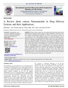
doxazosin reduced the risk of symptomatic progression (defined as a 4-point increase in AUA-SI) by 45% relative to placebo, but it did not reduce the overall long-term risk of AUR or BPH-related surgery; the only effect was the delay in time to AUR (7, 35). Similar results came out in the Alfuzosin Long-Term Efficacy and Safety Study (ALTESS), where only a trend towards a lower incidence of BPH-related surgery was seen in the alfuzosin group, even if statistically non significant when compared with the placebo group (35). A logical explanation for this would be the observation that, to date, large-scale studies have not demonstrated a significant effect of alphablockers on PV. Interestingly, data from a ‘real-life’ practice study suggest that men not responding to alfuzosin treatment (IPSS stable or worsening, and bother score > 3 under treatment) have a greater risk of AUR or BPH-related surgery. First-line treatment with alfuzosin might therefore help to select patients at risk of BPH progression and to optimise their management (26). Side effects The adverse events (AEs) most commonly observ e d with alpha-blockers at a significantly higher frequency than with placebo are dizziness and postural hypotension, although there may be diff e rences in the rates of such events between individual agents within the class (39). Alfuzosin and tamsulosin are better tolerated than terazosin and doxazosin, especially for cardiovascular AEs (39). Studies have demonstrated a higher incidence of abnormal ejaculation with tamsulosin compared with placebo (36-39). 5alpha-reductase inhibitors 5ARIs prevent the conversion of testosterone to dihydrotestosterone (DHT), the primary androgen involved in prostate development. Two 5ARIs are available for the treatment of BPH: dutasteride and finasteride. Finasteride inhibits only the type 2 isoenzyme of 5AR, while dutasteride is a dual inhibitor of both 5AR type 1 and type 2. Treatment with dutasteride results in a greater degree and consistency of DHT suppression compared with finasteride (94.7 ± 3.3% with 0.5 mg dutasteride vs 70.8 ± 18.3% suppression observed with 5 mg finasteride) (40). Current EAU guidelines for the management of BPH recommend 5ARIs for the treatment of bothersome LUTS in men with a prostate volume > 30-40 ml, when there is no absolute indication for surgery (4). Others have proposed that 5ARIs should be considered as first-line medical treatment in men with symptomatic, progressive BPH as indicated by a prostate volume ≥ 30 ml and/or PSA ≥ 1.5 ng/ml (41). Clinical effectiveness Finasteride In the 4-year PLESS study (n=3,040), finasteride treatment significantly improved symptom scores (2.6 points vs 1.0 for placebo; p < 0.001) (17) (Figure 1). More recently, the MTOPS study showed that finasteride reduced the risk of symptomatic progression (defined as an increase in the AUA-SI of ≥ 4 points) by 30% compared with placebo (p = 0.016) (7). Archivio Italiano di Urologia e Andrologia 2009; 81, 1
3
F. Pinto, M. Racioppi, E. Sacco, A. Totaro, A. Brescia, A. Volpe, M. Gardi, P.F. Bassi
Figure 1. Effect of finasteride or placebo on symptom scores (on the Quasi-AUA Symptom Scale). Reproduced from The PLESS study (17).
Symptom Score
Placebo
Finasteride
Year Placebo 1438 Finasteride 1437
1296 1314
1101 1153
961 1047
In the PLESS study, after 4 years, finasteride reduced the relative risk of AUR by 57% (7% of men receiving placebo and 3% of those receiving finasteride; p < 0.001) and surgery by 55% (10% of men receiving placebo and 5% of those treated with finasteride, p < 0.001) (17). Similar results were shown at 4.5 years in the MTOPS study with
853 965
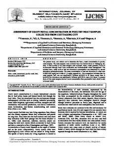
a significant reduction in the risk of AUR and need for surg e ry in patients receiving finasteride monotherapy (p < 0.001 versus placebo) (7). Dutasteride Phase III studies with dutasteride showed a 4.5point improvement in symptoms at 2 years, compared with 2.3 points in the placebo group (p < 0.001) with significant improvements in symptoms from 6 months in the majority of patients (42). At the end of a 2-year open-label extension of these studies, improvements in symptom scores increased to 6.4 points (p < 0.001 vs 2 years of dutasteride treatment) ( F i g u re 2). Indeed, continuous improvement in symptoms was seen out to 4 years of treatment with dutasteride (43). These studies have also shown a reduced relative risk of AUR (57%) and s u rgical intervention (48%) compared with placebo at 2 years (both p < 0.001 versus placebo); this reduction was maintained to 4 years during open-label phases of the studies (42, 43). The explanation of these effects is the action on PV. Finasteride reduces prostate volume by 1819% compared with a 14-24% increase with placebo (7, 17). In phase III studies, dutasteride demonstrated a 26% reduction in PV relative to placebo (42, 43).
Side effects Sexual dysfunction is the most commonly reported side effect, although it is worth noting that there is a relatively high prevalence of sexual dysfunction in untreated men with BPH (44). Furthermore, side effects decrease during prolonged treatment (17, 45). Another consequence of
Figure 2. Effect of dutasteride on mean change in AUA-SI scores from baseline over 48 months (43)..
Double blind
Open label placebo/dutasteride (P/D)
dutasteride/dutasteride (D/D)
Treatment month *p < 0.001 between treatment groups; †p < 0.001 for differences for both treatment groups from month 24.
4
Archivio Italiano di Urologia e Andrologia 2009; 81, 1
Progression risk factors and subsequent medical management of symptomatic benign prstatic hyperplasia
5ARI therapy is the PSA reduction, even if it is enough to double PSA values from 6 months onwards in 5ARI-tre a ted men, and establishing a new baseline PSA level, in order to pre s e rve the clinical utility of the PSA test in prostate cancer detection (46, 47). Data from the PCPT show that treatment with finasteride may have actually enhanced the sensitivity of PSA for detecting all prostate cancers and high-grade disease due to pre f e rential suppression of PSA related to BPH (48). Combination therapy The rationale for 5ARI/alpha-blocker combination therapy is based on the evidence that alpha-blockers offer rapid symptomatic relief without targeting the underlying disease process, while 5ARIs provide mid- and longterm symptom relief as well as a reduction in the risk of progression events such as AUR and BPH-related surgery. These complementary effects provide greater and more durable benefits than either monotherapy and is a recommended option in treatment guidelines (5). Initial studies, assessing finasteride in combination with terazosin, doxazosin or alfuzosin, failed to establish a benefit for combination therapy over placebo or alpha-blocker monotherapy in terms of improving LUTS or Qmax (49-51) because of the short duration (6 months to 1
year), during which time a significant response to finasteride therapy was unlikely to occur. In contrast, the MTOPS study showed that the risk of long-term clinical progression was reduced by 66% with combination therapy (p < 0.001 versus placebo) and to a greater extent than with either finasteride or doxazosin monotherapy (34% and 39%, respectively) (7). The risks of AUR and need for BPH-related surg e ry over the 4-year study were significantly reduced with combination therapy and finasteride monotherapy (both p < 0.001) but not with doxazosin monotherapy (7). A recent analysis of the MTOPS data concluded that men with prostate volumes of ≥ 25 ml and a PSA of ≥ 1.5 ng/ml may benefit from combination therapy (52). The CombAT study is currently examining the effects on BPH of the dual 5ARI dutasteride, both as monotherapy and in combination with tamsulosin (53). In comparison with the MTOPS study, men recruited into the CombAT study are deemed to be at higher risk of BPH progression, with baseline PV of ≥ 30 ml and PSA levels of ≥ 1.5 ng/ml. A pre-planned analysis of CombAT at 2 years has shown sustained symptom improvement with combination therapy, and a significantly greater degree of improvement after 12 months compared with either dutasteride or tamsulosin monotherapy (Figure 3) (54, 55). Additional results were
Figure 3. Change in IPSS following treatment with dutasteride, tamsulosin or dutasteride/tamsulosin in combination (54).
Baseline Study month Combination (n = 1575)
Dutasteride (n = 1592)
Tramsulosin (n = 1582)
*p < 0.001 for combination vs dutasteride; †p < 0.001 for combination vs tamsulosin; ‡p = 0.18 for combination vs tamsulosin; §p = 0.032 for combination vs tamsulosin.
Archivio Italiano di Urologia e Andrologia 2009; 81, 1
5
F. Pinto, M. Racioppi, E. Sacco, A. Totaro, A. Brescia, A. Volpe, M. Gardi, P.F. Bassi
improvements in quality of life (55) and Qmax for combination therapy compared to monotherapy at all time points from month 6 to month 24 (each p ≤ 0.006) (at month 24, mean changes from baseline were +2.4 ml/sec with combination therapy, +1.9 ml/sec with dutasteride and + 0.9 ml/sec with tamsulosin) (55). Drug-related AEs were more common with combination therapy than with either monotherapy (particularly erectile dysfunction, ejacu l a t o ry disorders and decreased libido), although withdrawals due to AEs were low in all groups (55). Studies on the discontinuation of alpha-blocker and continuation of 5ARI showed that symptom relief was maintained after alpha-blocker withdrawal in the majority of patients with moderate symptoms, whereas those with severe symptoms were more likely to experience worsening (56, 57). However, more recent results from two large, randomised and with longer follow-up studies (MTOPS and CombAT) demonstrate the benefits of longterm combination treatment over monotherapy for improving symptoms (7, 55).
CONCLUSIONS BPH is a chronic, complex disease that is pro g ressive in many men. Although symptom deterioration is the most f requently occurring progression event, patients are often m o re concerned about pro g ression to events such as AUR or BPH-related surgery. Men with enlarged prostates (> 30 ml) are at particularly high risk of disease progression and should receive the most effective management strategy available, derived from current guidelines and existing evidence. Although accurate PV measurement is difficult using DRE, elevated PSA is an accurate predictor of an e n l a rged prostate (once prostate cancer has been ruled out) and it can be used as a helpful tool to estimate prostate volume. A PSA threshold of ≥ 1.5 ng/ml should be used to identify patients at high risk of BPH progre ssion. Other dynamic variables such as symptom scores and PVR volume may also be helpful in predicting those men at risk of suffering AUR. Given the significant pain, discomfort, economic and emotional burden associated with AUR and prostate surgery, it is important to consider therapeutic approaches that reduce the risk of such progression events while also achieving symptom relief. Alpha-blockers provide rapid symptom relief that is maintained in the long-term, 5ARIs have been shown to provide continued symptom improvements (to 4 years with dutasteride) while also reducing the risk of progression to AUR or BPH-related surgery. Although 5ARIs reduce serum PSA levels by approximately 50% after 6 months, they do not mask PSA changes; doubling the PSA measurement in treated men preserves the utility of the test for detecting prostate cancer. In men with risk factors for BPH progression (including prostate volume ?30 ml and serum PSA ≥ 1.5 ng/ml) and an IPSS of ≥ 7 associated to moderate to sever bothers 5ARI monotherapy or combination therapy are recommended treatment options to minimise the risk of progression while in men with low risk factors for BPH progression (prostate volume ≤ 30 ml and serum PSA ≤ 1.5 ng/ml) an alpha-blocker monotherapy can be proposed.
6
Archivio Italiano di Urologia e Andrologia 2009; 81, 1
CombAT is the first study to show benefit in improving BPH symptoms for combination therapy over the alphablocker, tamsulosin, in the first 9 months of treatment and its findings will assist primary care physicians when making treatment decisions. Ultimately, patients should be presented with a re a s o nable estimate of their baseline risk of pro g ression along with the benefits and risks of medical therapy and the need for long-term treatment so that an informed decision can be made. Efficient and effective allocation of treatment according to severity and risk factors will result in fewer patients being treated with minimal benefits.
REFERENCES 1. Roehrborn CG. Benign prostatic hyperplasia: an overview. Rev Urol 2005; 7 (suppl. 9):S3-14. 2. Speakman MJ, Kirby RS, Joyce A, et al. Guideline for the primary care management of male lower urinary tract symptoms. BJU Int 2004; 93:985-90. 3. Boyle P, Robertson C, Mazzetta C, et al. The prevalence of lower urinary tract symptoms in men and women in four centres. The UrEpik Study. BJU Int 2003; 92:409-14. 4. Carballido Rodriguez J, Badia Llach X, Gimeno Collado A, et al. (Validity of tests for initial diagnosis and its concordance with final diagnosis in patients with suspected benign prostatic hyperplasia.) Actas Urol Esp 2006; 30:667-74. 5. Madersbacher S, Alivizatos G, Nordling J, et al. EAU guidelines on assessment, therapy and follow-up of men with lower urinary tract symptoms suggestive of benign prostatic obstruction (BPH guidelines). Eur Urol 2004; 46:547-54. 6. Emberton M, Andriole GL, de la Rosette J, et al. Benign prostatic hyperplasia: a progressive disease of aging men. Urology 2003; 61:267-73. 7. McConnell JD, Roehrborn CG, Bautista OM, et al. The long-term effect of doxazosin, finasteride, and combination therapy on the clinical progression of benign prostatic hyperplasia. N Engl J Med 2003; 349:2387-98. 8. Jacobsen Girman CJ, Jacobson DJ, et al. Long-term (92-month) natural history of changes in lower urinary tract symptom severity. BJU Int 2000; 86:248-9. 9. Jacobsen SJ, Jacobson DJ, Girman CJ, et al. Natural history of prostatism: risk factors for acute urinary retention. J Urol 1997; 158:481-7. 10. Pickard R, Emberton M, Neal DE. The management of men with acute urinary retention. Br J Urol 1998; 81:712-20. 11. Choong S, Emberton M. Acute urinary retention. BJU Int 2000; 85:186-201. 12. Desgrandchamps F, De La Taille, Doublet JD. The management of acute urinary retention in France: a cross-sectional survey in 2618 men with benign prostatic hyperplasia. BJU Int 2006; 97:727-33. 13. Flanigan RC, Reda DJ, Wasson JH, et al. 5-year outcome of surgical resection and watchful waiting for men with moderately symptomatic benign prostatic hyperplasia: a Department of Veterans Affairs cooperative study. J Urol 1998; 160:12-6. 14. Kawakami J, Nickel JC. Acute urinary retention and surgery for benign prostatic hyperplasia: the patient's perspective. Can J Urol 1999; 6:819-22.
Progression risk factors and subsequent medical management of symptomatic benign prstatic hyperplasia
15. Teillac P. (Benign prostatic hyperplasia: Patients' perception of medical treatment and their expectations. Results of a French survey involving patients treated with finasteride). Therapie 2002; 57:473-83. 16. Emberton M, Marberger M, de la Rosette J. Understanding patient and physician perceptions of benign prostatic hyperplasia in Europe: The Prostate Research on Behaviour and Education (PROBE) Survey. Int J Clin Pract 2007; (Epub ahead of print). 17. McConnell JD, Bruskewitz R, Walsh P, et al. The effect of finasteride on the risk of acute urinary retention and the need for surgical treatment among men with benign prostatic hyperplasia. Finasteride long-term efficacy and safety study group. N Engl J Med 1998; 338:557-63. 18. Roehrborn CG, Boyle P, Bergner D, et al. Serum prostate-specific antigen and prostate volume predict long-term changes in symptoms and flow rate: results of a four-year, randomized trial comparing finasteride versus placebo. PLESS study group. Urology 1999; 54:662-9.
32. MacDonald R, Wilt TJ, Howe RW. Doxazosin for treating lower urinary tract symptoms compatible with benign prostatic obstruction: a systematic review of efficacy and adverse effects. BJU Int 2004; 94:1263-70. 33. AUA Practice Guidelines Committee. AUA guideline on management of benign prostatic hyperplasia (2003). J Urol 2003; 170:530-47. 34. Djavan B, Chapple C, Milani S, et al. State of the art on the efficacy and tolerability of alpha1-adrenoceptor antagonists in patients with lower urinary tract symptoms suggestive of benign prostatic hyperplasia. Urology 2004; 64:1081-8. 35. Roehrborn CG. Alfuzosin 10 mg once daily prevents overall clinical progression of benign prostatic hyperplasia but not acute urinary retention: results of a 2-year placebo-controlled study. BJU Int 2006; 97:734-41.
19. Anderson JB, Roehrborn CG, Schalken JA, et al. The progression of benign prostatic hyperplasia: examining the evidence and determining the risk. Eur Urol 2001; 39:390-9.
36. Chapple CR, Wyndaele JJ, Nordling J, et al. Tamsulosin, the first prostate-selective alpha 1a-adrenoceptor antagonist. A meta-analysis of two randomized, placebo-controlled, multicentre studies in patients with benign prostatic obstruction (symptomatic BPH). European tamsulosin study group. Eur Urol 1996; 29:155-67.
20. Roehrborn CG. Accurate determination of prostate size via digital rectal examination and transrectal ultrasound. Urology 1998; 51 (Suppl 1):19-22.
37. Lepor H. Long-term evaluation of tamsulosin in benign prostatic hyperplasia: placebo-controlled, double-blind extension of phase III trial. Tamsulosin investigator group. Urology 1998; 51:901-6.
21. Roehrborn CG, Girman CJ, Rhodes T, et al. Correlation between prostate size estimated by digital rectal examination and measured by transrectal ultrasound. Urology 1997; 49:548-57. Chute CG, Panser LA, Girman CJ et al. The prevalence of prostatism: a population-based survey of urinary symptoms. J Urol 1993; 150:85-9.
38. Lepor H. Phase III multicenter placebo-controlled study of tamsulosin in benign prostatic hyperplasia. Tamsulosin investigator group. Urology 1998; 51:892-900.
22. Applewhite JC, Matlaga BR, McCullough DL, et al. Transrectal ultrasound and biopsy in the early diagnosis of prostate cancer. Cancer Control 2001; 8:141-50.
39. Schulman CC, Cortvriend J, Jonas U, et al. Tamsulosin: 3-year long-term efficacy and safety in patients with lower urinary tract symptoms suggestive of benign prostatic obstruction: Analysis of a European, multinational, multicenter, open-label study. European tamsulosin study group. Eur Urol 1999; 36:609-20.
23. Mochtar CA, Kiemeney LA, van Riemsdijk MM, et al. Prostatespecific antigen as an estimator of prostate volume in the management of patients with symptomatic benign prostatic hyperplasia. Eur Urol 2003; 44:695-700.
40. Clark RV, Hermann DJ, Cunningham GR, et al. Marked suppression of dihydrotestosterone in men with benign prostatic hyperplasia by dutasteride, a dual 5alpha-reductase inhibitor. J Clin Endocrinol Metab 2004; 89:2179-84.
24. Rhodes T, Girman CJ, Jacobsen SJ, et al. Longitudinal prostate growth rates during 5 years in randomly selected community men 40 to 79 years old. J Urol 1999; 161:1174-9.
41. Bartsch G, Fitzpatrick JM, Schalken JA, et al. Consensus statement: the role of prostate-specific antigen in managing the patient with benign prostatic hyperplasia. BJU Int 2004; 93 (Suppl 1):27-9.
25. Roehrborn CG, Malice M, Cook TJ, et al. Clinical predictors of spontaneous acute urinary retention in men with LUTS and clinical BPH: a comprehensive analysis of the pooled placebo groups of several large clinical trials. Urology 2001; 58:210-6.
42. Roehrborn CG, Boyle P, Nickel JC, et al. Efficacy and safety of a dual inhibitor of 5-alpha-reductase types 1 and 2 (dutasteride) in men with benign prostatic hyperplasia. Urology 2002; 60:434-41.
26. Emberton M. Definition of at-risk patients: dynamic variables. BJU Int 2006; 97 Suppl 2:12-5.
43. Debruyne F, Barkin J, van Erps P, et al. Efficacy and safety of long-term treatment with the dual 5 alpha-reductase inhibitor dutasteride in men with symptomatic benign prostatic hyperplasia. Eur Urol 2004; 46:488-94.
27. Sarma AV, Jacobson DJ, McGree ME, et al. A population based study of incidence and treatment of benign prostatic hyperplasia among residents of Olmsted County, Minnesota: 1987 to 1997. J Urol 2005; 173:2048-53. 28. Burnett AL, Wein AJ. Benign prostatic hyperplasia in primary care: What you need to know. J Urol 2006; 175:S19-24. 29. Lowe FC. Phytotherapy in the management of benign prostatic hyperplasia. Urology 2001; 58 (Suppl 6A):71-7. 30. Schwinn DA. The role of ·1-adrenergic receptor subtypes in lower urinary tract symptoms. BJU Int 2001; 88 Suppl 2:27-34. 31. MacDonald R, Wilt TJ. Alfuzosin for treatment of lower urinary tract symptoms compatible with benign prostatic hyperplasia: a systematic review of efficacy and adverse effects. Urology 2005; 66:780-88.
44. Thompson IM, Goodman PJ, Tangen CM, et al. The influence of finasteride on the development of prostate cancer. N Engl J Med 2003; 349:215-24. 45. Schulman C, Pommerville P, Hofner K, et al. Long-term therapy with the dual 5alpha-reductase inhibitor dutasteride is well tolerated in men with symptomatic benign prostatic hyperplasia. BJU Int 2006; 97:73-9. 46. Andriole GL, Guess HA, Epstein JI, et al. Treatment with finasteride preserves usefulness of prostate-specific antigen in the detection of prostate cancer: results of a randomized, double-blind, placebo-controlled clinical trial. Urology 1998; 52:195-201. 47. Andriole GL, Marberger M, Roehrborn CG. Clinical usefulness of serum prostate-specific antigen for the detection of prostate can-
Archivio Italiano di Urologia e Andrologia 2009; 81, 1
7
F. Pinto, M. Racioppi, E. Sacco, A. Totaro, A. Brescia, A. Volpe, M. Gardi, P.F. Bassi
cer is preserved in men receiving the dual 5alpha-reductase inhibitor dutasteride. J Urol 2006; 175:1657-62. 48. Thompson IM, Chi C, Ankerst DP, et al. Effect of finasteride on the sensitivity of PSA for detecting prostate cancer. J Natl Cancer Inst 2006; 98:1128-33. 49. Kirby RS, Roehrborn C, Boyle P, et al. Efficacy and tolerability of doxazosin and finasteride, alone or in combination, in treatment of symptomatic benign prostatic hyperplasia: the pro s p e c t i v e European doxazosin and combination therapy (PREDICT) trial. Urology 2003; 61:119-26. 50. Lepor H, Williford WO, Barry MJ, et al. The efficacy of terazosin, finasteride, or both in benign prostatic hyperplasia. Veterans Affairs cooperative studies benign prostatic hyperplasia study group. N Engl J Med 1996; 335:533-39. 51. Debruyne FM, Jardin A, Colloi D, et al. Sustained-release alfuzosin, finasteride and the combination of both in the treatment of benign prostatic hyperplasia. European Alfin study group. Eur Urol 1998; 34:169-75. 52. Kaplan SA, McConnell JD, Roehrborn CG, et al. Combination therapy with doxazosin and finasteride for benign prostatic hyperplasia in patients with lower urinary tract symptoms and a baseline total prostate volume of 25 ml or greater. J Urol 2006; 175:217-20. 53. Siami P, Roehrborn CG, Barkin J, et al. Combination therapy with dutasteride and tamsulosin in men with moderate-to-severe benign prostatic hyperplasia: the CombAT (Combination of Avodart and Tamsulosin) trial rationale and study design. Contemp Clin Trials 2007; 28:770-9. 54. Roehrborn C, Siami P, Barkin J, et al. The effects of dutasteride, tamsulosin, and combination therapy on lower urinary tract symptoms and Qmax in men with BPH and prostatic enlargement: twoyear results from the Combination of Avodart® and Tamsulosin (CombAT) study. Urology 2007; 70 (Suppl 3A):19. 55. Roehrborn C, Siami P, Barkin J, et al. The effects of dutasteride, tamsulosin and combination therapy on lower urinary tract symptoms in men with benign prostatic hyperplasia and prostatic e n l a rgement: 2-year results from the CombAT study. J Urol 2008; 179:616-21. 56. Baldwin KC, Ginsberg PC, Roehrborn CG, et al. Discontinuation of alpha-blockade after initial treatment with finasteride and doxazosin in men with lower urinary tract symptoms and clinical evidence of benign prostatic hyperplasia. Urology 2001; 58:203-9.
8
Archivio Italiano di Urologia e Andrologia 2009; 81, 1
57. Barkin J, Guimarães M, Jacobi G, et al. Alpha-blocker therapy can be withdrawn in the majority of men following initial combination therapy with the dual 5-reductase inhibitor dutasteride. Eur Urol 2003; 44:461-6. Correspondence Francesco Pinto, MD Department of Urology, Catholic University School of Medicine Largo F. Vito 1, 00168 Rome
[email protected] Marco Racioppi, MD Department of Urology, Catholic University School of Medicine Largo F. Vito 1, 00168 Rome Emilio Sacco, MD Department of Urology, Catholic University School of Medicine Largo F. Vito 1, 00168 Rome Angelo Totaro, MD Department of Urology, Catholic University School of Medicine Largo F. Vito 1, 00168 Rome Antonio Brescia, MD Department of Urology, Catholic University School of Medicine Largo F. Vito 1, 00168 Rome Andrea Volpe, MD Department of Urology, Catholic University School of Medicine Largo F. Vito 1, 00168 Rome
[email protected] Mario Gardi, MD Department of Urology, Catholic University School of Medicine Largo F. Vito 1, 00168 Rome Pier Francesco Bassi Professor & Director Department of Urology, Catholic University School of Medicine Largo F. Vito 1, 00168 Rome
[email protected]
ORIGINAL PAPER
Minute focus of prostate cancer on needle biopsy: Correlation with radical prostatectomy specimen. Emanuele Montanari1, Alberto Del Nero1, Giacomo Gazzano2, Barbara Mangiarotti1, Paolo Bernardini1, Fabrizio Longo1, Giovanni Cordima1, Emanuele Itri1 Department of Urology, San Paolo Hospital, University of Milan; Department of Pathology, San Paolo Hospital, University of Milan
1 2
Objectives: To determine if the presence of a single minute neoplastic lesion defined as a lesion ≤ 0.5 mm in length and Gleason score ≤ 6 at biopsy is a reliable predictor of Summary the presence of a potentially clinically insignificant carcinoma at radical prostatectomy. Patients and Methods: We searched in our series of 151 consecutive patients submitted to radical retropubic prostatectomy from September 2003 to April 2007 for patients with a single minute focus of cancer at prostate biopsy. In all bioptic samples we calculated the total length of cores, length and percentage of neoplastic areas and Gleason grade. Total PSA and PSA density was obtained in all patients. Potentially clinically insignificant cancers at radical prostatectomy were defined as those with a tumor volume ≤ 0.5 cc, Gleason score ≤ 6 and organ confined disease. The clinical and pathological characteristics of patients with minute prostatic lesion were compared with other prostate cancers by using the 2-sample t-test and chi square test. Results: In 18 (11.9%) patients the prostate biopsy showed a single neoplastic focus of ≤ 0.5 mm in length and Gleason score of ≤ 6. At definitive histological analysis of the RRP specimen only 5 patients (27.7%) presented a neoplasia potentially clinically insignificant. These patients on the preoperative criteria didn’t show any statistically significant difference from the group with clinically significant neoplastic lesion at radical prostatectomy as far as prostate volume, total PSA, PSA density and total length of bioptic core. Conclusion: The weak correspondence between the presence of neoplastic lesions of minimal entity at prostate biopsy and potentially clinical insignificant carcinoma at radical prostatectomy has also been confirmed by our data: only 30% of patients with a single minute focus of well differentiated prostate cancer at biopsy showed at definitive pathology a potentially clinically insignificant cancer. Moreover the parameters we considered as possible predictive factors of clinically insignificant carcinoma did not demonstrate to be reliable criteria in order to identify these patients. KEY WORDS: Prostate; Biopsy; Microfocal; Prostate cancer; Radical prostatectomy. Submitted 10 January 2008; Accepted 11 April 2008
INTRODUCTION The widespread use of serum prostate specific antigen (PSA) and more aggressive prostate biopsy protocols have increased the detection of small volume organ confined prostate tumours. In the literature, up to 30% of all cases of PSA-detected prostate cancer (clinical stage T1c) ultimately result potentially clinically insignificant with a volume ≤ 0,5 cc and Gleason score ≤ 7 (1-3). During the past decade several studies have attempted to create models to preoper-
atively predict potentially clinically insignificant prostate cancer but the sensitivity is disappointing, with reported range between 33% and 77%. At this moment no clear consensus has been reached re g a rding the best pre d i ctive model for insignificant prostate cancer and there is evidence that a small volume of cancer at biopsy is not necessarily a small cancer in radical prostatectomy specimen (4-6). In addition, the clinical significance of finding a single Archivio Italiano di Urologia e Andrologia 2009; 81, 1
9
E. Montanari, A. Del Nero, G. Gazzano, B. Mangiarotti, P. Bern a rdini, F. Longo, G. Cordima, E. Itri
minute focus of adenocarcinoma at prostate biopsy (0.5 mm or less in length and Gleason score ≤ 6) has not been clarified yet (5, 14). We revised a consecutive series of patients who underwent radical prostatectomy at our Department to determine if the presence of a minute cancer focus at biopsy is predicting an insignificant cancer in the RRP specimen and whether there were factors that could be useful in identifying these cases preoperatively.
PATIENTS
AND METHODS
We reviewed our series of 151 consecutive patients submitted to radical retropubic prostatectomy in the period September 2003-April 2007 to identify those patients with a single minute focus of cancer at prostate biopsy. Minute focus of cancer at biopsy was defined as the presence of 1 single neoplastic focus measuring 0,5 mm or less of well-differentiated (Gleason score ≤ 6) adenocarcinoma. All biopsies were performed at our Department using a 12-core protocol and evaluated by one experienced pathologist. Prostate biopsies were done under US control with a 6.5 MHz.transrectal probe. Needle biopsies were obtained using an automatic biopsy gun with a 18 gauge cutting biopsy needle. The prostate cores were taken from each prostatic lobe at 4 consecutive levels, the base, upper and lower mid zones and apex, including far-lateral peripheral zone, lateral peripheral zone, peripheral zone and transition zone. The following parameters were analized in detail: total length of cores, length and percentage of neoplastic cores and Gleason grade. Total PSA and free/total PSA ratio were obtained in all patients on an ambulatory basis prior to biopsy. PSA density was calculated by dividing PSA by the weight of the prostate at prostatectomy. Prostatectomy specimens were weighed, inked on their surfaces and processed according to the Stanford protocol using serial transverse sections at 3 mm intervals from the apex to the base. The tumour was graded according to the Gleason grading system and staged using TNM classification. The tumour volume was evaluated using the cylinder method: the surface of the tumour multiplied by its height, calculated by the number of sections where it was seen, multiplied by the thickness of each section. Potentially clinically insignificant cancers at radical prostatectomy were defined as those with a tumor volume ≤ 0.5 cc, Gleason score ≤ 6 and organ confined disease as defined by Epstein and Walsh (1). The clinical and pathological characteristics of patients with minute prostatic lesion were compared with other prostate cancers by using the 2-sample t-test and !2-test.
10
minute focus, with dimension ≤ 0,5 mm in length and Gleason score ≤ 6. Table 2 shows the different characteristics between the patients with minute lesion and those with bioptically more extensive neoplastic lesions. Statistically significant diff e rences between the two groups of patients appeared only with regards to total PSA (6.92 ng/ml versus 9.46 ng/ml. P = 0.01) and PSA density (0.12 versus 0.21 p = 0.002), while the total length of the bioptic core and prostate volume did not show statistically significant differences. At definitive histological analysis post radical prostatectomy of the 18 patients with neoplastic minute lesion, the average prostate volume was 51.17 cc (SD 24.59) and the average tumor volume was 1.2 cc (range: 0.3-3.6 cc). In 5 of 18 patients Gleason score was increased from 6 to 7. A focal positive margin was present in 3 cases: at the prostate base in two cases and apex in the other. Potentially clinically insignificant tumors were present only in 5 patients out of 18 (27.7%). These patients on the preoperative criteria didn’t show any statistically significant difference from the group with clinically significant neoplastic lesion at radical prostatectomy as far as prostate volume, total PSA, PSA density and total length of bioptic core.
DISCUSSION An increasing number of patients are diagnosed with a potentially clinically insignificant prostate cancer. Radical prostatectomy can be interpreted as a very aggressive treatment in these cases. The data provided by the European Screening Program (9) resulting in 40-50% of possible over treatments, confirm that this phenomenon is not related to a limited number of patients. In two series a single well differentiated neoplastic focus at final pathology was found in 5% of patients (10, 11). On the other hand, while the appearance of more bioptic samples resulting positive for neoplasia or high Gleason Table 1. Characteristics of patients in the whole series. Median (SD) No pts Age
151 65.5 (44-76)
No of biopsies
10.2 (2.46)
Lenght of biopsy
14.54 (4.54)
Prostate volume
51.72 (31.08)
Total PSA
9.11 (6.84)
PSA density
0.20 (0.17)
RESULTS
Bioptic Gleason score
6.33 (0.88)
The characteristics of 151 patients who underwent radical prostatectomy during the study period are shown in Table 1. 35 patients (23.1%) presented 1 single neoplastic focus at biopsy. Among these, in 18 cases (average age 67.2 yrs, range: 54-74 yrs), corresponding to 11.9% of the general case histories, the neoplastic lesion presented as
Pathological stage:
Archivio Italiano di Urologia e Andrologia 2009; 81, 1
pT2
110
pT3a
30
pT3b
9
pT4
2
Minute focus of prostate cancer on needle biopsy: Correlation with radical prostatectomy specimen
Table 2. Characteristics of patients with minute lesion at biopsy compared to group with more than one positive bioptic core. Group 1 (mean-SD)
Group 2 (mean-SD)
p
9.88-2.78
9.95-2.37
NS
Length of biopsy (cm)
11.52-4.79
14.97-4.45
NS
Prostate volume
51.17-24.59 47.77-34.78
Number of biopsies
NS
Total PSA
6.92 -2.86
9.46-7.75
0.01
PSA density
0.12 - 0.05
0.21-0.20
0.002
Group 1 = patients with minute lesions at biopsy Group 2 = patients with more than one positive bioptic core p = statistical significance
grades usually correlates with high risk of neoplastic extracapsular extension, the appearance of a lesion of minimal entity does not always correlate with a clinically insignificant carcinoma at definitive histology. Analysis of our experience revealed that in about 12% of the cases, therapeutic decision has to be assumed on the basis of the presence at prostate biopsy of a well-differentiated neoplastic lesion (Gleason score < 6 ) with minimal extension (0.5 mm). D’Amico et al. (12) demonstrated that 92% of patients with one microscopic focus of prostate cancer at biopsy specimen had, in reality, a cancer involving at least half a lobe at radical prostatectomy. Thorson et al. (13), analysed post prostatectomy specimens in 50 patients with bioptic minimal cancer showing that only 18% of these presented pathological insignificant cancer, defined as organ confined tumour with Gleason grade score of 4 or 5 and less than 0.2 ml. volume. Allan et al. (14), evaluated 54 patients with a single neoplastic focus less than 0.5 mm in length at biopsy. Clinically insignificant cancer was observed in 67% of the patients at radical prostatectomy. Boccon Gibod et al. (5) analysed 56 patients with one single focus of less than 3 mm with Gleason score < 6 out of 610 biopsies and demonstrated that only 29% of the patients met the criteria of clinically insignificant prostate cancer. Moreover, they didn’t observe any difference in the preoperative criteria between patients with microscopic lesion and the entire group. Several models proposed in the literature to predict clinically significant disease at radical prostatectomy showed an extremely variable range of sensitivity, from 33% to 77 and no clear consensus has been reached regarding the best predictive model for insignificant prostate cancer. Anast et al. (4) showed in their experience that the best model for prediciting insignificant prostate cancer was less than 10% tumour as the greatest percentage of carcinoma in one core and a biopsy Gleason score less than 7, but the sensitivity reported for this model was only 51.7%. On the contrary, Lee et al. (16 ) demonstrated that involved core length alone cannot be used to identify patients with clinically insignificant disease and Di Marco et al. (17) reported only a 48% chance of having a potentially insignificant tumor if less
than 5% of biopsy surface area is positive for cancer, less than 20% biopsy cores are positive for cancer with Gleason score < 6 and PSA level less than 10 ng/ml. Ochiai et al. (18) recently proposed a model based on a combination of tumor length, Gleason score and prostate volume, showing a positive predictive value of 70% and a diagnostic accuracy of 66.5%. Moreover Gleason score concordance in the bioptic samples and radical prostatectomy specimens improves increasing the prostate zones that are probed and/or the total amount of neoplastic tissue available for the pathologist, ranging between 28% when using a sextant biopsy scheme and 72% with more extended prostate biopsy protocols (19-24). As a consequence the concordance percentage may be lower when the bioptic neoplastic lesion showed a very low volume with higher risk to undergrade the tumor. In our series the Gleason bioptic score was confirmed after radical prostatectomy in 84/151 patients (55.6%), but in 35 patients with a single neoplastic focus at biopsy, the Gleason score was confirmed in 17/35 patients (48.5%). Similarly Boccon Gibod et al. reported an upgrading rate of 26% at prostate saturation biopsy in the reevaluation of microfocal prostate cancer (25) but an upgrading rate of 66% in radical prostatectomy series (5). The weak correspondence between the presence of neoplastic lesions of minimal entity at prostate biopsy and clinically insignificant carcinoma at radical prostectomy is also confirmed in our data. Among the 18 patients with lesions of minimal entity at biopsy, a potentially clinically insignificant neoplasia was confirmed at definitive histological analysis in only 5 cases (27.7%). Our data are evaluable for a number of reasons: the patients were diagnosed in a limited time span of about 3.5 years, without bias represented by a significant migration stage as it is observed in larger series of ten years enrollment and cancer was diagnosed through a standardized and constant biopsy protocol. Moreover a single pathologist, carried out the pathological evaluation, both of the biopsy and of the prostate gland after radical prostatectomy, eliminating, therefore, the inter-observational variability.
CONCLUSION The presence at prostatic biopsy of a minute lesion corresponds to a clinically insignificant neoplasia only in one fourth of the cases. At the moment there is no possibility to diagnose preoperatively the smaller group of patients with minimal neoplastic lesion at biopsy and a possibly clinically insignificant cancer at prostatectomy. This evidence has to be discussed thoroughly with the patient who must be informed on the possibility of an over or under treatment by radical prostatectomy.
REFERENCES 1. Epstein JI, Walsh PC, Carmichael M, Brendler CB. Pathologic and clinical findings to predict tumor extent of nonpalpable (stage T1c) prostate cancer. JAMA 1994; 271:368. 2. Carter HB, Sauvageot J, Walsh PC, Epstein JI. Prospective evaluation of men with stage T1c adenocarcinoma of the prostate. J Urol 1997; 157:2206. Archivio Italiano di Urologia e Andrologia 2009; 81, 1
11
E. Montanari, A. Del Nero, G. Gazzano, B. Mangiarotti, P. Bern a rdini, F. Longo, G. Cordima, E. Itri
3. Chan TY, Chan DY, Lecksell K, Stutzman RE, Epstein JI. Does increased needle biopsy sampling of the prostate detect a higher number of potentially insignificant tumours? J Urol 2001; 166:2181. 4. Anast JW, Andriole GL, Bismar TA, Yan Y, Humphrey PA. Relating biopsy and clinical variables to radical prostatectomy findings: can insignificant and advanced prostate cancer be predicted in a screening population? Urology 2004; 64:544. 5. Boccon-Gibod LM, Dumonceau O, Toublanc M, Ravery V, BocconGibod LA. Microfocal prostate cancer: A comparison of biopsy and radical prostatectomy specimen features. Eur Urol 2005; 48:895. 6. Roemeling S, Roobol MJ, Postma R, et al. Management and survival of screen-detected prostate cancer patients who might have been suitable for active surveillance. Eur Urol 2006; 50:475. 7. True LD. Surgical pathology examination of the prostate gland. Practice survey by the American Society of clinical pathologists. Am J Clin Pathol 1994; 102:572. 8. Cantrell BB, DeKlerk DP, Eggleston JC, Boitnott JK, Walsh PC. Pathological factors that influence prognosis in stage A prostatic cancer: the influence of extent versus grade. J Urol 1981; 125:516. 9. Draisma G, Boer R, Otto SJ, et al. Lead times and overdetection due to prostate specific antigen screening: estimates from the European randomized study of screening for prostate cancer. J Nat Cancer Inst 2003; 95:868. 10. Hoedemaeker RF, van der Kwast TH, Schroder FH. The clinical significance of a small focus of well-differentiated carcinoma at prostate biopsy. BJU International 2003; 92(2):92. 11. Truskinovsky AM, Sanderson H, Epstein JI. Characterization of minute adenocarcinomas of prostate at radical prostatectomy. Urology 2004; 64:733. 12. D’Amico AV, Wu Y, Chen MH, Nash M, Renshaw AA, Richie JP. Pathologic findings and prostate specific antigen outcome after radical prostatectomy for patients diagnosed on the basis of a single microscopic focus of prostate carcinoma with a Gleason score ≤ 7. Cancer 2000; 89:1810. 13. Thorson P, Vollmer RT, Arcangeli C, Keetch DW, Humphrey PA. Minimal carcinoma in prostate needle biopsy specimens: diagnostic features and radical prostatectomy follow up. Mod Pathol 1998; 11:543. 14. Allan RW, Sanderson H, Epstein JI. Correlation of minute (0.5 mm or less) focus of prostate adenocarcinoma on needle biopsy with radical prostatectomy specimen: role of prostate specific antigen density. J Urol 2003; 170:370. 15. Humphrey PA, Vollmer RT. Percentage carcinoma as a measure of prostatic tumor size in radical prostatectomy tissues. Mod Pathol 1997; 10:326. 16. Lee AK, Doytchinova T, Chen M, et al. Can the core length involved with prostate cancer identify clinically insignificant disease in low risk patients diagnosed on the basis of a single positive core ?. Urol Oncol 2003; 21:123. 17. Di Marco DS, Blute ML, Zincke H, et al. Multivariate models to predict clinically important outcomes at prostatectomy for patients with organ-confined disease and needle biopsy Gleason scores of 6 or less. Urol Oncol 2003; 21:439. 18. Ochiai A, Trpkov K, Yilmaz A, Donnelly B, Babaian RJ. Validation of a prediction model for low volume/low grade cancer: Application in selecting patients for active surveillance. J Urol 2007; 177:907.
12
Archivio Italiano di Urologia e Andrologia 2009; 81, 1
19. King CR. Patterns of prostate cancer biopsy grading: trends and clinical implications. Int J Cancer 2000; 90:305. 20. Babaian RJ, Toi A, Kamoi K, et al. A comparative analysis of sextant and an extended 11-core multisite directed biopsy strategy. J Urol 2000; 163:152. 21. Gore JL, Shariat SF, Miles BJ, et al . Optimal combinations of systematic sextant and laterally directed biopsies for the detection of prostate cancer. J Urol 2001; 165:1554. 22. San Francisco IF, DeWolf WC, Rosen S, Upton M, Olumi AF. Extended prostate needle biopsy improves concordance of Gleason grading between prostate needle biopsy and radical prostatectomy. J Urol 2003; 169:136. 23. Emiliozzi P, Maymone S, Paterno A, et al. Increased accuracy of biopsy Gleason score obtained by extended needle biopsy. J Urol 2004; 172:2224. 24. Mian BM, Lehr DJ, Moore CK, et al. Role of prostate biopsy schemes in accurate prediction of Gleason scores. Urology 2006; 67:379. 25. Boccon-Gibod LM, Longchamp NB, Toublanc M, Boccon-Gibod LA, Ravery V. Prostate saturation biopsy in the reevaluation of microfocal prostate cancer. J Urol 2006; 176:961.
Correspondence Emanuele Montanari, MD Associate Professor Department of Urology - Ospedale S. Paolo Via De Rudini - 20100 Milano (Italy)
[email protected] Alberto Del Nero, MD Urologist Department of Urology - Ospedale S. Paolo Via De Rudini - 20100 Milano (Italy) Giacomo Gazzano, MD Pathologist Department of Pathology - Ospedale S. Paolo Via De Rudini - 20100 Milano (Italy) Barbara Mangiarotti, MD Urologist Department of Urology - Ospedale S. Paolo Via De Rudini - 20100 Milano (Italy) Paolo Bernardini, MD Urologist Department of Urology - Ospedale S. Paolo Via De Rudini - 20100 Milano (Italy) Fabrizio Longo, MD Urologist Department of Urology - Ospedale S. Paolo Via De Rudini - 20100 Milano (Italy) Giovanni Cordima, MD Resident Department of Urology - Ospedale S. Paolo Via De Rudini - 20100 Milano (Italy) Emanuele Itri, MD Resident Department of Urology - Ospedale S. Paolo Via De Rudini - 20100 Milano (Italy)
ORIGINAL PAPER
Transrectal-HIFU as primary minimally-invasive option for localized prostate cancer. Is spinal anaesthesia cost-effective? A single centre experience in over 100 patients. Paolo Grosso1, Leonardo D’Urso2, Devis Collura2, Raffaella Citro1, Maria Teresa Grassano1, Rosanna Macchiarulo1, Luca Rivalta1, Cristina Valz1, Giovanni Muto2, Enrica Guglielminotti1 S.C. Anestesia A, Presidio Ospedaliero S. Giovanni Bosco ASL 4 – Torino; S.C. Urologia, Presidio Ospedaliero S. Giovanni Bosco ASL 4 – Torino
1 2
Introduction: The management of Prostate cancer (PC), since PSA testing has been introduced in the clinical practice, has been significantly spoiled by a “leading-time Summary bias” effect. As a consequence, this has brought to a dramatic diagnosis anticipation at the 4th-5th decade of life in sexually active and otherwise asymptomatic men. Standard options as radical prostatectomy or EBRT are hampered by a significant negative impact on patient’s QoL. More recently several alternative minimally-invasive ablative treatment modalities have been proposed with promising results. Among these, TR-HIFU (Trans-Rectal High Intensity Focused Ultrasound) is playing a growing role in the treatment of localized low-intermediate risk PC, although long-term oncologic outcome are still awaited. In order to achieve an optimal result, a specific TR-HIFU’s requirement is given by an unchanging target throughout the whole procedure. Therefore, the ideal anaesthesia should be either minimally-invasive and allow to get a motionless target up to 3-4 hours. A retrospective evaluation of efficacy and safety of a spinal anaesthesia in this patien’s setting was done. Material and Methods: 107 patients with localized prostate cancer treated in our institution from October 2004 to December 2007 with TR-HIFU procedure received a subarachnoidal anaesthesia with combined administration of 0,5% normobaric racemic bupivacaine (15 to 17,5 mg.) and sufentanil 5 µg. Results: This technique allowed covering the whole TR-HIFU procedure (analgesia and motor blockade up to 4-5 hours). It was well tolerated by patients who only rarely required additional sedative or analgesics. A low anaesthesia-related side effects rate, as arterial hypotension, nausea and vomiting, and no severe side effects of intrathecal opioids, as deep sedation, bradycardia, myosis, bradypnoea and oxygen desaturation, occurred. Intraoperative employment of sedatives and postoperative need of analgesics was low. Conclusions: Using a low-dose intrathecal sufentanil an effective spinal block either on the sensitive and motor pathways was provided. Patients’ tolerance to the procedure was good and the side-effect rate low. No adverse reactions to intrathecal sufentanil 5 µg were observed. In our experience TR-HIFU can be performed with neuraxial block in most of the cases and it’s associated to a favorable cost-benefit rate. KEY WORDS: Prostate cancer, TR-HIFU, Intrathecal sufentanil, Opioids, Subarachnoidal anaesthesia. Submitted 31 May 2008; Accepted 15 December 2008
INTRODUCTION Trans-Rectal HIFU (High Intensity Focused Ultrasounds) represents an effective and safe treatment option for localized prostate cancer alternative to surgery and EBRT. It has been conceived as a minimally invasive treatment,
that can be performed in a day-surgery setting. Although it’s available on the european market since 1996, it still suffers from the lack of long-term outcomes providing strong evidences supporting its inclusions in the internaArchivio Italiano di Urologia e Andrologia 2009; 81, 1
13
P. Grosso, L. D’Urso, D. Collura, R. Citro, M.T. Grassano, R. Macchiarulo, L. Rivalta, C. Valz, G. Muto, E. Guglielminotti
tional evidence-based guidelines. Nevertheless, safety profile and associated side effects seem to be encouraging. A Sonablate® 500 device (Focus Surgery, Inc., Indianapolis, USA) was used for this study since October 2004. In order to keep a minimally invasive profile TRHIFU was neither associated to TransUrethral Resection of the Prostate (TURP) carried out in the same operative session nor to general anaesthesia. F rom the anaesthetic point of view, TR-HIFU differs f rom the most common urological pro c e d u res in the lower urinary tract for its specific re q u i rements. Basically, Operative Time (OT) is highly dependent on multiple variables, the main given by prostate size. The p rostate size significantly impacts on OT as it re p resents the result of all individual lesions created by visuallyd i rected TR-HIFU with Sonablate® 500. The number of cycles (on the average three: anterior, midgland and posterior) depend on the gland volume. Of utmost importance and critical determinants for a proper conduct of the therapy are that the patient must be motionless throughout the whole pro c e d u re and a proper pre o p e rative bowel preparation as well as. Any patient’s movement as well as the presence of faeces or air in the re ctum will turn into target shift from the pre o p e r a t i v e t reatment planning to the therapeutic phase. This variable is as important as the operator experience and can actually spoil the outcome either in terms of safety and oncologic ones. As matter of fact, if during any tre a t m e n t phase the target is lost (e.g. because of probe’s displacement, patient’s movements due to postural and/or psychological discomforts, coughing, hiccup), the current treatment’s cycle must be stopped and the system need to be reset. As a consequence, the probe’s reposition in order to get an optimal coupling with the prostate, the new treatment planning and treatment cycle will re s u l t in a significant increase of operative times. An additional variable contributing to lengthen operative time is given by the suprapubic tube placement requiring bladder filling, pre f e r red over a transurethral catheter in order to avoid any iatrogenic trauma on a fresh heathinduced peri-urethral oedema. On this basis, in the TR-HIFU setting the optimal choice of anaesthesia technique should be based on the following considerations: 1) TR-HIFU is a minimally invasive pro c e d u re even less invasive than a prostate biopsy and most endoscopic urological pro c e d u res: general anaesthesia’s advantages must to be counterbalanced with its additional invasivity in the decision-making process; 2) Optimal motor blockade is necessary to allow a p rolonged lithotomic position of 4-5 hours in o rder to allow the optimal coupling between the p robe and the target. The motor blockade must therefore cover the pelvic girdle and floor, lumbar paravertebral and abdominal wall’s muscles. This could not be provided by an epidural (either single or continuous) block; 3) No selective spinal block is required. Here is reported our experience with spinal anaesthesia in over 100 patients treated with the TR-HIFU for clinically localized prostate cancer.
14
Archivio Italiano di Urologia e Andrologia 2009; 81, 1
MATERIALS
AND METHODS
Technical notes on the device, mechanism of action and procedure: The Sonablate® 500 consists of an ultrasound power generator, a transrectal probe with a double focal length (3 and 4 cm), an articulated arm, and a cooling system (Sonachill®). The Sonablate® 500 transrectal probe uses a 4 MHz ultrasound transducer for imaging the prostate as well as for thermal ablation (at the focus site the energy density is in the range 1300-2200 W/cm 2). At level of the target site sequential ablative lesions are created, each one of a dimension of 3 x 3 x 12 mm or 3 x 3 x 18 mm of tissue depending on the focal length used, respectively of 3 and 4 cm. Tissue destruction by HIFU results from both mechanical and thermal effects (1). Both mechanisms contribute to the transformation of ultrasound energy into tissue heating with consequent coagulative necrosis within the focal zone (1). Tissue’s cavitation in the microenvironment creates hyperechoic changes on the real-time Bmode ultrasound images . Before starting the TR-HIFU, the bladder is filled up through an urethral catheter and a suprapubic tube is placed. Thereafter, the patient is set in a lithotomic position and the probe is inserted transrectally and set fixed through an articulated supporting arm once the target has been located. A dedicated software allows transrectal ultrasound 3D imaging, virtual “slicing” of the prostate and sequential treatment according to planning.
ANAESTHESIA
AND
PATIENTS
According to the considerations done in the intro d u ction, a spinal (subarachnoidal) anaesthesia was pref e r red either for its low invasivity and in order to get a long-acting spinal block. An association between a local anaesthetic and an opioid was used taking into account that according to Italian drug regulations only morphine and sufentanil are re g i s t e red for the subarachnoidal use (4-6). We used 0,5% racemic bupivacaine as it provides a good motor block and has a favourable cost/benefit profile, as well. In addition, sufentanil was used either for its favourable features in terms of onset (5 min. for sufentanil, 20-30 min. for morphine), length of action (4-6 hours for sufentanil, 20-30 hours for morphine) and lack of late side-effects. Besides, an association of morphine and sufentanil, even using low doses (e.g. 60 µg of morphine and 2,5 µg of sufentanil rather than 150-200 µg or 5-7,5 µg in single administrations) doesn’t seem to improve analgesic effect, or reduce severe side-effects’ occurrence, as also recently reported (9). So, our final preference fell on the association of 0,5% racemic normobaric bupivacaine with sufentanil 5 µg, preferably at L3-L4 level. 107 patients with a mean age of 70 years (range 55-78), ASA classes II and III, were admitted to our hospital with a diagnosis of localized prostate cancer and scheduled for a TR-HIFU with curative intent. The patients didn’t receive any premedication in the ward. A light sedation with Midazolam 1 to 1,5 mg. i.v. was given after in the OR and under vital parameters monitoring.
Transrectal-HIFU as primary minimally-invasive option for localized prostate cancer
Vascular filling up was provided through infusion of 500 mL of saline. Subarachnoidal blocks were performed with patients in a sitting position at L3-L4 intervertebral space using atraumatic spinal conical-tip needles (Whitacre needles 25 or 27 G.). A subcutaneous local anaesthesia was given using 2% Xylocaine 2 mL. Sufentanil (Fentatienil®, Angelini, 50 µg/mL) has been diluted with saline to obtain an 5µg/mL opioid dilution and 1 mL added to 15 or 17,5 mg. of racemic normobaric bupivacaine 5mg/mL (0,5%). The overall injected volume into the liquoral space was 4 to 4,5 mL. The occurrence of early arterial hypotension (decrease of MAP > 20% from basal values) was treated with fillingup solutions (Ringer lactate or hydroxyethyl starch 6%). In unresponsive cases, some fractionated i.v. boluses of ephedrine (2 mg every 5 minutes) was done; patient’s positioning has been delayed in these cases. Postoperative analgesia was usually performed with acetaminophen i.v. 4 g/24 hours, beginning with the first dose of 1g close to the pro c e d u re conclusion; in the event of early onset of postoperative pain, tramadol i.v. bolus 1 mg/kg followed by an infusion of tramadol 300 mg/24 hours was added to acetaminophen. Prudentially, we always avoided any i.v. administration of fentanyl. Droperidol 0,625-1,25 mg i.v. was employed in PONV (post-operative nausea and vomiting) prevention.
RESULTS In the 107 undergone TR-HIFU, mean Operative Time (OT) was 181 ± 56 min. However, the overall procedure’s mean OT, including also patient positioning and suprapubic tube placement, was 243 ± 63min. Mean motor blockade length was 230 ± 41min. Length of analgesia was 285 ± 38 min allowing to cover the entire procedure length. In 15 patients (14%) early hypotension, related to subarachnoid blockade, occurred. Only in two patients boluses of ephedrine, in addition to filling-up solutions, were needed. No PONV episodes were registered. In 3 cases (2,8%) the procedure was suspended because of patients’ movements and restarted after sedation with boluses of midazolam. Intraoperative boluses of midazolam were necessary in 16 cases (16,9%). In 11 patients (10,2%) pain (VAS 4-6) was observed during the last hour of the procedure and was treated with i.v. tramadol 1 mg/kg. In one case conversion to narcosis was needed because of pain and/or a discomfort feeling onset in the last third of procedure. The main known adverse effects of intrathecal opioids, including re s p i r a t o ryfailure, oversedation, nausea, vomiting and pruritus were not observed intra and postoperatively.
DISCUSSION
AND
CONCLUSIONS
In the last two decades, intrathecal association of local anaesthetics and opioids has been a common practice in anesthesiology. Sufentanil is available on the market from 1993.
Intrathecal opioids provide a prolonged and potentiated analgesic effect in comparison with local anesthetics alone. Spinal anaesthesia with local anaesthetics in combination with opioids was chosen for the TR-HIFU’s procedure for its low invasivity and for the long-acting analgesia and motor blockade. The employed dose of opioid drug demonstrated its safety. All the procedures were completed and conversion to general anaesthesia was necessary only in one patient. Patients’ sedation with boluses of midazolam successfully covered the procedure’s length, being well tolerated as well. Postop analgesic use was very low. On the other hand neuraxial administration of opioids may be associated to adverse side effects as nausea, vomiting, itching and oversedation with bradypnoea and oxygen desaturation. Low-dose intrathecal sufentanil 5 µg, (range from 2,5 to 12,5 µg) is commonly used by anesthetists as it provides a long-acting analgesia with a single injection at the time of surgery, improving post-operative analgesia as well. The characteristic high sufentanil liposolubility limits cranial diffusion of the drug, mainly associated to nausea, vomiting and respiratory distress. Pharmacokinetics of sufentanil exclude late (6-8 hours after the administration).respiratory adverse side effects This technique met patients’ comfort need as well. In our experience TR-HIFU technique can be safely performed under neuraxial block, with exclusion of general contraindications. Further technical improvements in the TR-HIFU technology are under way and they surely will allow to get a significant mean OT drop in next future. If so, in our opinion opioids should be maintained while lowering the relatively high dose of local anaesthetic.
REFERENCES 1. Uchida T, Ohkusa H, Nagata Y, Hyodo T, Satoh T, Irie A. Treatment of localized prostate cancer using high-intensity focused ultrasound. British Journal of Urology 2006; 97:56-61. 2. Illing RO, Leslie TA, Kennedy JE, Calleary JG, Ogden CW, Emberton M. Visually directed high-intensity focused ultrasound for organ-confined prostate cancer: a proposed standard for the conduct of therapy. British Journal of Urology 2006; 98:11871192. 3. Campbell DC, Carman WR, Datta S. The addiction of bupivacaine to intrathecal sufentanil for labor anaesthesia. Anaesth Analg 1995; 81:305-309. 4. De Filippo S, Lambiase G, De Robertis E, Ebano A, Palomba R. Impiego del sufentanil per via subaracnoidea in one-day surgery. ALR 7(S 1):238-239. 5. Fournier R, Gamulin Z, Van Gessel E. Respiratory depression after 5 micrograms of intrathecal sufentanil. Anaesth Analg 1998; 87:1377-8. 6. Rathmell JP, Lair TR, Nauman B. The role of intrathecal drugs in the treatment of acute pain. Anaesth Analg 2005; 101:S30-S43. 7. Cole PJ, Craske DA, Wheathley RG. Efficacy and respiratory e ffects of low-dose spinal morphine for postoperative analgesia following knee arthroplasty. British Journal of Anaesthesia 2000; 85: 233-7. Archivio Italiano di Urologia e Andrologia 2009; 81, 1
15
P. Grosso, L. D’Urso, D. Collura, R. Citro, M.T. Grassano, R. Macchiarulo, L. Rivalta, C. Valz, G. Muto, E. Guglielminotti
8. Karaman S, Kocabas S, Uyar M, Hayrazan S, Firat V. The effects of sufentanil or morphine added to hyperbaric bupivacaine in spinal anaesthesia for Caesarean section. Eur. Journal of Anaesthesiology 2006; 23:285-91.
Correspondence Paolo Grosso, MD Anesthesiologist - S.C. Anestesia A Presidio Ospedaliero S. Giovanni Bosco Piazza Donatore di sangue, 3 - 10154 Torino
[email protected] Leonardo D’Urso, MD Urologist - S.C. Urologia Presidio Ospedaliero S. Giovanni Bosco Piazza Donatore di sangue, 3 - 10154 Torino Devis Collura, MD Urologist - S.C. Urologia Presidio Ospedaliero S. Giovanni Bosco Piazza Donatore di sangue, 3 - 10154 Torino Raffaella Citro, MD Anesthesiologist - S.C. Anestesia A Presidio Ospedaliero S. Giovanni Bosco Piazza Donatore di sangue, 3 - 10154 Torino Maria Teresa Grassano, MD Anesthesiologist - S.C. Anestesia A Presidio Ospedaliero S. Giovanni Bosco Piazza Donatore di sangue, 3 - 10154 Torino Rosanna Macchiarulo, MD Anesthesiologist - S.C. Anestesia A Presidio Ospedaliero S. Giovanni Bosco Piazza Donatore di sangue, 3 - 10154 Torino Luca Rivalta, MD Anesthesiologist - S.C. Anestesia A Presidio Ospedaliero S. Giovanni Bosco Piazza Donatore di sangue, 3 - 10154 Torino Cristina Valz, MD Anesthesiologist - S.C. Anestesia A Presidio Ospedaliero S. Giovanni Bosco Piazza Donatore di sangue, 3 - 10154 Torino Giovanni Muto, MD Director - S.C. Urologia Presidio Ospedaliero S. Giovanni Bosco Piazza Donatore di sangue, 3 - 10154 Torino Enrica Guglielmotti, MD Director - S.C. Anestesia A Presidio Ospedaliero S. Giovanni Bosco Piazza Donatore di sangue, 3 - 10154 Torino
16
Archivio Italiano di Urologia e Andrologia 2009; 81, 1
9. Rutili A, Maggiani M, Bertelloni C, Molinari D. Persistent overdose caused by a very small dose of intrathecal morphine in an elderly patient undergoing vaginal hysterectomy: a case report. Minerva Anestesiol 2007; 73:433-6.
ORIGINAL PAPER
Tissue engineering in urinary bladder: Morphological and functional characterization. Danilo Zani1, Claudio Simeone1, Nicola Arrighi1, Alessandro Antonelli1, Alessandra Moroni1, Maura Ferrari2, Sergio Cosciani Cunico1 Divisione Clinicizzata di Urologia, Università degli Studi di Brescia; Reparto Substrati Cellulari, Istituto Zooprofilattico della Lombardia e dell’Emilia Romagna
1 2
Introduction: In the last years, tissue engineering has attracted lots of researchers, in urology too. This is due to the possibility to use this technique in several pathologies’ Summary therapies, which generally require reconstructive surgical solutions. Our work’s aim is to evaluate morphological and functional aspects of cultivated urothelial and detrusorial tissues, both in “monolayer growth” and on scaffolds, in order to understand the chance of using them in reconstructive surgery. Materials and methods: tissue cultures of detrusorial and urothelial cells have been obtained from animals (pigs, rabbits) and men. The urothelial nature of obtained cells has been demonstrated by traditional histological observation (Hematoxylin - Eosin), by immuno-fluorescence assay (specific for cyto-keratins antibodies), by immuno-histo-chemistry techniques (using specific cyto-keratins 7, 17, and 20 antibodies). Detrusorial tissue has been studied by using antibodies specific for alpha-actin. Results: urothelial and smooth muscle cells, when isolated and expanded in vitro, keep the typical characteristics of original tissue, as showed by classical histological observation (H-E), immuno-histo-chemistry and immuno-fluorence assays. These results were positive both in monolayer colonies and on scaffolds. In vitro results are encouraging and they demonstrate that it is possible to obtain in vitro vesical tissue that could have analogous characteristics to the original organ; even though clinical utilisation of this technique must be more investigated, both in vitro and in vivo. KEY WORDS: Smooth muscle; Urothelial cells; Scaffolds; Tissue engineering. Submitted 31 May 2008; Accepted 15 December 2008
INTRODUCTION It is quite known that in urological surgery bowel is used to rebuild segments of lower urinary tract. At the moment surgical solution is the best option for patients, although adverse events may occur. Aim of our observation is to study tissue engineering in urology as it could be a new option to prevent from typical problems. In particular, bladder rebuilding has been experimented by using extracellular matrix or vesical tissue (urothelial and detrusorial), which was generally cultivated on specific scaffolds. The first technique seems to be easier and closer to a clinical role, but it shows a shorter in vivo duration. The second researching line supposes a “total rebuilding of the organ” by using cell culture on biomaterials, and this tends to restrict the clinical use in a large number of cases (1). Another tissue engineering application has been proposed:
it is named “injective technique”, and it supposes to cultivate cells in vitro, to scatter them in a fluid matrix and finally to inject in the contest of bladder’s wall. The aim is to rebuild anatomical structures that have been losing their functions during patient’s life. This method could be used quite easily to treat diseases such as incontinence, vescico-ureteral reflux, OAB. In our experience gained from years of researching, we have improved our techniques of isolation, cultivation, and identification of urothelial and smooth muscle cells (2). Our object is to obtain tissue with characteristics as similar as possible to the structures that it is meant to substitute. This work is necessary in prospect of studying an in vivo application. Recently some re s e a rching groups have presented their preliminary results about bladder rebuilding in quite Archivio Italiano di Urologia e Andrologia 2009; 81, 1
17
D. Zani, C. Simeone, N. Arrighi, A. Antonelli, A. Moroni, M. Ferrari, S. Cosciani Cunico
young patients (3). We wonder if a similar work could be carried on in older patients (e.g. 65 years old), who a re more likely to be undergone urinary ways surg e ry.
MATERIALS AND METHODS Cells isolation and cultivation Bladders of healthy young pigs (2-3 months old, n = 6) and of New Zealand rabbits (2 months old, n = 10) have been drawn. Human tissue came from patients who underwent cystectomy for urothelial invasive neoplasm. Patients have been correctly informed and gave their consent to this utilisation. In particular, we took samples from 7 men and 3 women, whose average age was 64. We drew tissue fragments from the macroscopically healthy zone of the bladder and as far as possible from the lesions. In both cases urothelial and detrusorial tissue have been separated mechanically. We carried out tissue histological observation (H-E: hematoxylin-eosin) and we found out that fragments were free from neoplastic disease. Tumorigenity tests (both in vitro and in vivo) gave negative results. We used “enzymatic” technique to isolate and cultivate urothelial cells. Urothelial fragments were put in a fluid matrix (DMEM), and treated with protease IX (dispase, concentration: 0,05%) and trypsin (concentration: 0,1%).
1
This treatment lasted 2 hours at a temperature of 37° C. After that, the obtained fluid was grossly strained and poured into a well with culture solution (6 wells plate, Costar Corporation, Cambridge, MA, USA). The culture solution (CEC) was prepared as follows: 65% DMEM, 21% Ham’s F12, 10% FBS, Ampho B (1%) and PenStrepto (1%), apotransferrin (final concentration 5 µg/ml), T3 (1,36 ng/ml), hydro-cortisone (0,4 ng/ml), Cholera toxin (8 ng/ml), Epidermal growth factor (10 ng/ml). When cells appeared confluent (generally 5 days later), they were re-plated after trypsinisation in double space, and in the end they were frozen. Detrusorial tissue was cultivated by using two different techniques: explant and enzymatic method. The former is simpler: little fragments (1 mm2) were picked up and posed on collagen coated plates with a drop (20 µl) of FBS on it; that means the fragment has to be surrounded and covered by serum. The plates were then put in incubator (37° C and 5% CO2). Two hours later, was added culture solution consisting in the following: 43,5% of DMEM, 43,5% M199, FBS 10%, 1% PS, 1% Ampho B, 1%, Na+ piruvate. In proximity of the fragments, cellular growth has been observed. When cells were confluent, they were treated with trypsin and expanded analogously to urothelial tissue. The latter isolation technique is the same used for urothelial cells. Cells characterisation has been carried on by histological observation (H-E), microscopy, and immunohistochem-
2
4 5 Figure 1. Human urothelium on monolay e r: indirect immunofluorescence assay with antibodies against pancytokeratins. Figure 2. Human urothelium on monolay e r: immuno-histo-chemistry assay with antibodies against cytokeratin 20. Figure 3. Rabbit urothelium on scaffolds: H-E assays. Figure 4. Human urothelium on scaffolds: immuno-histo-chemistry assay with antibodies against cytokeratin 7. Figure 5. Human detrusor: H-E assay. Figure 6. Human detrusor: immuno-cyto-chemistry assay with antibodies against alpha-actin. Figure 7. Swine detrusor: immuno-histo-chemostry assay with antibodies against alpha-actin.
18
Archivio Italiano di Urologia e Andrologia 2009; 81, 1
3
6
7
Tissue engineering in urinary bladder: Morphological and functional characterization
i s t ryassays by using anti-smooth muscle antibodies or anti pan-cytokeratins. The concentration of antibodies was 1:50 for primary antibody and 1:300 for the secondary one (antimouse). Positioning on scaffolds and observation After trypsinisation, cells were put on scaffolds of ialuronic acid and fibroin at concentration of 3 x 10⁴/ cm2. The scaffolds were 9-cm2 large. For urothelial tissue, cells became confluent after 3-4 days, while smooth muscle cells required a longer period (between 15 and 30 days). Once confluent, cells growth has been evaluated and characterized as described above, i.e. by classical histological o b s e rvation (E-E), microscopical observation, by immunofluorescence assay by using anti- smooth muscle actin antibodies in myoblasts and anti-cytokeratin 7, 17, 20 in urothelial cells. Concentration of antibodies was the same as used during cellular characterization.
RESULTS Our experience demonstrated that is possible to obtain detrusorial and urothelial tissue, both in monolayer cultivation and on biomaterials. These results agreed with other works in literature. Cells that have been in vitro cultivated and expanded keep their typical characteristics, as showed by E-E colouring, immunohistochemistry assays, immunofluorence assays. Just after isolation, urothelial cells appeared alone and scattered. During the following 2-3 days, no proliferation was evident. Later, cells growth was clear and confluence was evident between four and eight days after plating. Classical histological assays showed proliferation for both human and animal materials. Other assays (immuno-histo-chemistry and immunofluorence one) were positive only for human tissue. Regarding detrusorial cells, after some resultless experiences with enzymatic technique, we decided to try to use the explant. Twenty days later, it was possible to see cells proliferation and to proceed to expansion. Histological f e a t u res were typical for smooth muscle tissue. Immunohisto-chemistry assay was positive both for human and swine lines. Used biomaterials showed their suitability for cellular cultivation.
DISCUSSION The term “tissue engineering” is quite recent and means the techniques using principles and methods of classical engineering to understand the relationship between function and structure of different tissues, and in order to develop new biological substitutes (4). In urology, three different future application techniques seem to be suitable: the first one supposes use of extracellular matrix, natural, synthetic or semi-synthetic differently combined; this matrix could be insert in vivo and become a scaffold on which cells could proliferate, aided by release of citokynes from matrix itself (5). The second research line supposes to use isolated and in vitro expanded cells (generally on scaffolds) (6). The third method consists of trying to apply techniques using cells that have been scattered in fluid matrix. In fact, it has been proposed to inject this solution in situ, typically in patients’ organs (7).
Goals are obviously different: the first and second research lines are aimed to obtain substitute that could be integrated in organism and become semi-artificial tissues with physiological functions (8-10); while the third one should be able to repair damage occurred during life or to restore tissues that show intrinsic lack of functions (7, 11). Interest in tissue engineering grows due to the fact that bladder reconstructive surgery is not free from adverse events; metabolic alterations, formations of calculi, fistulas and neoplastic development are described in literature, and could be obstacles for a durative solution (12-15). Therefore tissue engineering has been object of different researches, especially in animals (16-17). Extra-cellular matrix application appears quite interesting as it is easily used and it does not require numerous laboratory passages. Actually, use of cultivated cells (both urothelial and detrusorial ones) has showed better functional and morphological results (17-19). Our work's results show that it is possible to have cultivated cells, both in “monolayer culture s” than on scaffolds. Urothelial cells showed to keep original tissue features after being in vitro isolated and on scaffolds cultivated. That has been proved by histological observation and cyto-keratins expression. The positive expression of cyto-keratin 7 is typical of urothelial differentiation. In addiction, the positive answer to assays using cyto-keratin 17 and 20 antibodies shows that cells are growing also on different layers. Actually the immuno-histo-chemistry assay shows that the first one (ck17) is typical for deeper layers, the second one (ck20) for superficial layer (20-22). As far as smooth muscle cultivation is concerned, explant has showed a bigger yield and that is due to different resistance of muscular cells to trypsin. Unfortunately the cultivation in this case is longer and more difficult. Regardless to technique, muscular cells keep their features, as showed by classical observation and immunoisto-chemistry assays. Positive answer came from use of specific for actin antibodies. Unlike urothelial cells, swine tissue had a similar answer to human one and so we supposed a cross-reactivity between human and swine smooth muscle. Similar results have been obtained both in monolayer and on fibroin and ialuronic acid scaffolds (23-26). Finally, although some articles referred about some clinical use of this technique with quite good results, we think that it needs to be tested longer (27-28). In fact, numerous problems has to be solved such as: the research of new materials that could assure cellular differentiation and growth, the creation of reliable test for immunological answer or infective colonisation, and, last but not least, a bioethical and legislative framing.
CONCLUSION Continuous evolution in reconstructive surg e ry brings research to find new solutions. In the last years, tissue engineering has been very productive, also regarding substrates for urothelial and muscular cells development (29), and for extra-cellular matrix as structure for urethral, bladder and ureteral rebuilding (30). Even though this technique is still experimental and numerous problems need to be solved, it appears to be an effective solution. Archivio Italiano di Urologia e Andrologia 2009; 81, 1
19
D. Zani, C. Simeone, N. Arrighi, A. Antonelli, A. Moroni, M. Ferrari, S. Cosciani Cunico
REFERENCES 1. Chris G, Andersson KE, Atala A. The future of bladder research: molecular profiling, new drugs targets, gene therapy, and tissue engineering. Curr Urol Rep 2007; 8:95-9. 2. Simeone C, Zani D, Cristinelli L, Moroni A, Faini G, Tambuscio T, F e rrari M, Cosciani Cunico S. Bladder tissue engineering: a study hypotesis and preliminary results. Urodinamica 2004; 14:189-190. 3. Atala A. Tissue engineered autologous bladder for patients needing cystoplasty. Lancet 2006; 367:1241-6. 4. Williams DF (1987). Definition in biomaterials. Oxford: Elsevier. 5. Santucci R Barber T. Resorbable Extracellular matrix grafts in Urologic Reconstruction. Inter Braz J Urol 2005; 31:192-203. 6. Atala A. Regenerative Medicine and Urology. BJU 2003 (suppl); 92:58-67. 7. Yokohama T, Canchellor MB, Watanabe T, et al. Primary myoblast injection into urethra and bladder as a potential treatment of stress urinary incontinence and impaired detrusor contractility. Neurourol Urodynam 2000; 19:279-287. 8. Donati L. (1994) Tissue Engeneering: applicazioni e prospettive. Ed. Università degli Studi di Milano. 9. Mitchell ME. Bladder augmentation in children: where are we have been and where are we going? BJU 2003; 92 (Suppl 1):29-34.
22. Hutton KA, Trejdosiewicz LK, Thomas DF, Southgate J. Urothelial tissue culture for bladder reconstruction: an experimental study. J Urol 1993; 150(2 Pt 2):721-5. 23. Scriven SD, Booth C, Thomas DF, Trejdosiewicz LK, Southgate J. Reconstitution of human urothelium from monolayer cultures. J Urol 1997; 158(3 Pt 2):1147-52. 24. Zhang Y, Kropp BP, Moore P, Cowan R, Furness PD 3rd, Kolligian ME, Frey P, Cheng EY. Coculture of bladder urothelial and smooth muscle cells on small intestinal submucosa: potential applications for tissue engineering technology. J Urol 2000; 164(3 Pt 2):928-34. 25. Pattison M, Wurster S, Webster T, Habestroh. Three dimensional, nanostructured PLGA scaffolds for bladder tissue replacemnt application. Biomat 2005; 26:2491-2500. 26. Inouye K, Kurokawa M, Nishikawa S, Tsukada M. Use of Bombyx Mori Silk fibroin as a substratum for cultivation of animal cells. J Biochem Biophys Methods 1998; 37:159-164. 27. Patterson RF, Shalhav AL, Barret E, Cheng L, Ligeman JE, Sanghvi N. Multilayered small intestinal submucosa inferior to autolougous bowel for laparoscopic bladder augmentation at one year follow up: North Central section American Urology Association, Chicago, IL, 2002; 109.
10. Atala A. New methods of bladder augmentation. BJU 2000; 85 (Suppl 3):24-34.
28. Pope JC 4th, Davis MM, Smith ER Jr, Walsh MJ, Ellison PK, Rink RC, et al. The ontogeny of canine small intestinal submucosa regenerated bladder. J Urol 1997; 158:1105-10.
11. Wein A. Diagnosis and treatment of the overactive bladder. Urology 2003; 62 (Suppl 5b):20-27.
29. Desgranchamps F. Biomaterials in functional reconstruction. Curr Op Urol; 2000; 10:201-206.
12. Miyake H, Hara S, Eto H, Arakawa S, Kaidono S, Hara I. Significance of renal function in changes in acid-base metabolism after orthotopic bladder replacement: colon neobladder compared with ileal neobladder. Int J Urol 2004; 11(2):83-7.
30. Atala A. Tissue engineering for bladder substitution. World J Urol 2000; 18:364-370.
13. Simeone C, Zani D, Pezzotti G, Zanotelli T, Cosciani Cunico S. Studio delle alterazioni metaboliche nei pazienti sottoposti a neovescica ortotopica e reservoir urinario. Urologia 1994; 61:229-234.
Danilo Zani, MD Researcher - Urology Dpt., University of Brescia Piazzale Spedali Civili, 1 - 25123 Brescia (Italy)
[email protected]
14. Nippgen JB, Hakenberg OW, Manseck A, Wirth MP. Spontaneous late rupture of orthotopic detubularized ileal neobladders: report of five cases. Urology 2001; 58:43-6. 15. Slojewski M, Sikorski A, Golab A. Giant neobladder calculus. Scand J Urol Nephrol 2000; 34:139-40.
Correspondence
Claudio Simeone, MD Associate Professor Urology Dpt., University of Brescia Piazzale Spedali Civili, 1 - 25123 Brescia (Italy)
[email protected]
16. Gustafson CJ, Kratz G. Tissue engineering in Urology. Curr Opin Urol 2001; 11:275-279.
Nicola Arrighi, MD Urology Dpt., University of Brescia Piazzale Spedali Civili, 1 - 25123 Brescia (Italy)
17. Merguerian PA, Reddy PP, Barrieras DJ, Wilson GJ, Woodhause K, Bagli DJ, Mcloire GA Khoury AE. Acellular bladder matrix allograft in the regeneration functional bladders, evaluation of large segment (> 24 cmq) substitution in porcine model. Br J Urol 2000; 85:894-898.
Alessandro Antonelli, MD Urology Dpt., University of Brescia Piazzale Spedali Civili, 1 - 25123 Brescia (Italy)
18. Yoo JJ Meng J, Oberpenning F Atala A. Bladder augmentation using allogenic bladder submucosa seeded with cells. Urology 1998; 51:221-5.
Alessandra Moroni MD Urology Dpt., University of Brescia Piazzale Spedali Civili, 1 - 25123 Brescia (Italy)
19. Oberpenning F, Meng J, Yoo J, Atala A. De novo reconstitution of a functional mammalian urinary bladder by tissue engineering. Nature Biotec 1999; 17:149-155.
Maura Ferrari Reparto Substrati Cellulari Istituto Zooprofilattico della Lombardia e dell'Emilia Romagna
20. Campodonico F, Benelli R, Michelazzi A, Ognio E, Tondini C, Maffezzini M. Bladder cell colture on Small Intestinal Submucosa as Bioscaffold: Experimental Study on Engeneered Urothelial grafts. European Urology 2004; 46:531-537.
20
21. Veranic P, Romih R, Jezernik K. What determines differentiation of urothelial umbrella cells? Eur J Cell Biol 2004; 83:27-34.
Archivio Italiano di Urologia e Andrologia 2009; 81, 1
Sergio Cosciani Cunico, MD Professor of Urology Urology Dpt., University of Brescia Piazzale Spedali Civili, 1 - 25123 Brescia (Italy)
[email protected]
ORIGINAL PAPER
Azoospermia and severe oligospermia in testicular cancer. Luca Carmignani1, Franco Gadda2 , Alessandro Paffoni3, Giorgio Bozzini1, Robert Stubinsky1, Stefano Picozzi1, Francesco Rocco2 Department of Urology, University of Milan, IRCCS Policlinico San Donato, Milano; Department of Urology, University of Milan, IRCCS Ospedale Maggiore Policlinico di Milano, Milano; 3 Department of Fertility, University of Milan, IRCCS Ospedale Maggiore Policlinico di Milano, Milano 1 2
Objective: of the study is a prospective evaluation of the severe oligospermic and azoospermic patients at the moment of testicular cancer diagnosis and following curSummary ative therapies. Materials and methods: between January 2003 and December 2006 thirty-five testicular cancers were diagnosed at our institution. As soon as a testicular mass was diagnosed patients were requested to cryo conserve a semen specimen pre-orchiectomy. Overall 20 patients adhered to our recommendation. The remaining 15 patients did not follow this recommendation only due to strictly personal and psychological reasons. Results: ten patients presented severe oligospermia or azoospermia at the moment of testicular cancer diagnosis. Nine months following testicular tumour removal 9 recovered or improved their fertility passing form an azoospermia status to a mean 5.68 x 10⁶ spermatozoa/ml. One patient remained azoospermic. Discussion and conclusion: in 1999 Petersen stated that the presence of so many infertile patients at the time of testis tumoural diagnosis was linked with defined genetic alterations in the other testis. These alterations were linked with severe oligospermia or azoospermia. Our study evidences how a considerable percentage of azoospermic and oligospermic patients recover fertility following definite testicular cancer therapies even if these are potentially harmful for spermatogenesis. KEY WORDS: Azoospermia, Testicular cancer, Fertility. Submitted 18 September 2008; Accepted 12 January 2009
INTRODUCTION Testicular cancer incidence is increasing worldwide (1, 2). Recent reports highlight a strong association between subfertile men and a subsequently higher risk of testicular cancer (3-6).Further studies concerning such associations led some authors to speculate that infertility and testicular neoplasms share a common etiology with hypospadias and cryptorchidism, the so-called testicular dysgenesis syndrome (3). Genetic and environmental factors seem to be involved in the etiology mechanism. The symptom of male subfertility can represent a hidden testicular neoplasm (6). Thus, the assessment of the infertile couple currently entails relevant clinical aspects more than simply reaching conception. A significant percentage, between 10 and 30% of patients afflicted by testicular cancer, are azoospermic at the moment of diagnosis (7). For these patients re c o v e ry of fertility has important clinical as well as psychological
implications considering that testicular cancer is one of the most curable neoplastic diseases with a long life expectancy. Approximately 80% of azoospermic patients affected by testicular cancer will recover fertility over the years following the curative procedures (orchiectomy followed by radio and/or chemotherapy, this even if both radiotherapy and chemotherapy be notoriously harmful for spermatogenesis) (8). As such one is led to postulate the existence of a series of factors which are able to influence fertility negatively which are specific to the neoplastic disease rather than genetic alterations which may afflict the controlateral didimus. Objective of the study is a prospective evaluation of the s e v e re oligospermic and azoospermic patients at the moment testicular cancer diagnosis and following curative therapies. Archivio Italiano di Urologia e Andrologia 2009; 81, 1
21
L. Carmignani, F. Gadda, A. Paffoni, G. Bozzini, R. Stubinsky, S. Picozzi, F. Rocco
MATERIALS
Table 1.
AND METHODS
Between January 2003 and December 2006 Patients’ characteristics. we diagnosed 35 testicular cancers at our Institution. All patients underwent a Patient Age at Histopathological Adiuvant Oncologic U rologic visit, scrotal ultrasound and diagnosis diagnosis therapy(ies) follow- up haematologic tumour marker evaluation. At 1 37 seminoma RT NED the time of diagnosis these patients were 2 27 Mixed germinal CT + laterocerv. LND NED advised to undergo preoperative seminal 3 24 Seminoma RT NED liquid cryo conservation. Twenty patients 4 24 Seminoma RT NED accepted to deposit a seminal liquid sam5 39 Seminoma RT NED ple. Patients refusing the cryo conservation 6 22 Seminoma RT NED did so strictly due to personal or psycho7 38 Seminoma RT NED logical reasons. Contextual to cryo conser8 20 Carcino embrionary Watchful waiting NED vation a routine spermatic count with a 9 37 Seminoma RT NED quantitative and qualitative evaluation of the seminal liquid was performed in accor10 32 Mixed germinal Watchful waiting NED dance to WHO guidelines. Morphological evaluation was carried out at a magnification of x 400 under an inverted microscope tumour. One of the patient with mixed germinal neoplasia (Nikon TE200) equipped with Hoffman contrast system. came to our observation with laterocervical nodal involveS u rgical pro c e d u re was carried out with inguinal access ment and was sent, after orchiectomy, to undergo 4 coursand spermatic cord clamping. In tumours presenting es of PEB chemotherapy, obtaining a complete response dimensions less than 4 cm in length an intraoperati(Table 1). ve frozen section in oncological sterile field was perAny of the 7 patients presenting a pure seminoma underformed. went radiotherapy. Spermiogram values were obtained Nine months following orchiectomy, independently of the form and type of any subseTable 2. quent therapy, seminal liquid was re-evaluated both morphologically and quantitatively Spermatic count at diagnosis. and possible partner pregnancies/paternities Vol. Tot. conc. Tot. mob. Prog. Eosin Test Rate of were assessed. Mob. fisiol. sperm. Patients affected by seminomas, in whom 3,3 0 NA adjuvant radiotherapy was proposed under4,8 0,5 0,1 0,1 NV NA went a precautional cycle to the retroperi3,2