[Cell Cycle 8:1, 6-10; 1 January 2009]; ©2009 Landes Bioscience
Extra View
E.
Dyskerin, telomerase and the DNA damage response
BU T
BaiWei Gu,1 Monica Bessler1,2 and Philip J. Mason1,3,*
1Division of Hematology; Department of Medicine; Departments of 2Molecular Biology and Pharmacology and 3Genetics; Washington University School of Medicine; St. Louis, Missouri USA
The Genetics of Dyskeratosis Congenita
.D
O
NO
T
Dyskeratosis congenita is genetically diverse with autosomal recessive and dominant and X-linked pedigrees having been described (Table 1). The major X-linked form is due to mutations in the DKC1 gene which encodes the 57 kD protein dyskerin.4 Dyskerin is a highly conserved nucleolar protein that, as part of a specialized nucleolar RNP, catalyses the pseudouridylation of specific residues in newly synthesized ribosomal RNAs and spliceosomal snRNAs.5 Dyskerin also associates with telomerase,6 and since products of 4 other genes whose mutation causes DC also associate with telomerase it is thought that telomerase defects underlie the DC phenotype. These other genes are NOP10,7 NHP2 (recessive),8 TERC9 and TERT10 (dominant). NOP10 and NHP2 are associated with dyskerin in both the RNP complex and in telomerase while TERC is the telomerase RNA template and TERT is the telomerase reverse transcriptase. A sixth gene causing DC, TINF2,11,12 encodes a protein TIN2,13 that is not part of telomerase but is one of the protein components of shelterin, a protein complex associated with telomere DNA at the ends of chromosomes.14
OS
CI E
NC E
The bone marrow failure syndrome Dyskeratosis congenita (DC), though rare, has attracted a great deal of attention in the last few years because it is caused by mutations in genes whose products are involved in telomere maintenance. The disease presents with a variety of features that can all be due to failure of tissues that require constant renewal via stem cell activity. It is thought this is caused by defects in telomere maintenance leading eventually to cell cycle arrest or cell death caused by critically short telomeres. The most common form of DC is the X-linked form caused by mutations in DKC1 encoding the nucleolar protein, dyskerin. We recently reported a mouse model of the X-linked form of the disease in which females heterozygous for a mutation that copies a human pathogenic mutation showed a growth disadvantage in cells expressing the mutant dyskerin. This growth disadvantage, which was associated with an enhanced DNA damage response, was dependent on telomerase but appeared to be independent of telomere shortening. Here we discuss these results in terms of the role of dyskerin in telomere maintenance and the possible role that the DNA damage response plays in the pathogenesis of DC.
DI ST
RI
Key words: dyskeratosis congenita, telomere shortening, X-inactivation, growth advantage, p53, mouse model
Introduction
20
08
LA
ND
ES
BI
Telomere homeostasis is important in cancer, where nearly all tumors require increased levels of telomerase to counteract the telomere shortening that takes place with each cell division.1 Telomere maintenance may also be important in aging, since telomere shortening in somatic cells, which express no or little telomerase, means that these cells have a finite number of cell divisions before critically short telomeres drive the cells into senescence in a process known as replicative aging.2,3 The role of telomere shortening in organismal aging is controversial. In dyskeratosis congenita we have the opportunity to observe the effects of defective telomere maintenance in humans, and the role of telomere maintenance in cancer and aging.
©
*Correspondence to: Philip J. Mason; Department of Internal Medicine; Div. Hematology; Washington University School of Medicine; 660 S. Euclid Ave; Box 8125; St. Louis, Missouri 63110 USA; Tel.: 314.362.8814; Fax: 314.362.8826; Email:
[email protected] Submitted: 10/20/08; Accepted: 10/24/08 Previously published online as a Cell Cycle E-publication: http://www.landesbioscience.com/journals/cc/article/7265 6
Molecular Pathology Dyskeratosis congenita, though classically defined by a triad of mucocutaneous features (skin pigmentation abnormalities, leukoplakia and nail dystrophy) usually involves bone marrow failure which is often the cause of death. It is always associated with very short telomeres that are shorter than the first percentile of telomere lengths seen in the normal population.15 DC mutations have also been found in patients with aplastic anemia16 and pulmonary fibrosis17,18 leading to a less restricted definition of the disease. Expression of the disease, which correlates to some extent with the gene mutation, (Table 1) varies between very severe presentation in children below 1 to mild anemia in old age. It is generally accepted that the defects in DC are caused by increased rates of telomere shortening in adult stem cells leading to failure of cell renewal and thereby causing bone marrow failure, pulmonary fibrosis and the other manifestations of DC, which include an increased susceptibility to cancer. In X-linked DC there is good evidence that the mutations in dyskerin lead to destabilization of TERC and a decrease in telomerase activity.19 In this X-linked condition female carriers of the disease usually show 100% skewing of X-inactivation, at least in white blood cells.20,21 At an early stage of embryonic development in female placental mammals one X-chromosome is inactivated in every cell. It is random whether in any given cell the
Cell Cycle
2009; Vol. 8 Issue 1
DNA damage response in DC mouse model
Table 1 Characteristics of genes mutated in dyskeratosis congenita Types of mutation
Clinical presentation
References
X-linked recessive. Dyskeratosis congenita (DC) varying in severity. The more severe variant Hoyeraal-Hreidarrson syndrome (HH) characterized by intra-uterine growth retardation, immune deficiency and cerebellar hypoplasia.
4, 45
Autosomal dominant. Usually sporadic. DC, HH, AA and Revesz syndrome, severe bone marrow failure with retinopathy. Nearly always severe.
11, 12
TERC Telomerase RNA 3q21-q28 RNA component of All types, deletions, component telomerase—provides point mutations etc. template for synthesis of telomere repeats
Autosomal dominant. Wide variety of presentation including DC, aplastic anemia, pulmonary fibrosis, MDS. Variable penetrance.
16–18, 45, 46
TERT Telomerase reverse 5p15.33 Telomerase reverse All types, deletions, transcriptase transcriptase—synthesizes point mutations etc. telomere repeats
Autosomal dominant. Rarely autosomal recessive. Wide variety of presentation including DC, aplastic anemia, pulmonary fibrosis, MDS. Variable penetrance.
10, 17, 18, 43, 45, 47–49
Autosomal recessive. Mild dyskeratosis congenita, low penetrance of aplastic anemia.
7
Autosomal recessive. Both patients had classical dyskeratosis congenita
8
Mainly missense clustered at aa 280–291 with two thirds affecting residue 282. Some frameshifts.
NC E
.D
O
NO
T
TINF2 TIN2—TRF1 interacting 14q12 Component of shelterin factor 2 complex at telomeres
DI ST
DKC1 Dyskerin Xq28 Pseudouridine synthase. Mainly mis-sense. Component of H/ACA Common recurrent RNP and telomerase mutation 40% of complex X-linked cases is A353V. Null mutations probably lethal
CI E
NOP10 NOP10—Nucleolar 15q14-q15 Component of H/ACA 1 missense mutation described protein 10 RNP and telomerase in 1 family with 3 complex homozygous affected siblings 2 patients have been described—1 homozygous for a missense mutation and the other compound heterozygous for a missense and a nonsense mutation.
BI
OS
NHP2 NHP2—Non-histone Chr 5 Component of H/ACA chromosome protein 2 RNP and telomerase complex
ND
ES
paternal or maternal X is inactivated.22,23 Thereafter in carriers of a DKC1 mutation there will be 2 types of cell, one with wild type and one with mutant dyskerin, and cells with wild type dyskerin must outgrow those with mutant dyskerin leading to only one type of cell in the adult.
LA
A Mouse Model of X-linked DC
©
20
08
We have made a mouse model of X-linked DC24 by recreating in the mouse a mutation that causes DC in an X-linked family,25 a deletion of exon 15 that leads to a truncation of the dyskerin protein by 22 amino acids in human and 21 amino acids in mouse. This deletion had the advantage that we were able to distinguish between the wild type and mutant protein on Western blots. Male mice carrying the deletion, Dkc1D15, did not show any obvious phenotype but in female heterozygous mice, Dkc1D15/+, expression of the two proteins was unequal in older mice with greater expression of the wild type protein. We reasoned that this was most likely due to a growth advantage enjoyed by the cells expressing wild type dyskerin after random X-inactivation. This was supported by the fact that www.landesbioscience.com
E.
Function
BU T
Chromosomal location
RI
Gene Gene product
the differences in expression at the protein level were reflected in differences in mRNA levels. The growth advantage was greater in hematopoietic tissues such as spleen and bone marrow but was also evident in less proliferative tissues such as liver. Part of the rationale for our experimental approach was that mice would not be expected to show any phenotype due to telomere shortening in the first generation. This was based on the observations that mice that are null for telomerase RNA, (Terc-/-)26 or telomerase reverse transcriptase (Tert-/-)27 do not show a phenotype at first but after inbreeding for several generations show a phenotype with similarities to human DC. This is because laboratory mice have very long telomeres and several generations in the absence of telomerase are required before telomeres reach a critically short length. Surprisingly, however, when we tested Dkc1D15/+Terc-/- or Dkc1D15/+Tert-/- mice no growth advantage was evident. The growth advantage was also partially rescued in Dkc1D15/+p53-/- mice. Together these results suggested that cells with the Dkc1D15 mutation exhibit slower growth than wild type cells and that this was dependent on telomerase, was independent of telomere length and was mediated, at least in part via p53.
Cell Cycle
7
DNA damage response in DC mouse model
20
08
LA
ND
ES
BI
OS
CI E
E.
BU T
O
.D
NC E
Telomere maintenance and the DNA damage response are closely connected with many of the proteins involved in the DNA damage response also playing important roles in telomere maintenance.29 It is well known that critically short telomeres induce a p53 dependent DNA damage response that, if unresolved, may lead to cell cycle arrest and senescence or apoptosis.30 Indeed the accepted view of the pathogenesis of DC is that defects in telomerase lead to accelerated telomere shortening and eventually to short telomere induced senescence/apoptosis in stem cells. As well as telomere length defective telomere structure may also cause a DNA damage response.31 Moreover a transient DNA damage response is thought to occur at telomeres during replication.32 Since telomere elongation and replication are presumably tightly coupled33 it is likely that defects in elongation may enhance this response. Although dyskerin is thought to be involved chiefly in the assembly of the functional telomerase complex its presence in purified active telomerase34 may suggest it plays an important role in telomerase action. On this model mutant dyskerin may affect the access of telomerase to its substrate or the efficiency of telomerase activity and lead to a dysfunctional telomere. Another possibility is that telomerase has a cellular function not dependent on telomere length and it is this function that is affected by mutant dyskerin, leading to the DNA damage response. Suggestions that telomerase has a telomere length independent function in mice arise from observations that overexpression of telomerase can promote tumorigenesis in mice with perfectly long telomeres35,36 and conversely that lack of telomerase can suppress chemically induced carcinogenesis in long telomere mice.37 In addition in human cells hTERT was shown to contribute to tumorigenesis in the absence of any effect on telomere length maintenance.38 The mechanism of this length independent role of telomerase and its connection with DNA damage at telomeres remains to be determined.
NO
T
Dyskerin and Telomerase in the DNA Damage Response Pathway?
RI
The involvement of p53 in the reduced growth of cells with a mutant dyskerin implicates a DNA damage responsive cell cycle checkpoint in the phenomenon and indeed male Dkc1D15 MEF cells were shown to have an enhanced DNA damage response following etoposide treatment. Both wild type and mutant cells showed an increase in DNA damage foci containing g-H2AX, a phosphorylated histone that appears rapidly at sites of DNA damage,28 especially double stranded breaks. Furthermore many of the DNA damage foci co-localized with telomeres in the mutant but not the wild type cells. These findings, implicating a hitherto unknown pathway, raise many new questions, in particular:- What is the role of dyskerin and telomerase in the DNA damage response? Is the reduction in growth and increased DNA damage response important in the pathogenesis of X-linked DC?
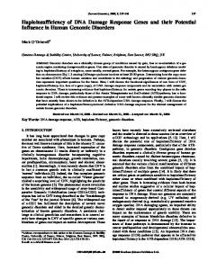
DC like phenotype was observed in male mice suggesting that the lower proliferation rate is not sufficient to impact on tissue renewal, at least in one generation. The assay we use to assess comparative proliferation rate, namely competitive growth of cells in a female mosaic after random X-inactivation may be very sensitive. Indeed if 32 cell divisions are required for the growth39 of an early embryo into an adult mouse then a 5% increase in the time between cell divisions will lead to an approximately 80% reduction in the number of the slower growing cells. Such a small reduction in growth rate, while easy to see in the competitive situation, may not affect the growth and development of males. Another factor here may be differences between mouse and humans in the detailed mechanisms of both telomere maintenance40,41 and stem cell function. Of the six genes so far identified as causing DC, two of them— NOP107 and NHP28— have been found to be very rare and present in one or a handful of families. The others fall into two groups in terms of presentation and penetrance. Mutations in TERT 10 and TERC15 have less than 100% penetrance and generally show anticipation with increased severity and earlier age of onset in later generations.42,43 This is likely due to the fact that later generations inherit shortened telomeres from the affected parent as well as the pathogenic mutation. Mutations in DKC1,4 and TINF2,11,12 however exert their effects in generation 1 where they usually cause severe aplastic anemia and dyskeratosis congenita of varying severity, with TINF2 mutations perhaps being on average more severe. The difference between these two groups may be that TERC and TERT mutations lead to telomere shortening with no consequences until a critically short telomere length is reached whereas DKC1 and TINF2 mutations in addition cause cell death via the p53 DNA damage pathway due to telomere defects independent of telomere length and thus exert their effects in generation 1. Supporting this idea is our finding of a DNA damage response in mice with a pathogenic Dkc1 mutation.24 While the TINF2 pathogenic mutations have not yet been shown to elicit a DNA damage response at telomeres other TIN2 mutations, that destabilize shelterin components TRF1 and TRF2 do induce DNA damage.44 All DC patients however have very short telomeres but whether short telomeres are always causative of the disease or are a consequence of increased replicative telomere erosion is not clear. Thus it is possible that the DNA damage caused by these mutations contributes to bone marrow failure by leading to increased rate of recruitment of stem cells and eventual stem cell exhaustion (Fig. 1).
DI ST
Mutant Dyskerin Affects the DNA Damage Response
©
Possible Importance of the DNA Damage Response in the Pathogenesis of X-Linked DC Is the induction of the DNA damage response an important factor in the pathogenesis of dyskeratosis congenita? In the Dkc1D15/+ heterozygous female mice the induction of the DNA damage response was associated with decreased proliferation. However no 8
Summary and Future Prospects In summary the finding of an increased DNA damage response in mice with a pathogenic mutation in Dkc1 has important implications for the role of dyskerin in telomere maintenance. Coupled with the recent finding of DC due to TINF2 mutations it suggests that failure to maintain telomere integrity in DC may arise by more than one pathway. It will now be important to work out precisely how dyskerin and TIN2 interact with other components of telomerase and of shelterin to bring about the exquisite control of telomere homeostasis needed to strike the essential balance between aging and malignancy. Acknowledgements
Work in the authors’ laboratories was supported by NCI grant R01 CA106995 to P.J.M and RFA-HL-04-008 to M.B.
Cell Cycle
2009; Vol. 8 Issue 1
OS
CI E
NC E
.D
O
NO
T
DI ST
RI
BU T
E.
DNA damage response in DC mouse model
BI
Figure 1. Model of pathogenesis of dyskeratosis congenita.
ES
References
©
20
08
LA
ND
1. Kim NW, Piatyszek MA, Prowse KR, Harley CB, West MD, Ho PL, et al. Specific association of human telomerase activity with immortal cells and cancer. Science 1994; 266:2011-5. 2. Hayflick L, Moorhead PS. The serial cultivation of human diploid cell strains. Exp Cell Res 1961; 25:585-621. 3. Harley CB, Futcher AB, Greider CW. Telomeres shorten during ageing of human fibroblasts. Nature 1990; 345:458-60. 4. Heiss NS, Knight SW, Vulliamy TJ, Klauck SM, Wiemann S, Mason PJ, et al. X-linked dyskeratosis congenita is caused by mutations in a highly conserved gene with putative nucleolar functions. Nat Genet 1998; 19:32-8. 5. Meier UT. The many facets of H/ACA ribonucleoproteins. Chromosoma 2005; 114:1-14. 6. Mitchell JR, Wood E, Collins K. A telomerase component is defective in the human disease dyskeratosis congenita. Nature 1999; 402:551-5. 7. Walne AJ, Vulliamy T, Marrone A, Beswick R, Kirwan M, Masunari Y, et al. Genetic heterogeneity in autosomal recessive dyskeratosis congenita with one subtype due to mutations in the telomerase-associated protein NOP10. Hum Mol Genet 2007; 16:1619-29. 8. Vulliamy T, Beswick R, Kirwan M, Marrone A, Digweed M, Walne A, et al. Mutations in the telomerase component NHP2 cause the premature ageing syndrome dyskeratosis congenita. Proc Natl Acad Sci USA 2008; 105:8073-8. 9. Vulliamy T, Marrone A, Goldman F, Dearlove A, Bessler M, Mason PJ, et al. The RNA component of telomerase is mutated in autosomal dominant dyskeratosis congenita. Nature 2001; 413:432-5. 10. Yamaguchi H, Calado RT, Ly H, Kajigaya S, Baerlocher GM, Chanock SJ, et al. Mutations in TERT, the gene for telomerase reverse transcriptase, in aplastic anemia. N Engl J Med 2005; 352:1413-24. www.landesbioscience.com
11. Savage SA, Giri N, Baerlocher GM, Orr N, Lansdorp PM, Alter BP. TINF2, a component of the shelterin telomere protection complex, is mutated in dyskeratosis congenita. Am J Hum Genet 2008; 82:501-9. 12. Walne AJ, Vulliamy TJ, Beswick R, Kirwan M, Dokal I. TINF2 mutations result in very short telomeres: Analysis of a large cohort of patients with dyskeratosis congenita and related bone marrow failure syndromes. Blood 2008. 13. Kim SH, Kaminker P, Campisi J. TIN2, a new regulator of telomere length in human cells. Nat Genet 1999; 23:405-12. 14. de Lange T. Shelterin: the protein complex that shapes and safeguards human telomeres. Genes Dev 2005; 19:2100-10. 15. Vulliamy TJ, Knight SW, Mason PJ, Dokal I. Very short telomeres in the peripheral blood of patients with X-linked and autosomal dyskeratosis congenita. Blood Cells Mol Dis 2001; 27:353-7. 16. Yamaguchi H, Baerlocher GM, Lansdorp PM, Chanock SJ, Nunez O, Sloand E, et al. Mutations of the human telomerase RNA gene (TERC) in aplastic anemia and myelodysplastic syndrome. Blood 2003; 102:916-8. 17. Armanios MY, Chen JJ, Cogan JD, Alder JK, Ingersoll RG, Markin C, et al. Telomerase mutations in families with idiopathic pulmonary fibrosis. N Engl J Med 2007; 356:1317-26. 18. Tsakiri KD, Cronkhite JT, Kuan PJ, Xing C, Raghu G, Weissler JC, et al. Adult-onset pulmonary fibrosis caused by mutations in telomerase. Proc Natl Acad Sci USA 2007; 104:7552-7. 19. Wong JM, Collins K. Telomerase RNA level limits telomere maintenance in X-linked dyskeratosis congenita. Genes Dev 2006; 20:2848-58. 20. Devriendt K, Matthijs G, Legius E, Schollen E, Blockmans D, van Geet C, et al. Skewed X-chromosome inactivation in female carriers of dyskeratosis congenita. Am J Hum Genet 1997; 60:581-7.
Cell Cycle
9
BU T RI DI ST T NO O .D
©
20
08
LA
ND
ES
BI
OS
CI E
NC E
21. Vulliamy TJ, Knight SW, Dokal I, Mason PJ. Skewed X-inactivation in carriers of X-linked dyskeratosis congenita. Blood 1997; 90:2213-6. 22. Lyon MF. Gene action in the X-chromosome of the mouse (Mus musculus L.). Nature 1961; 190:372-3. 23. Beutler E, Yeh M, Fairbanks VF. The normal human female as a mosaic of X-chromosome activity: studies using the gene for C-6-PD-deficiency as a marker. Proc Natl Acad Sci USA 1962; 48:9-16. 24. Gu BW, Bessler M, Mason PJ. A pathogenic dyskerin mutation impairs proliferation and activates a DNA damage response independent of telomere length in mice. Proc Natl Acad Sci USA 2008; 105:10173-8. 25. Vulliamy TJ, Knight SW, Heiss NS, Smith OP, Poustka A, Dokal I, et al. Dyskeratosis congenita caused by a 3' deletion: germline and somatic mosaicism in a female carrier. Blood 1999; 94:1254-60. 26. Blasco MA, Lee HW, Hande MP, Samper E, Lansdorp PM, DePinho RA, et al. Telomere shortening and tumor formation by mouse cells lacking telomerase RNA. Cell 1997; 91:25-34. 27. Erdmann N, Liu Y, Harrington L. Distinct dosage requirements for the maintenance of long and short telomeres in mTert heterozygous mice. Proc Natl Acad Sci USA 2004; 101:6080-5. 28. Sedelnikova OA, Pilch DR, Redon C, Bonner WM. Histone H2AX in DNA damage and repair. Cancer Biol Ther 2003; 2:233-5. 29. d’Adda di Fagagna F, Teo SH, Jackson SP. Functional links between telomeres and proteins of the DNA-damage response. Genes Dev 2004; 18:1781-99. 30. d’Adda di Fagagna F, Reaper PM, Clay-Farrace L, Fiegler H, Carr P, Von Zglinicki T, et al. A DNA damage checkpoint response in telomere-initiated senescence. Nature 2003; 426:194-8. 31. Denchi EL, de Lange T. Protection of telomeres through independent control of ATM and ATR by TRF2 and POT1. Nature 2007; 448:1068-71. 32. Verdun RE, Karlseder J. Replication and protection of telomeres. Nature 2007; 447:924-31. 33. Gilson E, Geli V. How telomeres are replicated. Nat Rev Mol Cell Biol 2007; 8:825-38. 34. Cohen SB, Graham ME, Lovrecz GO, Bache N, Robinson PJ, Reddel RR. Protein composition of catalytically active human telomerase from immortal cells. Science 2007; 315:1850-3. 35. Gonzalez-Suarez E, Samper E, Ramirez A, Flores JM, Martin-Caballero J, Jorcano JL, et al. Increased epidermal tumors and increased skin wound healing in transgenic mice overexpressing the catalytic subunit of telomerase, mTERT, in basal keratinocytes. Embo J 2001; 20:2619-30. 36. Artandi SE, Alson S, Tietze MK, Sharpless NE, Ye S, Greenberg RA, et al. Constitutive telomerase expression promotes mammary carcinomas in aging mice. Proc Natl Acad Sci USA 2002; 99:8191-6. 37. Gonzalez-Suarez E, Samper E, Flores JM, Blasco MA. Telomerase-deficient mice with short telomeres are resistant to skin tumorigenesis. Nat Genet 2000; 26:114-7. 38. Stewart SA, Hahn WC, O’Connor BF, Banner EN, Lundberg AS, Modha P, et al. Telomerase contributes to tumorigenesis by a telomere length-independent mechanism. Proc Natl Acad Sci USA 2002; 99:12606-11. 39. Wasserstrom A, Frumkin D, Adar R, Itzkovitz S, Stern T, Kaplan S, et al. Estimating cell depth from somatic mutations. PLoS Comput Biol 2008; 4:1000058. 40. Wright WE, Shay JW. Telomere dynamics in cancer progression and prevention: fundamental differences in human and mouse telomere biology. Nat Med 2000; 6:849-51. 41. Smogorzewska A, de Lange T. Different telomere damage signaling pathways in human and mouse cells. Embo J 2002; 21:4338-48. 42. Vulliamy T, Marrone A, Szydlo R, Walne A, Mason PJ, Dokal I. Disease anticipation is associated with progressive telomere shortening in families with dyskeratosis congenita due to mutations in TERC. Nat Genet 2004; 36:447-9. 43. Armanios M, Chen JL, Chang YP, Brodsky RA, Hawkins A, Griffin CA, et al. Haploinsufficiency of telomerase reverse transcriptase leads to anticipation in autosomal dominant dyskeratosis congenita. Proc Natl Acad Sci USA 2005; 102:15960-4. 44. Kim SH, Beausejour C, Davalos AR, Kaminker P, Heo SJ, Campisi J. TIN2 mediates functions of TRF2 at human telomeres. J Biol Chem 2004; 279:43799-804. 45. Vulliamy TJ, Dokal I. Dyskeratosis congenita: the diverse clinical presentation of mutations in the telomerase complex. Biochimie 2008; 90:122-30. 46. Vulliamy T, Marrone A, Dokal I, Mason PJ. Association between aplastic anaemia and mutations in telomerase RNA. Lancet 2002; 359:2168-70. 47. Vulliamy TJ, Walne A, Baskaradas A, Mason PJ, Marrone A, Dokal I. Mutations in the reverse transcriptase component of telomerase (TERT) in patients with bone marrow failure. Blood Cells Mol Dis 2005; 34:257-63. 48. Du HY, Pumbo E, Manley P, Field JJ, Bayliss SJ, Wilson DB, et al. Complex inheritance pattern of dyskeratosis congenita in two families with 2 different mutations in the telomerase reverse transcriptase gene. Blood 2008; 111:1128-30. 49. Marrone A, Walne A, Tamary H, Masunari Y, Kirwan M, Beswick R, et al. Telomerase reverse-transcriptase homozygous mutations in autosomal recessive dyskeratosis congenita and Hoyeraal-Hreidarsson syndrome. Blood 2007; 110:4198-205.
E.
DNA damage response in DC mouse model
10
Cell Cycle
2009; Vol. 8 Issue 1