Transform (DWT) are being used to denoise ECG signal and a ... wave follows QRS complex which indicates the ... time resolution at low and high frequencies.
International Journal of Engineering Trends and Technology (IJETT) – Volume 36 Number 5- June 2016
ECG Signal Denoising with Savitzky-Golay Filter and Discrete Wavelet Transform (DWT) Harjeet Kaur#1, Rajni*2 #
Research Scholar, ECE department, MRSPTU, INDIA Associate Professor, ECE Department, MRSPTU, INDIA
*
Abstract — Electrocardiogram (ECG) demonstrates the electrical activity of heart muscles over a period of time. The ECG is one of the extensively used physiological parameters for examination and diagnosis of cardiac diseases. The non-stationary ECG signal often gets contaminated with different noises. Hence, it is required to denoise the signal to provide accurate information to physicians. In this paper, Savitzky-Golay filter and Discrete Wavelet Transform (DWT) are being used to denoise ECG signal and a comparison is provided between two methods. The filter and DWT are applied on MITBIH arrhythmia database to check the robustness of proposed methods. Two parameters, signal to noise ratio (SNR) and mean squared error (MSE) are used for performance comparison.
repolarization of ventricles. Sometimes a conditional U wave is also present [7]. The amplitude of these peaks and duration of constituent intervals reveal clinically essential information [8]-[9]. An ECG wave with these components is given below in Fig. 1.
Keywords — Electrocardiogram, Savitzky-Golay Filter, Discrete Wavelet Transform, SNR, MSE. I. INTRODUCTION The Electrocardiogram is a consistent and dependable investigation tool which offers an extensive amount of information regarding functioning of heart [1]-[2]. The Electrocardiogram (ECG) represents heart’s electrical activity which is recorded with the help of ECG machine and electrodes [3]. The ECG signal is represented by sequential contraction and relaxation heart muscles. The human heart structure contains two upper chambers and two lower chambers known as atria and ventricles respectively. Under healthy cardiac conditions, cardiac cycle begins from Sino Atria node, the right atrium and travels from atria to Atrio ventricular node [4]. This path of beat propagation is traced accurately to ensure the regular activity of heart [5]. II. ECG WAVE AND NOISE The ECG is demonstrated in the form of different peaks and valleys. These peaks and valleys are depicted by symbols P, Q, R, S and T [6]. In ECG signal a heartbeat begins with P wave that corresponds to the depolarization of atria, upper two chambers in the heart structure. The P wave is followed by QRS complex that is formed by combing Q, R and S waves. The QRS complex illustrates the depolarization of ventricles. Then a T wave follows QRS complex which indicates the
ISSN: 2231-5381
Fig. 1 ECG waveform with characteristic features [10]
The utilitarian information that an ECG signal provides include [11]: Heart position and conduction disturbances Relative chamber size Heartbeat origin and propagation Effects of drugs on heart condition Change in electrolyte concentration Being an electrical signal, ECG is much prone to various types of noise [12]: Motion artifacts Baseline wandering Powerline interference Baseline drift is caused due to respiration, coughing or movement of patient [13]. It is the major cause of noise and can vary important signal characteristics [14]. Therefore, it needs to be removed first from ECG signal for further signal analysis. Powerline interference is caused by harmonics which are generated inside electrical appliance used to capture ECG signals [15]. The disturbance caused due power line interference can be removed with proper and careful utilization of hardware employed for recording ECG signals [15].
http://www.ijettjournal.org
Page 266
International Journal of Engineering Trends and Technology (IJETT) – Volume 36 Number 5- June 2016
The motion artefacts are caused due to electrode skin impedance. III. MATERIALS AND METHODS A. Database For analysing ECG signals, data is collected form Physionet site [16] under MIT-BIH arrhythmia database [17]. The arrhythmia database consists of 48 records. Each ECG signal is sampled at a frequency of 360Hz. All signals are sightly of 30 minutes duration. The ECG signals from this database contain different heart abnormalities with various kinds of noise [18]. B. Savitzky-Golay Filter Savitzky and Golay proposed a method leastsquares polynomial fitting for sample smoothing in [19]. In Savitzky-Golay (Sgolay) filters data is fitted onto a polynomial of given order [15]. The order is the degree of the polynomial and number of samples used for smoothing data is indicated by frame size. Sgolay filter has an important peak preserving property which is very useful in ECG signal analysis [20]. C. Discrete Wavelet Transform In the field of signal processing, Wavelet Transform (WT) has been used in a number of applications. The WT is an influential method that describes a signal in its time-frequency domain [21]. Recently, Discrete Wavelet Transform (DWT) becomes a popular tool to study non-stationary signals such as Electrocardiogram [3], [22]. When different components of ECG wave are subjected to multiresolution analysis they become clearly visible [23]. The DWT decomposes a signal in different frequency bands at different resolution hence called multiresolution analysis. The major advantage of DWT is its ability to provide good frequency and time resolution at low and high frequencies respectively [24]. In DWT, a signal is decomposed using pair of complementary filters [25] and down samplers as illustrated in Fig. 2.
as depicted in Fig. 2. In applications that are based on WT, selection of mother wavelet that matches with the characteristics of the signal under investigation is of great significance [22]. IV. METHODOLOGY The different steps involved in denoising ECG signals are given below: Generation and addition of random noise in the ECG signal Firstly, baseline drift and other noise present in the signal are removed by applying Sgolay filter of length equal to19 Then secondly denoising is done by using DWT with 10 levels of decomposition. Afterwards thresholding is employed to remove noise from ECG signal Finally, performance parameters i.e. SNR (in dB) and MSE are computed for two techniques. The SNR and MSE are calculated as given in Eqs. (1) and (2) respectively. (1)
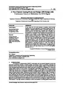
(2) where indicates original signal, is the denoised signal and M corresponds to number of samples in the signal of interest. V. RESULTS AND DISCUSSION The ECG signals used for denoising are taken from MIT-BIH arrhythmia database. The fifteen different ECG records: 100, 102, 103, 104, 105, 106, 107, 111, 112, 114, 117, 121, 123, 201 and 203 are utilized to verify the performance of Sgolay filter and DWT. In DWT method, signal is decomposed using Bior3.1 wavelet due to its high SNR and low MSE than other wavelets [24]. All the work is implemented on MATLAB software. The waveform of ECG sample number 100 contaminated with noise is shown in Fig. 3.
Fig. 2 Wavelet decomposition of a signal with DWT [22]
The first high pass filter (HPF) is the discrete mother wavelet and second low pass filter (LPF) is its mirror version. The coefficient of HPF is known as detail (d1) and coefficient of LPF is called as approximation (a1). The process continues as the approximation (a1) coefficient is decomposed further
ISSN: 2231-5381
Fig. 3 Noisy ECG sample number 100
Although each ECG signal is of 30 minutes but for simplification waveforms are shown for 10
http://www.ijettjournal.org
Page 267
International Journal of Engineering Trends and Technology (IJETT) – Volume 36 Number 5- June 2016
seconds. The resulting ECG waveforms of Sgolay and DWT filtering are depicted in Figs. 4 and 5 respectively.
VI. CONCLUSIONS This paper presents two techniques for denoising ECG signal and compares performance of these techniques in terms of SNR and MSE. The simulated results shown in Tables 1, illustrate highest value of SNR and lowest value of MSE for DWT in comparison with Sgolay filter. Hence from simulated results, it is evident that DWT is better than Sgolay method. REFERENCES [1]
[2] Fig. 4 Denoised ECG sample number 100 with Sgolay filter [3]
[4]
[5]
[6]
[7]
Fig. 5 Denoised ECG sample number 100 with DWT
The performance evaluation of proposed denoising methods in tabulated form is shown below in Table 1.
[8] [9]
[10]
TABLE I PERFORMANCE COMPARISON OF DENOISING TECHNIQUES
ECG Signal 100 102 103 104 105 106 107 111 112 114 117 121 123 201 203
Sgolay filter SNR MSE (in dB) 5.917 0.044 7.761 0.210 6.160 0.014 4.230 0.026 6.113 0.015 1.368 0.016 6.456 0.097 7.565 0.021 3.082 0.417 9.708 0.009 5.275 0.275 6.379 0.400 6.062 0.190 7.678 0.058 4.495 0.010
ISSN: 2231-5381
DWT SNR MSE (in dB) 7.452 0.002 10.220 0.116 8.269 0.006 9.403 0.005 8.357 0.001 3.768 0.006 10.164 0.024 9.545 0.006 6.466 0.007 10.866 0.008 8.650 0.008 9.828 0.002 8.847 0.003 9.504 0.002 6.438 0.006
[11]
[12]
[13]
[14]
[15]
[16] [17]
P. Bhardwaj, R. K. Choudhary, and R. Dayama, ―Analysis and classification of cardiac arrhythmia using ECG signals,‖ International Journal of Computer Applications, vol. 38, pp. 37-40, Jan. 2012. S. T. Prasad, and S. Varadarajan, ―ECG signal analysis: different approaches,” International Journal of Engineering Trends and Technology (IJETT), vol. 7, pp. 212-216, Jan. 2014. H. Y. Lin, S. Y. Liang, Y. L. Ho, Y. H. Lin, and H. P. Ma, ―Discrete wavelet transform based noise removal and feature extraction for ECG signals,‖ IRBM, vol. 35, pp. 351-361, 2014. Rajni, and I. Kaur, ―Electrocardiogram signal analysis- an overview,‖ International Journal of Computer Applications, vol. 84, pp. 22-25, Dec. 2013. H. Nagendra, S. Mukherjee, and V. Kumar, ―Application of wavelet techniques in ECG signal processing: an overview,‖ International Journal of Engineering Science and Technology (IJEST), vol. 3, pp. 7432-7443, 2011. R. J. Martis, U. R. Acharya, and H. Adeli, ―Current methods in electrocardiogram characterization,‖ Comput. Biol. Med., vol. 48, pp. 133-149, 2014. A. Josko, and R. J. Rak, ―Effective simulation of signals for testing ECG analyser,‖ IEEE Trans. Instrum. Meas., vol. 54, pp. 1019-1024, June 2005. L. Scharmroth, An Introduction to Electrocardiography, 7th ed., Wiley:India, 2009. N. Sooma, J. Singh, and M. Tiwari, ―Feature extraction of ECG signal using HHT algorithm,‖ International Journal of Engineering Trends and Technology (IJETT), vol. 8, pp. 454-460, Feb. 2014. D. Sadhukhan, and M. Mitra, ―R-peak detection algorithm for ECG using double difference and RR interval processing,‖ Procedia Technology, vol. 4, pp. 873-877, 2012. Rajni, and H. Kaur, ―Methods of electrocardiogram signal analysis to detect arrhythmia - a review,‖ in Proc. National Conference on Communication, Computing & Systems (NCCCS-2015), Aug. 2015, p. 1-5. B. Arvinti, M. Castache, D. Toader, M. Olten, and A. Isar, ―ECG statistical denoising in the wavelet domain‖ in ISETC, Nov. 2010, p. 307-310. J. Oster, O. Pietquin, M. Kraemer, and J. Felblinger, “Nonlinear bayesian filtering for denoising of electrocardiograms acquired in a magnetic resonance environment,‖ IEEE Trans. Biomed. Eng., Vol. 57, pp. 1628-1638, July 2010. M. V. R. Lele, and P. K. S. Holkar, ―Removal of baseline wander from ECG signal,” in Proc. international Conference on Recent Trends in engineering & Technology-2013, Feb. 2013, p. 60-65. I. kaur, and Rajni, ―Denoising of ECG signal using filters and wavelet transform,‖ in Proc. International Conference on Recent Trends in Electronics, Data Communication and Computing (ICRTEDC-2014), 2014, p.28-31. http://www.physionet.org/ A. L. Goldberger, L. A .N. Amaral, L. Glass, J. M. Hausdroff, P. C. Ivanov, R. G. Mark, J. E. Mietus, G. B. Moody, C. K. Peng, and E. H. Stanely, ―PhysioBank, Physio Toolkit and PhysioNet: components of a new
http://www.ijettjournal.org
Page 268
International Journal of Engineering Trends and Technology (IJETT) – Volume 36 Number 5- June 2016
[18]
[19]
[20]
[21]
research resource for complex physiologic signals,‖ Circulation, vol. 101 pp. e215-e220, 2000. R. J. Martis, U. R. Acharya, C. M. Lim, J. S. Suri, ―Characterization of ECG beats from cardiac arrhythmia using discrete cosine transform in PCA framework,‖ Knowl.- Based Syst., vol. 45, pp. 76-82, 2013. A. Savitzky, and M. J. E. Golay, ―Smoothing and differentiation of data by simplified least squares procedures,‖ Anal. Chem., vol. 36, pp. 1627–1639, 1964. N. Rastogi, and R. Mehra, ―Analysis of Savitzky- Golay filter for baseline wander cancellation in ECG using Wavelets‖, International Journal of Engineering Sciences & Emerging Technologies (IJESET), vol. 6, pp. 15-23, 2013. K. Daqrouq, and A. R. Al-Qawasmi, ―ECG Enhancement using Wavelet Transform,‖ WSEAS Transactions on Biology and Biomedicine, vol. 7, pp. 62–72, 2010.
ISSN: 2231-5381
M. AlMahamdy, and H. R. Riley, ―Performance study of different denoising methods for ECG signals,‖ Procedia Comput. Sci.,‖ vol. 37, pp. 325-332, 2014. [23] S. Banerjee, R. Gupta, and M. Mitra, ―Delineation of ECG characteristics features using multiresolution wavelet analysis method,‖ Measurement, vol. 45, pp. 474-487, 2012. [24] I. Kaur, Rajni, and G. Sikri, ―Denoising of ECG signal with different wavelets,‖ International Journal of Engineering Trends and Technology (IJETT), vol. 9, pp. 658-661, Mar. 2014. [25] S. Patel, and D.A. Datar, ―ECG data compression using wavelet transform,‖ International Journal of Engineering Trends and technology (IJETT), vol. 10, pp. 770-776, Apr. 2014. [22]
http://www.ijettjournal.org
Page 269