CLINICAL GASTROENTEROLOGY AND HEPATOLOGY 2012;10:593–597
EDUCATION PRACTICE A 42–Year–Old Woman With a New Diagnosis of Sclerosing Cholangitis ANDREA A. GOSSARD and KEITH D. LINDOR Division of Gastroenterology and Hepatology, Mayo Clinic, Rochester, Minnesota
Clinical Scenario
A
42-year-old woman is referred to you for severe pruritus, dark urine, and acholic stools. These symptoms have gradually increased during the past 7–10 days. She had previously been in good health with no history of hepatobiliary dysfunction. She complains of right upper quadrant discomfort and a general sense of malaise. Blood tests reveal a normal complete blood count, but her liver enzymes are elevated in a cholestatic pattern. Her alkaline phosphatase level is approximately 3 times the upper limit of normal (ULN) at 428 U/L, the aspartate aminotransferase level is 1.5 times the ULN at 116 U/L, and the total serum bilirubin is 6.4 mg/dL, with a direct bilirubin of 4.8 mg/dL. An antimitochondrial antibody is performed to evaluate for primary biliary cirrhosis and is negative. On examination, she appears unwell with obvious jaundice. She is afebrile, and there are no spider angiomas noted. She has generalized tenderness with palpation of the right upper quadrant of the abdomen. Abdominal imaging is performed with an ultrasound and reveals focal dilation of the intrahepatic bile ducts in the right and left hepatic lobes. Also noted is diffuse wall thickening of the distal hepatic and common bile duct. There is no obvious hepatic mass and no evidence of cholelithiasis or choledocholithiasis. The gallbladder and pancreas appear normal, and there is no splenomegaly or ascites.
The Problem Primary sclerosing cholangitis (PSC) is a chronic, cholestatic liver disease of unknown etiology. Potential causes include disordered immunoregulation, infections, or bacterial products with portal bacteremia. The disease leads to diffuse inflammation and fibrosis of the biliary tree, which ultimately causes biliary cirrhosis and portal hypertension. Patients with PSC might present with incidentally noted elevated liver enzymes in a cholestatic profile. Others present with symptoms of cholestasis including fatigue, jaundice, acholic stools, dark urine, hyperpigmentation, or pruritus. A subset will have evidence of cholangiocarcinoma (CCA) at the time of initial presentation. Fifteen percent to 44% of patients might be entirely asymptomatic (Table 1). Autoantibodies such as an antinuclear antibody or antineutrophil cytoplasmic antibody might or might not be present. The diagnosis of PSC is typically made in the setting of a cholestatic clinical picture and an abnormal cholangiogram. The evaluation of a patient with new cholestasis might include laboratory work, cross-sectional imaging, and a cholangiogram (Figure 1). Endoscopic retrograde cholangiopan-
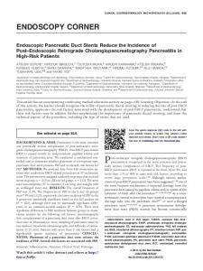
creatography (ERCP) has been the preferred method of evaluating for PSC; however, this procedure is not risk free. Potentially serious complications such as pancreatitis and bacterial cholangitis can occur in up to 10% of patients undergoing ERCP. Magnetic resonance cholangiography (MRCP) is a noninvasive method of evaluating the biliary tree and is increasingly being used. The MRCP image in Figure 2 shows changes of PSC. In the setting of PSC, the bile ducts typically demonstrate a “beaded” appearance when imaged. Often, there will be areas of significant stenosis alternating with focal bile duct dilation. The disease might be diffusely spread throughout the biliary tree or confined to the intrahepatic ducts (⬍25% of all PSC patients). Approximately 5% of patients with cholestatic changes will have a normal-appearing biliary tree cholangiographically but have changes histologically that are consistent with PSC, a condition termed small-duct PSC. It might be that some of these patients will eventually develop disease of the large bile ducts as well, but this progression is unpredictable. A liver biopsy might be helpful in confirming the diagnosis of PSC and in providing prognostic information, but it is not necessary in all cases. Histologic features might include periductal fibrosis, a paucity of bile ducts, and variable degrees of fibrosis encasing the intralobular bile duct (Figure 3). This is described as “onion skin” fibrosis. Often, however, only nondiagnostic histologic changes are seen. Secondary sclerosing cholangitis is characterized by a similar process but is typically due to causes such as longterm bile duct obstruction, surgical trauma, or infection. The majority of patients with PSC (75%) will have coexisting inflammatory bowel disease (IBD). Of these patients, 70% will have chronic ulcerative colitis. A subset of these will demonstrate evidence of IBD affecting the right side of the colon with rectal sparing. The diagnosis of IBD might or might not precede the diagnosis of PSC.
Abbreviations used in this paper: AASLD, American Association for the Study of Liver Diseases; CCA, cholangiocarcinoma; CT, computed tomography; ERCP, endoscopic retrograde cholangiopancreatography; IBD, inflammatory bowel disease; MRCP, magnetic resonance cholangiopancreatography; MRI, magnetic resonance imaging; PSC, primary sclerosing cholangitis; UDCA, ursodeoxycholic acid; ULN, upper limit of normal. © 2012 by the AGA Institute 1542-3565/$36.00 http://dx.doi.org/10.1016/j.cgh.2012.02.022
594
GOSSARD AND LINDOR
CLINICAL GASTROENTEROLOGY AND HEPATOLOGY Vol. 10, No. 6
Table 1. Clinical Presentation of PSC Symptoms
%
Asymptomatic Fatigue Pruritus Jaundice Hepatomegaly Abdominal pain Weight loss Splenomegaly Ascending cholangitis Hyperpigmentation Variceal bleeding Ascites
15–44 75 70 30–69 34–62 16–37 10–34 30 5–28 25 2–14 2–10
Management Strategies and Supporting Evidence Treatment There is no approved or widely accepted medical therapy for PSC. Multiple agents have been studied with largely negative results. Among these are ursodeoxycholic acid (UDCA) at varying doses (13–15 mg/kg/d to 28 –30 mg/kg/d), as well as immunosuppressive agents such as azathioprine, budesonide, methotrexate, mycophenolate mofetil, and tacrolimus. In addition, bezafibrate, colchicine, cladribine, cyclosporine, etanercept, infliximab, oral and transdermal nicotine, D-penicillamine, pentoxifylline, pirfenidone, and silymarin have all been studied. None have provided convincing evidence of benefit, although some have resulted in modest improvement in bio-
Figure 2. MRCP showing PSC.
chemistries. UDCA at doses of 10 –15 mg/kg/d has been associated with biochemical improvement and histologic improvement in small pilot studies. Larger studies have failed to corroborate this finding. Use of UDCA at doses of 28 –30 mg/kg/d has been associated with negative outcomes and is not recommended. There are data that show that use of UDCA might be associated with a decreased risk of colorectal neoplasia in patients with PSC and IBD. Continued research would be of value.
Managing Complications Patients with PSC are at an increased risk for bacterial cholangitis, which requires treatment with broad-spectrum antibiotics. There is little role for surgical management of PSC in the absence of malignancy or liver transplantation. Endoscopic management of symptomatic strictures, which are present in
Figure 1. Algorithm for evaluation of cholestatic presentation. AMA, antimitochondrial antibody.
Figure 3. Histologic evidence of PSC. (Note the concentric periductal fibrosis.)
June 2012
approximately 10% of newly diagnosed patients, might be accomplished with balloon dilatation or stenting of the bile ducts. Stenting should be reserved for those strictures not responsive to balloon dilation. Stenotic lesions are more often benign than malignant; nonetheless, brushings and biopsies should be performed when dominant strictures are discovered. Brush cytology has a disappointing sensitivity of between 18%– 40% in published studies. The specificity for CCA, however, is excellent at nearly 100%. Use of fluorescent in situ hybridization of cytologic samples might provide additional sensitivity, and more research is necessary in this regard. Use of digital image analysis was not found to be of value. Sonographic imaging of concerning strictures might be performed with endoscopic ultrasound and is helpful in guiding biopsy. Long-term stenting is not recommended because of stent obstruction or potential migration of the stent and the associated risk of biliary obstruction. If a patient is stented, removal or replacement of the stent is necessary after 3– 6 weeks in most cases. Percutaneous cholangiography might also be performed when strictures are not able to be accessed via ERCP. Occasionally, use of endoscopic ultrasound is necessary to direct therapeutic efforts. Liver transplantation has an excellent outcome for patients with PSC, with a 1-year survival rate approaching 97% and a 5-year survival of 85%– 88%. These patients are at an increased risk of rejection and infection when compared with patients transplanted for other liver diseases. As with many other liver diseases, the disease can and does recur in 2%– 40% of patients after transplantation. CCA is a most dreaded complication of PSC. Management of the patient with PSC should include cancer surveillance with imaging of the liver and biliary system every 12 months. Although there are few data to support this recommendation, referral to centers with experience in hepatobiliary surgery and liver transplantation might afford benefit to those diagnosed with early-stage CCA. Imaging might be done with abdominal ultrasound, computed tomography (CT), or magnetic resonance imaging (MRI). There is no effective medical therapy for CCA. Surgical resection might be curative, but the presence of PSC is usually a contraindication to partial hepatectomy because of the risk of de novo CCA developing with remaining at-risk epithelium in the absence of total hepatectomy. Stenting of malignant biliary obstructions, radiation therapy, photodynamic therapy, and chemotherapy might provide effective palliation for unresectable patients. Liver transplantation for early CCA is possible. Early-stage CCA is defined as a unicentric mass lesion of less than 3 cm in radial diameter and no intrahepatic or extrahepatic metastases. Typically, adjuvant agents such as radiotherapy and chemosensitization with 5-fluorouracil and brachytherapy, and capecitabine are necessary before liver transplantation for CCA. Overall 5-year survival rates are 70% for select patients with perihilar CCA who undergo this approach. Clearly, this is a significant improvement over typical CCA survival rates. This option is available at a limited number of transplant centers.
Primary Sclerosing Cholangitis and Inflammatory Bowel Disease Nearly 70% of patients with PSC will have comorbid IBD. In this setting, colon cancer screening with colonoscopy and biopsies should be done annually because the concurrent
NEW DIAGNOSIS OF SCLEROSING CHOLANGITIS
595
diagnosis of IBD with PSC increases the risk of colon cancer significantly. Four-quadrant surveillance biopsies should be obtained of the entire colon to adequately screen for early dysplastic changes because these lesions are often not polypoid.
Clinical Management Patients with PSC should be immunized for hepatitis A and B to decrease the risk of contracting a second liver condition. In addition, excessive use of alcohol is ill-advised. Monitoring for development of osteoporosis with bone mineral densitometry should be performed every 2–3 years in patients with advanced-stage liver disease and in women with PSC, particularly after the age of 50. Screening for esophageal varices with an upper endoscopy should be performed if there is evidence of advanced-stage disease and portal hypertension. Monitoring for fat-soluble vitamin deficiencies (A, D, E, and K) is also important. Serum vitamin A and E levels, along with vitamin D metabolites and prothrombin time, would be of value. Pruritus might develop in the patient with PSC, and effective treatments are available. Often, use of antihistamines such as diphenhydramine, 25–50 mg at bedtime, or bile acid sequestrants such as cholestyramine, 4-g packets twice daily, might provide relief. In other cases, rifampin at doses of 150 –300 mg twice daily is used with good results. Some patients will require multiple therapies.
Areas of Uncertainty The estimated 10-year survival for patients with PSC is approximately 65% in a population-based study, but there is wide variability in the natural history. Whereas some patients have particularly problematic disease with multiple symptomatic strictures requiring therapeutic ERCP, others have a relatively uneventful clinical course and seemingly slow disease progression. Ongoing randomized clinical trials to identify effective therapies are needed particularly for those with problematic disease. Although there are prognostic models, there is little consensus regarding an optimal model. The Mayo risk score uses age, bilirubin, aspartate aminotransferase, albumin, and history of variceal bleeding as prognostic parameters. The score stratifies patients into groups of low, intermediate, or high risk for negative outcomes. Other attempts at prognostication include the presence of dominant strictures. Patients with a history of dominant strictures had a reduced survival in the absence of liver transplantation. There is limited ability to predict individual patient outcomes on the basis of the prognostic models available to date. There is a 10%–15% lifetime risk of CCA in the setting of PSC. Although use of ultrasound is cost-effective with minimal risk to the patient, it might not be the most sensitive tool for detecting CCA. CT and MRI are not ideal either. In a large study, use of ultrasound, CT, and MRI yielded positive predictive values of 48%, 38%, and 40%, respectively. Use of serum carbohydrate antigen 19-9 testing might provide additional information; however, this level can be affected by infection and nonmalignant processes such as cholestasis, so it is of limited value. In fact, no study has demonstrated value of carbohydrate antigen 19-9 as a screening test for CCA in PSC. ERCP and MRCP net an overall positive predictive value for CCA of 23% and 21%, respectively. Use of intraductal ultrasound might improve the predictive value.
596
GOSSARD AND LINDOR
Published Guidelines The European Association for the Study of Liver Disease published practice guidelines for PSC in 2009. The American Association for the Study of Liver Diseases (AASLD) published practice guidelines for PSC in 2010. In patients with cholestatic biochemical profile, indirect (MRCP) or direct cholangiography (ERCP) is recommended for making the diagnosis of PSC. European Association for the Study of Liver Disease guidelines suggest that an ERCP be performed if the MRCP is normal, but suspicion for PSC in an IBD patient remains high. Routine liver biopsy for diagnosis of PSC in patients with typical cholangiographic findings is not recommended. In patients with a normal ERCP or MRCP, a liver biopsy to diagnose small-duct PSC might be warranted. In patients with disproportionately elevated aminotransferases, a liver biopsy is advisable to diagnose or exclude an autoimmune overlap syndrome. In all patients with possible PSC, measuring serum immunoglobulin G4 levels to exclude immunoglobulin G4 –associated sclerosing cholangitis is appropriate. In patients with increases in serum bilirubin and/or worsening pruritus, progressive bile duct dilatation on imaging studies, and/or cholangitis, an ERCP is recommended promptly to exclude a dominant stricture. In patients with dominant strictures from PSC, management with endoscopic dilatation with or without stenting is advisable. In patients with dominant strictures from PSC in whom an endoscopic approach is unsuccessful, biliary tract dilatation by percutaneous cholangiography with or without stenting should be considered. Performing brush cytology and/or endoscopic biopsy to exclude a superimposed malignancy at the time of endoscopic therapy for dominant strictures is recommended. In patients with dominant strictures refractory to endoscopic and/or percutaneous management, AASLD recommends surgical therapy for select patients without cirrhosis. Antimicrobial therapy with treatment of bile duct obstruction in dominant strictures is often necessary to effectively resolve cholangitis. Use of ciprofloxacin,
CLINICAL GASTROENTEROLOGY AND HEPATOLOGY Vol. 10, No. 6
500 mg twice a day for 10 –14 days, would be appropriate. In patients with recurrent bacterial cholangitis, AASLD recommends the use of prophylactic long-term antibiotics. An example of prophylaxis might include ciprofloxacin 500 mg twice a day for 2 weeks of every 2 months. This might be rotated with metronidazole if ciprofloxacin is not effective. In patients with refractory bacterial cholangitis, evaluation for liver transplantation might be warranted. In addition, recommendations with regard to complications associated with advanced-stage liver disease, comorbid IBD, pregnancy, and pediatric PSC are provided in these practice guidelines.
Recommendations for This Patient This patient would require ERCP (Figure 4). Endoscopic management of a presumed bile duct narrowing with balloon dilation would likely be necessary, with extraction of intraductal debris such as sludge or stones if present. If a dominant stricture is noted, brushings and biopsies of the area should be performed. Prophylactic antibiotics should be provided. If balloon dilation fails to adequately improve the stricture, temporary stenting would be appropriate. Typically, stents are left in place for 3– 6 weeks in PSC patients and are then either removed or exchanged. Follow-up blood work including liver enzymes and total and direct bilirubin would be helpful to ensure improvement after therapeutic ERCP. In the event the bilirubin fails to improve after several weeks or the patient demonstrates evidence of hepatic decompensation, consideration of orthotopic liver transplantation might be warranted.
Differential Diagnosis The differential diagnosis for this patient would include primary biliary cirrhosis, secondary sclerosing cholangitis, choledocholithiasis, and immunoglobulin G4 –associated cholangitis. Bile duct obstruction from mass lesions and CCA would need to be considered in a patient presenting with new symptomatic cholestasis. Suggested Reading
Figure 4. An ERCP.
1. Lindor KD, Kowdley KV, Luketic VA, et al. High-dose ursodeoxycholic acid for the treatment of primary sclerosing cholangitis. Hepatology 2009;50:808 – 814. 2. Yachimski P, Pratt DS. Cholangiocarcinoma: natural history, treatment, and strategies for surveillance in high-risk patients. J Clin Gastroenterol 2008;42:178 –190. 3. Baron TH. Photodynamic therapy: standard of care for palliation of cholangiocarcinoma? Clin Gastroenterol Hepatol 2008;6: 266 –267. 4. Tamura S, Sugawara Y, Kaneko J, et al. Recurrence of primary sclerosing cholangitis after living donor liver transplantation. Liver Int 2007;27:86 –94. 5. Fevery J, Verslype C, Lai G, et al. Incidence, diagnosis, and therapy of cholangiocarcinoma in patients with primary sclerosing cholangitis. Dig Dis Sci 2007;52:3123–3135. 6. Björnsson E, Angulo P. Cholangiocarcinoma in young individuals with and without primary sclerosing cholangitis. Am J Gastroenterol 2007;102:1677–1682. 7. Lazaridis KN, Gores GJ. Primary sclerosing cholangitis and cholangiocarcinoma. Semin Liver Dis 2006;26:42–51.
June 2012
8. Broomé U, Bergquist A. Primary sclerosing cholangitis, inflammatory bowel disease, and colon cancer. Semin Liver Dis 2006;26: 31– 41. 9. Boberg KM, Jebsen P, Clausen OP, et al. Diagnostic benefit of biliary brush cytology in cholangiocarcinoma in primary sclerosing cholangitis. J Hepatol 2006;45:568 –574. 10. Sinakos E, Saenger AK, Keach J, et al. Many patients with primary sclerosing cholangitis and increased serum levels of carbohydrate antigen 19-9 do not have cholangiocarcinoma. Clin Gastroenterol Hepatol 2011;9:434 – 439. 11. Stanich PP, Björnsson E, Gossard AA, et al. Alkaline phosphatase normalization is associated with better prognosis in primary sclerosing cholangitis. Dig Liver Dis 2011;43:309 –313. 12. Molodecky NA, Kareemi H, Parab R, et al. Incidence of primary sclerosing cholangitis: a systematic review and meta-analysis. Hepatology 2011;53:1590 –1599. 13. Naitoh I, Zen Y, Nakazawa T, et al. Small bile duct involvement in IgG4-related sclerosing cholangitis: liver biopsy and cholangiography correlation. J Gastroenterol 2011;46:269 –276. 14. Goldberg DS, French B, Thomasson A, et al. Current trends in living donor liver transplantation for primary sclerosing cholangitis. Transplantation 2011;91:1148 –1152. 15. Sinakos E, Lindor K. Treatment options for primary sclerosing cholangitis. Expert Rev Gastroenterol Hepatol 2010;4:473– 488. 16. Oh HC, Kim MH, Lee KT, et al. Clinical clues to suspicion of IgG4-associated sclerosing cholangitis disguised as primary scle-
NEW DIAGNOSIS OF SCLEROSING CHOLANGITIS
17. 18.
19. 20.
21.
597
rosing cholangitis or hilar cholangiocarcinoma. J Gastroenterol Hepatol 2010;25:1831–1837. Karlsen TH, Schrumpf E, Boberg KM. Primary sclerosing cholangitis. Best Pract Res Clin Gastroenterol 2010;24:655– 666. Angulo P, Grandison GA, Fong DG, et al. Bone disease in patients with primary sclerosing cholangitis. Gastroenterology 2011;140: 180 –188. Alba LM, Angulo P, Lindor KD. Primary sclerosing cholangitis. Minerva Gastroenterol Dietol 2002;48:99 –113. Brandsaeter B, Schrumpf E, Bentdal O, et al. Recurrent primary sclerosing cholangitis after liver transplantation: a magnetic resonance cholangiography study with analyses of predictive factors. Liver Transplant 2005;11:1361–1369. Razumilava N, Gores GJ, Lindor KD. Cancer surveillance in patients with primary sclerosing cholangitis. Hepatology 2011;54: 1842–1852.
Reprint requests Address requests for reprints to: Andrea Gossard, CNP, Mayo Clinic 200 First Street West, Rochester, Minnesota 55905. e-mail:
[email protected]; fax: (507) 284-0538. Conflicts of interest The authors disclose no conflicts.