Antigone Skopelitou, Michael Mykoniatis. Department of Experimental Pharmacology, Medical School, University of Athens, Athens, Greece. Cadmium is a ...
Effect of Cadmium on Liver Regeneration after Partial Hepatectomy in Rats Alexandra Margeli, Stamatios Theocharis, Spyridon Skaltsas, Antigone Skopelitou, Michael Mykoniatis Department of Experimental Pharmacology, Medical School, University of Athens, Athens, Greece Cadmium is a nonabundant element that is widely distributed throughout the biosphere and its toxic effects are becoming potentially more serious due to industrialization. It has been reported that cadmium might interact with nucleic acid biosynthesis. In this study we examined the effect of cadmium administration, either 24 hr before or simultaneously to partial hepatectomy, on the liver regenerative process in rats, at different time intervals. The rate of DNA synthesis was suppressed markedly in the cadmium pretreated group and the first peak of liver regeneration was delayed, compared to the simply partially hepatectomized one. The administration of cadmium simultaneously to partial hepatectomy, caused a marked decrease of the rate of DNA biosynthesis, compared to the pretreatment.The rate-determining enzyme thymidine kinase was suppressed in the liver of both cadmium-treated groups. Biochemical parameters and histological findings were also coestimated. The above data suggest that either preor simultaneous administration of cadmium, suppressed the liver regenerative process, probably due to the inhibition of thymidine kinase. - Environ Health Perspect 102(Suppl 3):273-276 (1994). Key words: cadmium, liver regeneration, rats, partial hepatectomy, 3HTdR incorporation, thymidine kinase
Introduction Cadmium (Cd) is a highly toxic element that is present in food and water and is accumulated in liver and kidney. It is known that Cd is one of the most harmful heavy metals able to induce renal, hepatic and testicular injury (1). Parenteral administration of a soluble Cd salt in rats, causes a rapid accumulation of Cd in the liver. Hepatic necrosis has been reported in rats and mice after acute exposure to Cd (2,3). The enzyme thymidine kinase (TK, E.C. 2.7.1.21), responsible for the phosphorylation of deoxythymidine and its subsequent incorporation into DNA, has been involved in the inhibition of DNA synthesis in Cd-treated cell cultures (4). We have shown that the enzyme TK is inhibited in the liver of Cd-treated rats (5). The liver regenerative process following partial hepatectomy (PH), with resection of two thirds of the hepatic mass, is a well established model of rapidly dividing cells. Experimentally, regeneration can be induced by any acute treatment that will remove or kill a large percentage of hepatic mass. Loss of parenchyma rapidly induces a wave of cell proliferation so that the total mass of liver is restored to normal (6). This paper was presented at the Second International Meeting on Molecular Mechanisms of Metal Toxicity and Carcinogenicity held 10-17 January 1993 in Madonna de Campiglio, Italy. Address correspondence to Dr. Stamatios Theocharis, Department of Experimental Pharmacology, Medical School, University of Athens, 115 27 Goudi, Athens, Greece.
Environmental Health Perspectives
In this experimental study we examined the effects of Cd administration on liver regeneration, after PH, at different time intervals. Cd was administered prior or simultaneously to PH. The regenerative process was estimated by the rate of DNA biosynthesis and the enzymatic activity of TK, in the liver. Biochemical and histopathological changes were also coestimated.
Materials and Methods Two hundred and eight male Quinster rats weighing 180 to 220 g each, obtained from the Hellenic Pasteur Institute, Athens, Greece, were used in this study. Animals had free access to food and water ad libitum, were kept in an air-conditioned room 21°C, with a photo-period of 12 hr light/12 hr dark and handled with human care. The rats were randomly selected and assigned to four main experimental groups. Experiments were started between 7:00 and 9:00 A.M. and the animals were fasted for 12 hr before any manipulation. They were subsequently subjected to 70% partial hepatectomy, according to Higgins and Anderson technique (7), or sham operation. The four main groups of rats were considered as follows: Group sham: operation consisting of gentle manipulation of the liver, under light ether anesthesia. Group I: PH as mentioned above. Group II: PH and intraperitoneal (ip) administration of 2.5 mg CdCI2/kg of body weight (bw), (cadmium chloride pure, CdCl2 + 2H20, E. Merck, Darmstadt) 24 hr prior to PH. Group III: PH and simultaneous ip.
administration of 1.0 mg CdCl2/kg bw, (Cadmium chloride pure, CdCl2+2H20, E. Merck, Darmstadt). The animals were sacrificed at 0, 12, 24, 36, 48, 60, 72, and 96 hr postoperatively, under ether anesthesia. All groups of rats were injected ip with tritium deoxythymidine (3HTdR: Amersham UK: 25 pCi/ 100 g, bw), 1 hr before sacrifice. Blood samples were collected via cardiac puncture. The samples were allowed to clot and the serum was removed by centrifugation at 10OOg for 10 min. All sera were sterile, hemolysis free and were kept at 4°C prior to assay on a Technicon RA1000 random access chemistry analyzer (Technicon Instruments Corporation, Tarrytown, NY). Immediately after exsanguination the livers were removed, cleaned, and weighed. Portions of livers were kept frozen at -80°C in order to be analyzed for their DNA content and their TK activity, while another portion was separated and immersed in 10% formalin solution for further histological examination. Liver portions were homogenized in ice-cold deionized water, and the DNA was extracted from the tissue (8). The determination of DNA in the tissue residue was based on the reaction of deoxyribose with diphenylamine (9). The specific activity of DNA was calculated from the radioactivity measured by a liquid scintillation counter (Wallac LKB 1211 Rackbeta, Sweden) and the amount of DNA that was determined colorimetrically. Results were expressed as
273
MARGELI ETAL.
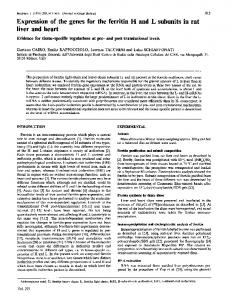
counts per minute incorporated per ug of IU/L cpm/ig DNA 400 DNA (cpm/pgDNA). The enzymatic activity of TK was 350 l Group I * Group I assayed in the supernatant fractions of liver 300 *Group 11 i Group portions after homogenization and cenEl Group III EGroup 250 trifugation at 105000g for 1 hr at 4°C with 200 a Beckman model L5-75 Ultracentrifuge, according to the method described by 150 Kahn et al. (10). Duplicate aliquots of each 100 sample were spotted onto DEAE cellulose 50 discs and placed in scintillation vials. The discs were counted for their radioactivity 0 12 48 00 72 96 0 24 36 content in a liquid scintillation counter hours after PH. 0 12 24 36 48 60 72 96 (Wallac LKB 1211 Rackbeta, Sweden). hours after P.H. The protein content of each sample was Figure 1. The rate of 3H thymidine incorporation in the determined by the method of Lowry et al. liver of groups 1, II, IlIl examined at different time intervals. Figure 3. Serum AST activity in the three main groups 1, II, (11). The activity of the enzyme was 3H-thymidine incorporation is expressed as cpm per pg III, examined at different time intervals. AST activity is expressed as IU/L. Values represent mean ± SD. expressed as counts per minute incorpo- hepatic DNA. Values represent mean ± SD. rated, per 1 min, per milligram of protein (cpm/min/mg protein). Biochemical evaluation of liver func- (Figure 1). The peak of DNA biosynthesis tion was estimated by measuring serum was observed at 48 hr in this group, 24 hr IU/L enzyme activities of aspartate aminotrans- later than that of group I. TK activity of ferase (AST), alanine aminotransferase group II, was increased mainly at 24, 36, (ALT) and alkaline phosphatase (ALP) and 48 hr (p