ANTICANCER RESEARCH 24: 3961-3964 (2004)
Effect of Estrogen, Tamoxifen and Epidermal Growth Factor on the Transcriptional Regulation of Vascular Endothelial Growth Factor in Breast Cancer Cells JEONG EON LEE1, KI-WOOK CHUNG4, WONSHIK HAN1, SUNG-WON KIM3, SEOK WON KIM1, HYUK JAI SHIN1, JI YEOUN BAE2 and DONG-YOUNG NOH1,2 1Department
of Surgery and Research Institute, Seoul National University College of Medicine, 28 Yongon-dong, Chongno-gu, Seoul 110-744; 3Department of Surgery, Seoul National University Bundang Hospital, 300 Gumi-dong, Bundang-gu, Seongnam-Si, Gyeonggi-do, 463-707; 4Center for Breast Cancer, National Cancer Center, 809 Madu 1-dong, Ilsan-gu, Goyang-si, Gyeonggi-do 411-769, Korea
2Cancer
Abstract. Background: VEGF (Vascular Endothelial Growth Factor) is a key factor of angiogenesis and high tissue VEGF levels are related to a poor prognosis in breast cancer. Materials and Methods: By semi-quantitative RT-PCR, we determined the relative expressions of VEGF mRNA in MCF-7 (both ER-·+ and ER-‚+ (mainly ER-·+), PR+, bcl-2+, EGFR-) and MBMDA-231 (only ER-‚+, PR-, EGFR-) breast cancer cells which were treated with estrogen, tamoxifen and EGF (Epidermal Growth Factor). Results: In MCF-7 cell lines, estrogen induced the expression of VEGF mRNA while tamoxifen reduced its expression. Estrogen and tamoxifen did not confer any significant effect on MB-MDA-231 cells and EGF showed no significant effect on MCF-7 or MB-MDA-231. Conclusion: Reduced VEGF mRNA expression of MCF-7 cells treated with tamoxifen may be related to the antagonistic effect of tamoxifen on ER-positive breast cancer, and this antagonistic effect may be related to ER-·. Angiogenesis plays an essential role in breast cancer. VEGF (Vascular Endothelial Growth Factor), also called vascular permeability factor, is a potent endothelial stimulator of proliferation, migration and tubular organization in
Correspondence to: Dong-Young Noh, M.D., Ph.D., Cancer Research Institute and Department of Surgery, College of Medicine, Seoul National University, 28 Yongon-dong, Chongnogu, Seoul 110-744, Korea. Tel: 82-2-760-2921, Fax: 82-2-766-3975, e-mail:
[email protected] Key Words: Breast cancer, MCF-7, MB-MDA-231, estrogen receptor (ER), estrogen, tamoxifen, VEGF.
0250-7005/2004 $2.00+.40
angiogenesis (1, 2). The level of VEGF expression is strongly correlated with intratumoral microvessel density, metastasis and poor prognosis (3). A significantly lower rate of response to first-line endocrine therapy was observed in patients with high VEGF levels, although its levels did not predict the response rate of first-line chemotherapy (4). VEGF mRNA and/or protein expression levels are low in the normal human mammary gland and are markedly higher in primary and metastatic breast cancer. In addition, high tumor VEGF levels increase the risk of recurrence, and are associated with shorter progression-free survival and post-relapse overall survival (5). Human VEGF genes contain eight exons and seven introns. VEGF proteins result from the alternative splicing of VEGF pre-mRNA from a single gene. There are several VEGF isoforms, of 121, 165, 189 and 206 amino acids (6). An additional isoform composed of 145 amino acids is expressed in placental cells and in various carcinoma cells of the female reproductive tract (7). Among the VEGF variants, VEGF121 and VEGF165 are the most commonly expressed in breast cancer tissue. These isoforms differ with respect to their heparin-binding capacity. VEGF121 is easily diffusible, whereas the others have heparin-binding regions which can bind to cell surfaces and the extracellular matrix (2). Estrogen is a major risk factor for the development of breast cancer and a modulator of angiogenesis in breast cancer tissue. However, the regulatory mechanisms of VEGF related to estrogen on breast cancer cells have not been defined. Several studies have reported that estrogen induces VEGF at both the mRNA and protein level in breast cancer cells (8-11), but Hyder et al. (12) found no VEGF mRNA or protein induction in any human breast cancer cells, including MCF-7 and MD-MBA-231 treated with estrogen. A recent
3961
ANTICANCER RESEARCH 24: 3961-3964 (2004) study showed down-regulated expressions of the mRNA of VEGFs in estrogen-treated MCF-7 cells (13). Acting as both an estrogen agonist and antagonist, tamoxifen is the most frequently used drug in endocrine therapy of breast cancer (11). It has been suggested that tamoxifen acts through functional estrogen response elements in the VEGF promoter gene (14), and that exposure to tamoxifen increases VEGF mRNA expression in MCF-7 cells (8-11). However, another study did not find such an effect (13). Still it is unclear whether tamoxifen up- or down-regulates VEGF. In addition to the conventional estrogen receptor-· (ER-·), another isoform, estrogen receptor-‚ (ER-‚), responds to estrogen and anti-estrogens differently (15). ER-· seems to mediate estrogen-dependent growth and the development of the mammary gland (16). The effect of ER-‚ remains controversial. It has been suggested that VEGF is a target gene for ER-· and ER-‚ in breast cancer cells, and that the agonist or antagonist effect of tamoxifen is related to the ER subtype (17). Studies on the effects of estrogen and tamoxifen on VEGF expression have been performed with respect to expressing ER-· or ER-‚. MCF-7 cells have both ER-· and ER-‚ but express mainly ER-·, while MD-MBA-231 cells express only ER-‚. Several reports have compared the effect of estrogen and/or tamoxifen with respect to ER-· or ER-‚ by using MCF-7 and MBA-231 cells (8, 12, 18), transfected MCF-7 and MD-MBA-231 cells (17) or transfected Ishikawa cells (19-21). There is a controversy about the effects of estrogen and tamoxifen. To investigate the effects of estrogen and tamoxifen on the expression of VEGF mRNA in different breast cancer cells according to their ER subtype profile, we chose the MCF-7 and MD-MBA-231 cell lines and have examined the effect of EGF on VEGF expression in these two cell lines.
Materials and Methods Cell culture. The human mammary carcinoma cell lines MCF-7 and MB-MDA-231 were obtained from the Korean Cell Line Bank. They were plated in 75-cm2 flasks (Falcon) and cultured in medium A (RPMI 1640 with 2 mM glutamine, 1% penicillin–streptomycin (100 U/ml), 5% fetal bovine serum) in a 37ÆC incubator under 5% CO2. To avoid the uncontrolled effects of endogenous and exogenous estrogen, the culture medium was changed for medium B (phenol red-free RPMI 1640 with 2 mM glutamine, 1% penicillin-streptomycin (100 U/ml), 5% dextran-coated charcoalstripped fetal bovine serum), and cells were cultured overnight prior to treatment with the experimental concentrations of estrogen, tamoxifen, or EGF. Semi-quantitative RT-PCR. Total RNA was extracted from the cell lysates with TRIzol reagent (Gibco- BRL Technologies, Maryland, USA). The RNA pellet was dissolved in diethyl pyrocarbonate (DEPC)-treated H2O to contain approximately 0.5 Ìg/ml to 1.0 Ìg/ml, and stored at -70ÆC. The quantity and quality of the RNA preparations were determined by absorbance at 260 and 280 nm. One microgram of total RNA sample was reverse transcribed using the first-strand cDNA synthesis kit (Roche Applied Science, Mannheim,
3962
Germany) with random primer p(dN)6, according to the manufacturer’s instructions. The cDNA was purified using phenol : chloroform : isoamyl alcohol (25 : 24 : 1) solution and dissolved in sterile distilled water. RT-PCR was performed on each cell line before treatment and after 1, 3, 12, or 24 hours of treatment with 1 nM estrogen, 1 ÌM tamoxifen or 10 ng/ml EGF. For PCR amplification, the following two primers were used: forward primer nucleotides: 5’-CTGGATCCATGGCAGAAGGAGGAG-3’ (Exon 2); reverse primer nucleotides: 5’-GAATTCAGACCGCCTCGG CTTGTC-3’ (Exon 8). Amplification conditions used were 94ÆC for 30 seconds, 61ÆC for 1 minute and 72ÆC for 1 minute; 35 cycles. Taq polymerase was obtained from TaKaRa Shuzo Co Ltd. (Tokyo, Japan). PCR products were electrophoresed in 2% agarose gel and stained with ethium bromide (Sigma Chemical Company, Missouri, USA) to identify mRNAs of VEGF121 and VEGF165. Data are represented as mean±SEM from three separate experiments. Quantification of autoradiographs. The density of the gel image was quantified and digitized using the Biological Image Processing System 3.0 (Biomedlab Co., Seoul, Korea). The optical density of bands was analyzed with background subtraction using ImageQuant software. The density of ‚-actin from the same cDNA was used to determine relative densities. Statistical analysis. Independent t-test was used to compare the mean values of relative expression to control between different experimental groups.
Results Semi-quantitative RT-PCR and 2% gel electrophoresis of the products from the MCF-7 breast cancer cells showed a timedependent increase in the expression of both VEGF121 and VEGF165 mRNAs after treatment with 1 nM of estradiol. For MCF-7 cells treated with 1 mM tamoxifen, time-dependent reductions in the expressions of VEGF121 and VEGF165 mRNAs were observed. No change with respect to control was observed in EGF-treated MCF-7 cells over 48 hours. Densitometry showed similar results to RT-PCR. A comparison of the relative densities of VEGF mRNA showed a ratio of 1: 2.94 (control vs. 48 hours later) after treatment with estradiol (p=0.018). The relative density of VEGF mRNA was down-regulated with a ratio of 1: 0.65 (control vs. 48 hours later) after tamoxifen treatment (p=0.024). No significant change in transcriptional activity of VEGF was observed by EGF treatment (p=0.318) (Figure 1). In the case of the MB-MDA-231 breast cancer cell line, no significant change with respect to control was observed in the expression of VEGF mRNA following treatment with estradiol (1 nM), tamoxifen (1 ÌM), or EGF (10 ng/ml) (p=0.431, 0.331 and 0.283, respectively) (Figure 2).
Discussion Most studies agree that estrogen induces the expression of VEGF in MCF-7 cells which express predominantly ER-· (811, 17, 18). We also showed a time-dependent increase of the
Lee et al: Transcriptional Regulation of VEGF in Breast Cancer Cells
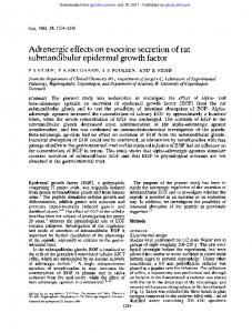
Figure 1. (A) VEGF expression in 2% gel electrophoresis of the RT-PCR products in MCF-7 cells treated with estradiol (1 nM), tamoxifen (1 ÌM), or EGF (10 ng/ml). For MCF-7 cell lines treated with 1 ÌM tamoxifen, we observe time-dependent reductions in the expressions of VEGF mRNA. (B) Densitometry measurement of in vitro relative expressions of VEGF in MCF-7 cells treated with estradiol (1 nM), tamoxifen (1 ÌM), or EGF (10 ng/ml). The relative densities of VEGF mRNA show a ratio of 1: 2.94 after treatment with estradiol (p=0.018) and 1: 0.65 after tamoxifen treatment (p=0.024) (control vs. after 48 hours).
expression of VEGF mRNA in MCF-7 cells. However, it has also been reported that estrogen does not affect the VEGF level, or even reduces its expression in MCF-7 cells in vitro (12, 13). In MB-MDA-231 cells which express only ER-‚, estrogen did not induce VEGF mRNA (11, 17, 18) or even reduced VEGF protein expression (8). According to our study, estrogen did not affect the transcriptional activity of VEGF mRNA in MB-MDA-231 cells. Buteau-Lozano et al. (17) reported that estrogen increases VEGF mRNA in ER-‚-coexpressing MCF-7 cells and ER-·-coexpressing MB-MDA-231 cells. From these results, we believe that estrogen plays an important role in the induction of VEGF in the presence of ER-·, while it does not in the case of ER-‚-only-expressing breast cancer cells. This means that endogenous ER-‚ does not support estrogen to induce VEGF production, although estrogen induces VEGF expression in the presence of both ER-· and ER-‚. Tamoxifen has both agonistic and antagonistic activity, depending on the tissue type, and its effects have not been clearly defined in breast cancer cells. Tamoxifen seems to induce the expression of VEGF in MCF-7 cells (9-11), or have no effect (8, 13). Unexpectedly, in our experiment with
Figure 2. (A) VEGF expression in 2% gel electrophoresis of the RT-PCR products in MD-MBA-231 cells treated with estradiol (1 nM), tamoxifen (1 ÌM), or EGF (10 ng/ml). (B) Densitometry measurement of in vitro relative expressions of VEGF in MD-MBA-231 cells treated with estradiol (1 nM), tamoxifen (1 ÌM), or EGF (10 ng/ml). There is no significant change with respect to control.
tamoxifen in MCF-7 cells, we observed a reduction in VEGF mRNA expression, which may explain the antagonistic effect of tamoxifen on ER-positive breast cancer. Tamoxifen did not increase the VEGF mRNA level in MB-MDA-231 cells (8, 18). Tamoxifen showed no effect on the transcriptional activity of VEGF mRNA in MB-MDA-231 cells in our study. In an experiment on ER-·-transfected MD-MBA-231 cells and on ER-‚-transfected MCF-7 cells, tamoxifen did not induce VEGF expression (17). Other experiments on the ER subtypes have been conducted. Although different clonal cells may respond in different ways to estrogen and tamoxifen, it is interesting to compare results in breast cell lines with those in other tissue clonal cells. Ishikawa cells originated from human endometrial cancer cells and have less than 100 functional ER/cell. Ishikawa cells were temporarily transfected with plasmids expressing ER-· or ER-‚ so as to impart the ER subtypes selectively, and were then used to investigate the effects of estrogen and tamoxifen (19-21). Estrogen induced VEGF expression in ER-·-transfected Ishikawa cells (19-21). Interestingly, estrogen induced VEGF expression in studies with ER-‚-expressing Ishikawa cells (19), while it did not induce VEGF mRNA (11, 17, 18) or
3963
ANTICANCER RESEARCH 24: 3961-3964 (2004) significantly reduced VEGF protein expression (8) in MBMDA-231 cells. These findings suggest that estrogen may induce VEGF expression in both mammary and endometrial cells in the presence of ER-·. In the presence of ER-‚, estrogen may induce VEGF expression in endometrial cells but not in mammary cells. On the other hand, tamoxifen induced VEGF expression in ER-·-transfected Ishikawa cells, but showed no significant change in ER-‚-transfected Ishikawa cells (21). The responses of VEGF expression in MCF-7 cells treated with tamoxifen are diverse; tamoxifen may induce (9-11) or does not affect (8, 13) or reduce VEGF expression, as our result. These findings suggest that tamoxifen may act as an estrogen agonist in endometrial cells in the presence of ER-·, though the action of tamoxifen is not defined in mammary cells. In the presence of ER-‚, tamoxifen does not induce VEGF expression in both mammary and endometrial cells. EGF is known to be a paracrine factor in breast cancer growth. Epidermal Growth Factor (EGF) has been suggested to influence breast cancer cells by modulating the estrogen receptor (22). In our experiment, we did not identify a definite effect of EGF on VEGF expression in either MCF-7 or MB-MDA-231 cells. It is difficult to identify a definite action cascade involving estrogen and tamoxifen through ER-· or ER-‚. A new model of breast cell line selectively expressing ER-· or ER-‚ would be a valuable tool to investigate the detailed roles of the ER subtypes in mammary cells.
Acknowledgements This work was supported by a research grant from the Cancer Research Institute, Seoul National University College of Medicine, Korea (2001) and S.N.U Research Fund 2001.
7
8
9
10
11
12
13 14
15
16
17
18
References 19 1
2 3
4
5
6
Locopo N, Fanelli M and Gasparini G: Clinical significance of angiogenic factors in breast cancer. Breast Cancer Res Treat 52: 150-173, 1998. Ferrara N and Davis-Smith T: The biology of vascular endothelial growth factor. Endocr Rev 18: 4-25, 1997. Weidner N, Semple JP, Welch WR and Folkman J: Tumor angiogenesis and metastasis – correlation in invasive breast carcinoma. NEJM 324: 1-8, 1991. Manders P, Beex LV, Tjan-Heijnen VC, Span PN and Sweep CG: Vascular endothelial growth factor is associated with the efficacy of endocrine therapy in patients with advanced breast carcinoma. Cancer 98: 2125-2132, 2003. Foekens JA, Peters HA, Grebenchtchikov N, Look MP, Meijer-van Gelder ME, Geurts-Moespot A, van der Kwast TH, Sweep CG and Klijn JG: High tumor levels of vascular endothelial growth factor predict poor response to systemic therapy in advanced breast cancer. Cancer Res 61: 5407-5414, 2001. Ferrara N: Vascular endothelial growth factor: molecular and biological factor. Curr Top Microbiol Immunol 237: 1-30, 1999.
3964
20
21
22
Poltorak Z, Cohen T, Sivan R, Kandelis Y, Spira G, Vlodavsky I, Keshet E and Neufeld G: VEGF145, a secreted vascular endothelial growth factor isoform that binds to extracellular matrix. J Biol Chem 272: 7151-7158, 1997. Coradini D, Pellizzaro C, Speranza A and Daidone MG: Hypoxia and estrogen receptor profile influence the responsiveness of human breast cancer cells to estradiol and antiestrogens. Cell Mol Life Sci 61: 76-82, 2004. Takei H, Lee ES and Jordan VC: In vitro regulation of vascular endothelial growth factor by estrogens and antiestrogens in estrogenreceptor positive breast cancer. Breast Cancer 9: 39-42, 2002. Garvin S and Dabrosin C: Tamoxifen inhibits secretion of vascular endothelial growth factor in breast cancer in vivo. Cancer Res 63: 8742-8748, 2003. Ruohola JK, Valve EM, Karkkainen MJ, Joukov V, Alitalo K and Harkonen PL: Vascular endothelial growth factors are differentially regulated by steroid hormones and antiestrogens in breast cancer cells. Mol Cell Endocrinol 149: 29-40, 1999. Hyder SM, Huang JC, Nawaz Z, Boettger-Tong H, Makela S, Chiappetta C and Stancel GM: Regulation of vascular endothelial growth factor expression by estrogens and progestins. Environ Health Perspect 108 Suppl 5: 785-790, 2000. Bogin L and Degani H: Hormonal regulation of VEGF in orthotopic MCF7 human breast cancer. Cancer Res 62: 1948-1951, 2002. Hyder SM, Nawaz Z, Chiappetta C and Stancel GM: Identification of functional estrogen response elements in the gene coding for the potent angiogenic factor vascular endothelial growth factor. Cancer Res 60: 3183-3190, 2000. Kuiper GGJM, Enmark E and Huikko MP: Cloning of a novel estrogen receptor expressed in rat prostate and ovary. Proc Natl Acad Sci USA 138: 5925-5930, 1996. Krege JH, Hodgin JB, Couse JF, Enmark E, Warner M, Mahler JF, Sar M, Korach KS, Gustafsson J-A and Smithies O: Generation and reproductive phenotypes of mice lacking estrogen receptor ‚. Proc Natl Acad Sci USA 95: 15677-15682, 1998. Buteau-Lozano H, Ancelin M, Lardeux B, Milanini J and PerrotApplanat M: Transcriptional regulation of vascular endothelial growth factor by estradiol and tamoxifen in breast cancer cells: a complex interplay between estrogen receptors · and ‚. Cancer Res 62: 4977-4984, 2002. Maity A, Sall W, Koch CJ, Oprysko PR and Evans SM: Low pO2 and ‚-estradiol induce VEGF in MCF-7 and MCF-7-5C cells: relationship to in vivo hypoxia. Breast Cancer Res Treat 67: 51-60, 2001. Mueller MD, Vigne JL, Minchenko A, Lebovic DI, Leitman DC and Taylor RN: Regulation of vascular endothelial growth factor (VEGF) gene transcription by estrogen receptors · and ‚. Proc Natl Acad Sci USA 97: 10972-10977, 2000. Ali SH, O'Donnell AL, Balu D, Pohl MB, Seyler MJ, Mohamed S, Mousa S and Dandona P: Estrogen receptor-· in the inhibition of cancer growth and angiogenesis. Cancer Res 60: 7094-7098, 2000. Mueller MD, Pritts EA, Zaloudek CJ, Dreher E and Taylor RN: Regulation of vascular endothelial growth factor by tamoxifen in vitro and in vivo. Gynecol Obstet Invest 55: 119-124, 2003. Stoica GE, Franke TF, Moroni M, Mueller S, Morgan E, Iann MC, Winder AD, Reiter R, Wellstein A, Martin MB and Stoica A: Effect of estradiol on estrogen receptor-alpha gene expression and activity can be modulated by the ErbB2/PI 3-K/Akt pathway. Oncogene 22: 7998-8011, 2003.
Received May 24, 2004 Revised September 3, 2004 Accepted October 4, 2004