Acton Lane, London NW10 7NS. ^Department of Anaesthesia, University of Leeds, 24. Hyde Terrace, Leeds LS2 9LN. Correspondence to C.T. with morphine 10 ...
Br. J. Anaesth. (1989), 63, 411-417
EFFECT OF PROPOFOL ON THE AUDITORY EVOKED RESPONSE AND OESOPHAGEAL CONTRACTILITY C. THORNTON, K. M. KONIECZKO, A. B. KNIGHT, B. KAUL, J. G. JONES, C. J. DORE AND D. C. WHITE This study had two objectives: to examine the effect of propofol anaesthesia on the auditory evoked response (AER) as part of a series of studies [1—4] to develop a technique for measuring "depth of anaesthesia", and to compare changes in the AER produced by propofol with changes in oesophageal contractility, a technique which has been claimed [5] to measure depth of anaesthesia. Changes in contractility of the lower portion of the oesophagus have been proposed as a measure of depth of anaesthesia. The contractions studied have been of two types: spontaneous contractions (SLOC), and provoked contractions (PLOC) produced by stimulating the oesophagus; the frequency of both has been related inversely to concentration of anaesthetic administered [5]. In the absence of an absolute standard against which to compare either the AER or oesophageal contractility, we have compared these with each other. PATIENTS AND METHODS
SUMMARY Six patients were anaesthetized with 70% nitrous oxide in oxygen supplemented by infusion of propofol 40, 80, 120, 160 and 200 fig kg-1 min-' sequentially in successive 10-min periods. Auditory evoked response (AER) and lower oesophageal contractility (LOC) were monitored. The AER findings were consistent with those noted in previous studies of i.v. agents. Early cortical waves showed attentuation of Pa and Nb amplitude (? < 0.01) and increase in Pa and Nb latency (P < 0.01; P < 0.05) with increasing blood concentrations of propofol. Brainstem waves were not affected significantly. LOC, provoked and spontaneous, showed no consistent relationship with blood concentration of propofol. The two variables AER and LOC were not related.
with morphine 10 mg and atropine 0.6 mg i.m. was followed by induction of anaesthesia with 1 Six patients aged 18-45 yr gave informed thiopentone 2-4 mg kg" i.v. The trachea was administration of pancuconsent to the study, which was approved by intubated following 1 Harrow District Ethics Committee and the Com- ronium 0.1 mg kg" i.v. and the lungs ventilated mittee on Safety of Medicines. Premedication with 70% nitrous oxide in oxygen. Seven to 10 min after induction, propofol was infused i.v. in five equal 10-min steps, starting at 40 ug kg"1 C. THORNTON, M.SC. ; K. M. KONIECZKO, F.F.A.R.C.S. ; A. B. min"1 and with a final rate of 200 ug kg"1 min"1. KNIGHT*,F.F.A.R.C.S. ;B. KAULT,M.D.,M.B.,B.S.;J. G. JONES:):, M.D., F.F.A.R.C.S., F.R.C.P.; C. J. DORE, B.SC; D. C. WHITE, In two patients (PI and P5) in whom there was a M.B., B.S., D.A., F.F.A.R.C.S.; Clinical Research Centre and delay between the end of the study and the Northwick Park Hospital, Watford Road, Harrow, Middlesex beginning of surgery, the recovery of the AER HA1 3UJ. Accepted for Publication: March 18, 1989. was monitored while the patient's lungs were Present addresses: * Anaesthetic Department, Hillingdon Hospital, Uxbridge, ventilated with 70 % nitrous oxide in oxygen. Middx UB8 3QW. Ventilation was adjusted to maintain the endt Department of Anaesthetics, Central Middlesex Hospital, tidal carbon dioxide concentration (HewlettActon Lane, London NW10 7NS. Packard 47210A infra-red analyser) within the ^Department of Anaesthesia, University of Leeds, 24 range 4.5-5.5 kPa. Body temperature was moniHyde Terrace, Leeds LS2 9LN. tored using a thermistor placed in the oesophagus Correspondence to C.T. Patients and anaesthesia
412
at the level of the aortic arch. Systemic arterial pressure and heart rate were recorded at 5-10 min intervals. Auditory evoked response The technique was essentially the same as that described previously [6], except for the use of purpose built EEG amplifiers with the same amplification and filter characteristics. Averages (n = 2048) of the auditory evoked response to click stimulation at 6 Hz were obtained before anaesthesia, after induction of anaesthesia before the start of the infusion of propofol, and at end of each infusion period. To monitor recovery after discontinuation of the infusion, AER responses were obtained over 3-min epochs. Oesophageal contractions Following intubation of the trachea, the ANTEC oesophageal monitor was used to record oesophageal contractions as described by Evans and White [7]. The oesophageal pressure probe was introduced into the lower one-third of the oesophagus (probe tip 35 cm from the lips). Spontaneous lower oesophageal contractions (SLOC) and provoked lower oesophageal contractions (PLOC) are identical in appearance, the latter distinguished from the former by occurring after a pressure stimulus set to occur automatically.
BRITISH JOURNAL OF ANAESTHESIA following induction of anaesthesia just before the addition of the test agent was given a time or a concentration of zero. The averaged AER were recorded over the final 5.7 min of each 10-min period. Regression analysis was used to calculate a slope for each patient, and analysis of variance was carried out on the slopes to compare the effect of propofol with that of other drugs and saline from previous studies [1—4] and to test for dose relationships. (Data from these studies were reanalysed to include the post-induction data point.) Oesophageal contraction variables examined were: mean height (mm Hg) of provoked oesophageal contractions (PLOC) and frequency of spontaneous oesophageal contractions (SLOC). For PLOC, the values corresponding to the AER sample period were averaged to give the mean PLOC height, and for the SLOC they were expressed as frequency per 5 min. Mean PLOC height and SLOC frequency were plotted against blood concentration of propofol and the mean slopes of these variables tested for a significant difference from zero. The Pa and Nb latencies and amplitudes were plotted also against mean PLOC height and SLOC frequency and the significance of the mean slopes tested to see if these variables were related. RESULTS
Anaesthetic concentration Venous blood samples were taken from the arm opposite to that receiving the infusion, just before starting the propofol and in the 6th, 8th and 10th min of each infusion period. In two patients in whom recovery was monitored, a sample was taken in the middle and at the end of this period. Blood concentrations of propofol were measured by Dr E. Douglas of I.C.I, according to the method of Plummer [8].
Propofol and AER Brainstem response Propofol resembled the other two i.v. agents, etomidate and Althesin, in that the mean slopes of the brainstem latencies and interpeak intervals against time were not significantly different from saline at the P < 0.05 level (table I). On the other hand, the inhalation agents had effects consistently different from those of saline on the III and V latencies, and interpeak intervals.
Analysis of data We examined brainstem waves (I, III and V latencies; I—III, I-V and III-V interpeak intervals; III and V amplitude) and early cortical waves (Pa and Nb latencies; Pa and Nb amplitudes). AER variables for each patient were plotted against time (midpoint of the averaging period) and against propofol concentration (mean of the blood samples obtained in the 6th, 8th and 10th min of each infusion period). The datum point
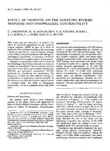
Early cortical response With increasing blood concentrations of propofol, there were progressive increases in latency and reductions in amplitude of waves Pa and Nb similar to those observed with halothane, enflurane, isoflurane, etomidate and Althesin, but not in the patients given saline (fig. 1). As with the other anaesthetic agents, the changes in Pa and Nb latency and amplitude reversed when the propofol infusion was stopped. Figure 2 shows the early cortical AER in one
413
PROPOFOL AND DEPTH OF ANAESTHESIA TABLE I. Brainstem latency and interpeak interval regressions against time. The pooled estimate of the between-patient SD of the variable was derived from an analysis of variance and used for the comparison between anaesthetic agents and saline. Means significantly different from that of saline: *P* (6 to 15)
9*'k (4 to 14) 9*'k (5 to 14) 0 ( - 4 to 3)
P2
P1
0.80.4-
0.20.12I 1 1 1 1 1 2.0-, 1.6P3 1.2- • •
1
1
1 1
P4
0.2-
-
0.122.0-1
(-16 (-37 (-40 (-17 (-22 (-17
to to to to to to (1 to
-33) -49) -52) -32) -39) -33) -18)
(-10 -20 -36*** (-28 _45*** (-38 -27** (-19 -36*** (-27 -22* (-13 -9
to to to to to to (1 to
-29) -43) -51) -35) -44) -31) -19)
Dose relationships. The changes with propofol, as with the other anaesthetic agents, were related linearly to concentration (fig. 3). The mean slopes of Pa and Nb latency and amplitude against concentration were significantly different from zero (P < 0.05) in all cases, except for the effects of Althesin on Nb latency (table III). Infigure3 and table III the propofol concentration is expressed in ED50 units, which are units of equipotency (their derivation is discussed later). This does not affect the significance of the difference of the mean slope from zero. Comparison of anaesthetics. On the basis of these
* •
0.4-
c (0
-25* -43*** _47*** -25** -31** -26* -9
Nb
•
"D
iplit'
1
-
3 . 0.8