Section of Immunobiology,1 Department of Pediatrics,3 and Howard Hughes Medical ...... Melvin, A. J., M. E. McGurn, S. J. Bart, C. Gibson, and D. B. Lewis. 1995 ...
MOLECULAR AND CELLULAR BIOLOGY, Jan. 1997, p. 199–208 0270-7306/97/$04.0010 Copyright q 1997, American Society for Microbiology
Vol. 17, No. 1
Differential Transcription Directed by Discrete Gamma Interferon Promoter Elements in Naive and Memory (Effector) CD4 T Cells and CD8 T Cells ´ N,1 THOMAS M. AUNE,1,2 LAURIE A. PENIX,3 MERCEDES R. RINCO
AND
RICHARD A. FLAVELL1,4*
Section of Immunobiology,1 Department of Pediatrics,3 and Howard Hughes Medical Institute,4 Yale University School of Medicine, New Haven, Connecticut 06520, and Division of Rheumatology and Immunology, Department of Medicine, Vanderbilt University School of Medicine, Nashville, Tennessee 372322 Received 5 April 1996/Returned for modification 24 May 1996/Accepted 20 September 1996
Acquisition of the ability to produce gamma interferon (IFN-g) is a fundamental property of memory T cells and enables one subset (T helper 1 [TH1]) to deliver its effector functions. To examine regulation of IFN-g gene expression in a model system which recapitulates TH1 differentiation, we prepared reporter transgenic mice which express the luciferase gene under the control of proximal and distal regulatory elements (prox.IFNg and dist.IFNg) from the IFN-g promoter. Memory T cells, but not naive T cells, secreted IFN-g and expressed both prox.IFNg and dist.IFNg transcriptional activities. Naive T cells required priming to become producers of IFN-g and to direct transcription by these elements. While both CD41 and CD81 T cells produced IFN-g, only CD41 T cells expressed prox.IFNg transcriptional activity. Induction of transcriptional activity was inhibited by known antagonists of effector T-cell populations. Cyclosporin A inhibited transcriptional activity directed by both elements in effector T cells. Elevated cyclic AMP inhibited transcriptional activity directed by prox.IFNg in primed CD41 T cells but enhanced transcriptional activity directed by dist.IFNg in primed CD81 T cells. Taken together, these data show that prox.IFNg and dist.IFNg transcriptional activities mirror IFN-g gene expression in naive and memory CD41 T cells but suggest that differences exist in regulation of IFN-g gene expression in CD41 and CD81 T-cell subsets. ATF/CREB family transcription factors (6). Further, hypomethylation or methylation of a CpG dinucleotide within the proximal element appears to correlate with expression of the endogenous gene in human T cells and murine T-cell clones (21, 42). An upstream silencer element (2251 to 2214 bp), as well as additional upstream activating elements, have also been described (41). To investigate the role of these AP-1/cyclic AMP (cAMP) response elements (CRE)/ATF-like elements in the transcriptional regulation of IFN-g gene expression during development of effector function in normal T cells, we have prepared reporter transgenic mice which express the luciferase gene under the control of the proximal or distal element from the immediate 59 promoter of the IFN-g gene. The results show that memory but not naive CD41 T cells direct transcriptional activity under the control of these elements. T cells can be primed, in vitro, to express the reporter gene under the control of either of these regulatory elements, just as they are primed to express the endogenous IFN-g gene. The effects of exogenous cytokines or pharmacological inhibitors upon transcriptional activity are delineated and compared with effects on endogenous IFN-g secretion.
Naive T cells produce interleukin-2 (IL-2) following challenge with antigen and antigen-presenting cells (APC) or following exposure to polyclonal activators, such as anti-CD3 (a-CD3) antibodies, but produce little or no gamma interferon (IFN-g) or IL-4 (8, 17, 25, 35). However, naive T cells can differentiate into producers of IL-4 or IFN-g under appropriate conditions (3, 8, 15, 17–19, 25, 32, 33, 35–37). Stimulation of naive T cells with antigen and APC, or with polyclonal activators, in the presence of IL-4 yields T cells which, upon rechallenge, produce IL-4 as their predominant cytokine and do not produce IFN-g. In contrast, stimulation with antigen and APC or with polyclonal activators in the presence of IL-2 and IL-12 yields T cells which, upon rechallenge, produce predominantly IFN-g and do not produce IL-4. This priming process allows naive T cells to differentiate into effector T cells which possess the ability to transcribe new genes, a hallmark of the development of acquired immunity (12, 13, 22, 23, 33). The molecular mechanisms by which naive T cells activate effector genes, such as the IL-4 or IFN-g gene, are poorly understood. One possible explanation is that the cytokines present during primary stimulation induce expression of specific nuclear factors which selectively activate IL-4 or IFN-g gene transcription. Within the 59-flanking region of the IFN-g gene, several important regulatory sites have been identified on the basis of transient transfection assays (1, 4–6, 26, 41, 42). Two promoter elements, a proximal (270 to 247 bp, prox.IFNg) and a distal (298 to 272 bp, dist.IFNg) element, which are critical for activation-specific expression of a reporter construct in Jurkat T cells, have been identified (26). Both elements contain sequences which bind both AP-1 and
MATERIALS AND METHODS Generation of transgenic mice. Plasmids containing a head-to-tail (59-to-39) dimer of prox.IFNg (270 to 247 bp from the transcription start site) and tetramer of dist.IFNg (298 to 272 bp) were generated by subcloning prox.IFNg or dist.IFNg synthesized with terminal BglII (59) and BamHI (39) restriction sites into the pIFN-39 basal promoter construct described previously (26). The sequence of each construct was confirmed by DNA sequencing. These regulatory elements and minimal promoter were then subcloned from the PEQ3 b-galactosidase vector by XhoI digestion, blunting with Klenow enzyme, and subsequent SalI digestion. A very short 59 polylinker sequence from PEQ3 (59-TCG ACC TGC AGG CAA GCT TAC GTA GAT CT-39; SalI, PstI, HindIII, and SnaBI sites) was included in both constructs. The resulting fragments containing either the proximal or distal element were gel purified and ligated into plasmid PRL-Luc
* Corresponding author. Mailing address: Section of Immunobiology, Yale University School of Medicine, 310 Cedar St., FMB 412, New Haven, CT 06520. 199
200
AUNE ET AL.
MOL. CELL. BIOL.
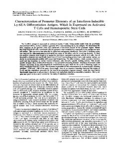
FIG. 1. Design of the prox.IFNg-Luc and dist.IFNg-Luc fusion genes used for microinjection. The HpaI digestion fragment contained the luciferase gene coding region ligated to a simian virus 40 (SV40) splice site and polyadenylation site, driven by the human IFN-g minimal promoter and either two copies of the proximal element or four copies of the distal element inserted in the correct orientation as described in Materials and Methods and Results. The sequences of these elements are noted on the right, with factors capable of binding these elements identified (6, 26, 28).
(7) which had been prepared by HindIII digestion, blunting with Klenow enzyme, and digestion with XhoI to remove the prolactin promoter (Fig. 1). The 2.8-kb HpaI fragment isolated from these plasmids (prox.IFNg-Luc and dist.IFNg-Luc, respectively) was injected into fertilized C57BL/6 3 CBA/N F2 eggs, and transgenic mice were generated as previously described (14). Founder animals were identified by Southern blot analysis of tail DNA, using the 32P-labeled HindIII-EcoRI fragment from plasmid PRL-Luc. Founder animals used in these studies have been backcrossed with B10.BR mice for at least three generations. Cell preparation and culture. Spleen cells, lymph node (LN) cells, or pooled spleen and LN cells were harvested from normal or transgenic animals. Measurement of transcriptional activity was done with freshly isolated naive or memory T cells from mice at 10 to 12 weeks of age. At this age, mice from our colony contain an approximately equal distribution of CD44lo (naive) and CD44hi (memory) T cells. Examination of transcriptional activity was done with effector T cells following in vitro priming from mice which were 5 to 6 weeks of age. At this age, mice from our colony contain T cells which are primarily ;90% CD44lo. Erythrocytes were lysed by briefly suspending cells in a hypotonic solution followed by returning cells to normal tonicity. CD31, CD41, or CD81 T cells were purified by negative selection. Ia-expressing cells and natural killer (NK) cells were removed by incubation with an anti-IE, IA (a-IE, IA) monoclonal antibody (MAb) (m5/115; Yale University Hybridoma Facility) and an (a-NK cell MAb (NK 1.1; American Type Culture Collection [ATCC]). Either an a-CD4 MAb (GK-1.5; ATCC) or an a-CD8 MAb (TIB 105; ATCC) was used to deplete CD41 or CD81 T cells, respectively. Cells were incubated for 30 min at 48C, washed, and further incubated with goat a-mouse and a-rat immunoglobulin G bound to magnetic beads (Collaborative Research) for 30 min at 48C. Cells bound to beads were removed with a magnet. Average purity of CD41 or CD81 T cells was ;90 to 95%, as determined by analysis with a Becton Dickinson FACStarPLUS cell sorter. CD41 T cells were further separated into naive and memory cell populations by cell sorting. T cells were labeled with fluorescein isothiocyanate-conjugated a-CD44 MAb and biotin-conjugated a-CD45RB MAb, followed by phycoerythrin-avidin (Pharmingen). CD44hi CD45RBlo cells were used as memory T cells, and CD44lo CD45RBhi cells were used as naive T cells. To prepare APC, erythrocyte-depleted splenocytes from nontransgenic littermates or B10.BR mice were depleted of CD41 and CD81 T cells by negative selection with a-CD4 and a-CD8 MAbs and were irradiated at 2,000 rads from a 137Cs source. Reagents used to stimulate lymphocytes were as follows: a-CD3 (clone 1452C11; ATCC), 1 mg/ml; IL-2, 10 U/ml; IL-4, 1,000 U/ml; IL-12, 3.5 ng/ml; concanavalin A (ConA), 2.5 mg/ml; phorbol myristate acetate (PMA), 20 nM; and ionomycin, 500 nM. Recombinant IL-2 was a gift from Biogen, Inc., recombinant murine IL-4 was a gift from DNAX, and recombinant murine IL-12 was a gift from Genetics Institute, Inc. Immobilized a-CD3 was prepared by adding 0.5 to 1 ml of MAb 2C11 (10 mg/ml) in 0.1 M sodium bicarbonate (pH 9.6) to a 24- or 48-well tissue culture plate for 3 to 6 h at 378C or overnight at 0 to 48C. Culture plates were washed thoroughly before use. The IFN-g enzyme-linked immunosorbent assay (ELISA) was performed with antibodies from Pharmingen according to the manufacturer’s procedures. The sensitivity of the ELISA was from 0 to 300 U of IFN-g per ml. IFN-g levels were measured from culture fluids from cells derived from either prox.IFNg-Luc or dist.IFNg-Luc mice. IFN-g levels in cultures of cells from both lines of mice were essentially equivalent under identical treatment conditions. Analysis of luciferase activity. Cells from various sources were cultured in complete RPMI 1640 medium with 10% fetal calf serum, 100 U of penicillin per ml, 100 U of streptomycin per ml, 2 mM L-glutamine, and 5 3 1025 M b-mercaptoethanol in 24- or 48-well tissue culture plates in volumes of 1 or 0.5 ml, respectively, at a density of 106/ml in the presence or absence of various stimuli
at 378C in 5% CO2 in air. Irradiated APC were used at a density of 5 3 105/ml of culture fluid. After the indicated periods of time, cultures were harvested, washed twice in phosphate-buffered saline, and suspended in 50 ml of lysis buffer (luciferase assay; Promega, Madison, Wis.) for 30 min at 258C. The supernatant was harvested by centrifugation, and 20-ml aliquots were assayed for luciferase activity with 100 ml of luciferase reagent (Promega) in a luminometer (Lumat LB9501) for 10 s. Cultures were in duplicate. Duplicate analyses of two aliquots from each cell lysate were performed, and the results were averaged. Results are expressed as the average of these readings per 106 cells with the standard error of the mean (SEM). The background measurement with luciferase reagent alone was subtracted from each reading. Results are expressed in relative light units (RLU). The absolute values obtained from individual readings ranged from 0 RLU (unstimulated cell lysates) up to 4,000 RLU (lysates from maximally stimulated cells).
RESULTS Generation of transgenic mice. Two elements, prox.IFNg and dist.IFNg, which are critical for induced expression of a reporter construct in transient transfection assays of the human Jurkat T-lymphoblastoid cell line have been identified within the immediate 59 flanking sequence of the human IFN-g gene (26). Transient transfection of reporter constructs containing two or four copies of the proximal or distal element, respectively, upstream of the minimal IFN-g promoter (Fig. 1) into Jurkat cells demonstrates that these elements are sufficient to direct transcriptional activity which is activation specific, sensitive to inhibition by cyclosporin A (CsA), and nearly as great in magnitude as that obtained with the entire 538-bp promoter (29). To determine the transcriptional activities of these elements in naive and primed effector or memory T cells, two sets of reporter transgenic mice containing a correctly oriented multimerized prox.IFNg dimer or dist.IFNg tetramer element upstream of the basal IFN-g promoter and a luciferase reporter gene were generated (Fig. 1). Two independent prox.IFNg-Luc lines have been identified, and levels of expression of transcriptional activity are comparable in the two lines. Three additional lines failed to express transcriptional activity. Five independent dist.IFNg-Luc lines have also been identified and analyzed. Transcriptional activities expressed in these five lines exhibited comparable qualitative properties. Our initial experiments indicated that these elements expressed transcriptional activity primarily in T cells (see below). To better assess the specificity of expression, other cells were also examined. Prox.IFNg and dist.IFNg transcriptional activities in primed, effector T cells were compared to transcriptional activity in B cells (purified by negative selection with a-CD4, a-CD8, a-NK 1.1, and a-Mac-1 MAbs), thymocytes, fibroblasts (explants of epidermis), and hepatocytes (obtained by collagenase diges-
VOL. 17, 1997
ACTIVITY OF IFN-g PROMOTER ELEMENTS IN PRIMARY T CELLS
201
tion of liver). Transcriptional activity was high in primed effector T cells but undetectable in B cells, thymocytes, fibroblasts, and hepatocytes following stimulation with either immobilized a-CD3 or PMA plus ionomycin (data not shown). Culture fluid supernatants harvested from B cells, thymocytes, fibroblasts, and hepatocytes did not contain detectable levels of IFN-g (data not shown). Cells in all cultures were .80% viable. Expression of prox.IFNg and dist.IFNg transcriptional activities in memory T cells. (i) Transcriptional activity in lymphoid populations. Initial experiments were performed to determine the levels of expression of prox.IFNg and dist.IFNg transcriptional activities in cells activated by various stimuli. Freshly isolated, pooled spleen and LN cells from 10- to 12week-old transgenic mice were stimulated with ConA, soluble a-CD3, immobilized a-CD3, PMA plus ionomycin, or IL-2 plus IL-12, harvested, and assayed for luciferase activity. Supernatant fluids were also harvested and analyzed for IFN-g levels. Only immobilized a-CD3 MAb, a signal which activates memory but not naive T cells (9), stimulated expression of prox.IFNg (Fig. 2A) and dist.IFNg (Fig. 2B) transcriptional activities. Comparable levels of transcriptional activity were expressed under the control of either the proximal or distal element. Unstimulated cells contained no detectable luciferase activity (not shown). Immobilized a-CD3, which stimulated the greatest level of transcriptional activity, also provided the most potent induction of IFN-g expression by spleen and LN cells (;250 U/ml) (Fig. 2C). By comparison, soluble a-CD3 failed to stimulate production of IFN-g (,20 U/ml). Other stimuli, such as ConA, PMA plus ionomycin, or IL-2 plus IL-12, stimulated spleen and LN cells to produce modest amounts of IFN-g (40 to 60 U/ml). While PMA plus ionomycin, ConA, or IL-2 plus IL-12 will stimulate both NK cells and T cells to produce IFN-g (3), immobilized a-CD3 will stimulate only memory T cells to produce IFN-g. Prox.IFNg and dist.IFNg transcriptional activities were clearly (Fig. 2A and B) expressed following stimulation with immobilized a-CD3 and were clearly not expressed following stimulation with PMA plus ionomycin or IL-2 plus IL-12. Multiple founders displayed this same expression pattern (not shown). This finding suggested that prox.IFNg and dist.IFNg transcriptional activities were expressed in the memory subset of T cells but not in NK cells. Examination of NK cell expression of prox.IFNg and dist.IFNg transcriptional activities will be the subject of a separate study. Examination of memory T-cell expression of prox.IFNg and dist.IFNg transcriptional activities was examined by isolation of CD4 T-cell subsets as outlined below. (ii) Transcriptional activity in purified CD41 T cells. Expression of the IFN-g promoter elements by purified CD41 T cells was examined following stimulation with soluble a-CD3 in the presence of APC or with immobilized a-CD3 (Fig. 3). Immobilized a-CD3, but not soluble a-CD3 plus APC, stimulated transcriptional activity directed by both the proximal and
FIG. 2. Stimulation of prox.IFNg and dist.IFNg transcriptional activities and IFN-g production in freshly isolated spleen and LN cells. Spleen and LN cells from transgenic mice were analyzed for expression of transcriptional activity directed by prox.IFNg (transgenic founder line 1 [Fig. 5]) (A) or dist.IFNg (transgenic founder line 1) (B) and for production of IFN-g (C) from 0 to 72 h after stimulation with immobilized (immob.) a-CD3, soluble (sol.) a-CD3 (1 mg/ml), ConA (2.5 mg/ml), PMA (P; 20 nM) plus ionomycin (I; 500 nM), or IL-2 (100 U/ml) plus IL-12 (3.5 ng/ml). Luciferase activity was analyzed as described in Materials and Methods. Averages of two readings each of duplicate cultures 6 SEM are shown. IFN-g levels (C) are averages of measurements of culture fluid from both prox.IFNg-Luc and dist.IFNg-Luc mice.
202
AUNE ET AL.
FIG. 3. Stimulation of transcriptional activity directed by the proximal or distal element in freshly isolated CD41 T cells by immobilized a-CD3. Purified CD41 T cells from prox.IFNg-Luc (transgenic founder line 1) and dist.IFNg-Luc (transgenic founder line 5) animals were cultured with immobilized (immob.) a-CD3 in the presence or absence of IL-2 or with soluble (sol.) a-CD3 and APC in the presence or absence of IL-2. After 48 h, cells were harvested and lysed. Lysates were analyzed for luciferase activity as described in Materials and Methods. Averages of two readings each of duplicate cultures 6 SEM are shown. Three separate experiments from two different founder lines yielded comparable results. IFN-g levels are the averages of measurements of culture fluids of T cells from the dist.IFNg-Luc mouse.
distal IFN-g promoter elements. Furthermore, addition of IL-2 enhanced expression of dist.IFNg transcriptional activity and slightly enhanced prox.IFNg transcriptional activity. IFN-g production was also greatest by CD4 T cells stimulated with immobilized a-CD3. Memory CD41 T helper 1 (TH1) cells proliferate and produce IFN-g following stimulation with immobilized a-CD3 but not following stimulation with soluble a-CD3 and APC (2, 9, 20, 34, 39). Naive CD41 T cells do not produce IFN-g following activation with either stimulus but do proliferate and produce IL-2 following activation with soluble a-CD3 and APC. This finding argues that activation of transcription by either the proximal or distal promoter element is a property of memory but not naive CD41 T cells. (iii) Expression of prox.IFNg and dist.IFNg transcriptional activities is a property of the CD44hi CD45Rblo memory subset of T cells. Naive and memory CD41 T cells have been defined by their levels of expression of CD44 and CD45RB (2, 9, 20, 34, 39). Naive CD41 T cells express low levels of CD44 and high levels of CD45RB, while memory CD41 T cells express high levels of CD44 and low levels of CD45RB. To investigate if the prox.IFNg or dist.IFNg element is preferentially activated in memory cells, CD41 T cells from transgenic mice were separated into naive (CD44lo CD45RBhi) and memory (CD44hi CD45RBlo) CD41 T-cell subsets by cell sorting. These two highly purified cell populations were stimulated with immobilized a-CD3 plus IL-2 or soluble a-CD3, IL-2, and APC for 48 h. Cell culture supernatant fluids were harvested and analyzed for endogenous IFN-g, and the cells were analyzed for transcriptional activity directed by the proximal and distal elements. These results are shown in Table 1. Only memory CD41 T cells stimulated with immobilized a-CD3 produced endogenous IFN-g and expressed transcriptional ac-
MOL. CELL. BIOL.
tivity under the control of the proximal or distal element. Naive CD41 T cells from both dist.IFNg-Luc or prox.IFNg-Luc transgenic mice failed to produce endogenous IFN-g or express transcriptional activity under either stimulation condition, and memory CD41 cells did not respond to soluble a-CD3 in the presence of APC with or without IL-2. Differentiation of T cells stimulates prox.IFNg and dist.IFNg transcriptional activities. (i) Kinetics of differentiation. To examine changes in expression of prox.IFNg and dist.IFNg transcriptional activities during priming of TH1 effector cells, purified CD31 T cells were activated with soluble a-CD3 and APC in the presence of IL-2 and IL-12. At various times, T cells were harvested and analyzed for expression of prox.IFNg and dist.IFNg transcriptional activities. T cells were also harvested, restimulated with immobilized a-CD3, and analyzed for expression of prox.IFNg and dist.IFNg transcriptional activities 24 h later. There was no detectable transcriptional activity directed by either element during soluble a-CD3 priming of these predominantly naive T cells from 5- to 6-week-old transgenic mice (not shown). When primed cells were restimulated with immobilized a-CD3, transcriptional activity was readily detected (Fig. 4). Both elements directed transcriptional activity following restimulation which was maximal in effector cells following 5 to 6 days of priming but was detectable following restimulation of T cells which had been primed for as little as 2 days. These kinetics of priming for expression of transcriptional activity were comparable to the kinetics of differentiation of T cells into producers of IFN-g (not shown). (ii) T cells from multiple founders express prox.IFNg and dist.IFNg transcriptional activities. Five different transgenic lines containing the dist.IFNg-Luc construct and two different transgenic lines containing the prox.IFNg-Luc construct have been established and analyzed. Although quantitative differences exist among the different founders, all show absence of expression of transcriptional activity during primary stimulation of CD31 T cells with soluble a-CD3 MAb and expression of transcriptional activity after rechallenge of primed CD31 T cells with immobilized a-CD3 (Fig. 5). The number of transgene copies in the different founders ranged from 5 to 30 per genome. The transgene copy number did not directly correlate with the level of expression of transcriptional activity. CD31 T cells from two of five founders which contained the prox.IFNgLuc transgene expressed measurable transcriptional activity following priming and restimulation. CD31 T cells from five of five founders which contained the dist.IFNg-Luc transgene expressed transcriptional activity following priming and restimulation. Results also show that effector T cells primed with soluble a-CD3, APC, and IL-2 express transcriptional activity following restimulation with ConA or PMA plus ionomycin,
TABLE 1. Transcriptional activities in naive and memory T cells T cellsa
Memory Naive
a-CD3
Immobilized Soluble 1 APC Immobilized Soluble 1 APC
IFN-gb (U/ml)
380 6 60 10 6 10 20 6 10 30 6 20
Relative luciferase activityc (mean 6 SEM) dist.IFNg-Luc
prox.IFNg-Luc
800 6 40 15 6 10 30 6 2 25 6 15
820 6 80 20 6 15 40 6 10 35 6 15
a T cells were purified by fluorescence-activated cell sorting (naive cells are CD41 CD44lo CD45RBhi and memory cells are CD41 CD44hi CD45RBlo) and stimulated with immobilized a-CD3 or soluble a-CD3 plus APC as described in Materials and Methods. b IFN-g levels were determined by ELISA. c Luciferase activity in cell lysates was determined as described in the text.
VOL. 17, 1997
ACTIVITY OF IFN-g PROMOTER ELEMENTS IN PRIMARY T CELLS
FIG. 4. Kinetics of priming of CD31 T cells for expression of transcriptional activity. Purified CD31 T cells from prox.IFNg-Luc (transgenic founder line 1) and dist.IFNg-Luc (transgenic founder line 5) mice were primed with a-CD3, IL-2, IL-12, and irradiated APC for the indicated periods of time. T cells were harvested and restimulated with immobilized a-CD3 for 24 h. Luciferase activity was analyzed as described in Materials and Methods. Averages of two readings each of duplicate cultures 6 SEM are shown.
two other stimuli known to stimulate production of IFN-g by effector T cells. (iii) Cytokine modulation of transcriptional activity in CD41 and CD81 T cells. IFN-g is produced by both CD41 and CD81 T cells. Purified CD41 and CD81 T cells from transgenic mice were stimulated, in vitro, with soluble a-CD3, APC, and selected cytokines. Primed cells were harvested, rechallenged with immobilized a-CD3, and then analyzed for prox.IFNg
203
and dist.IFNg transcriptional activities and for IFN-g production (Fig. 6). Priming, sufficient to induce transcriptional activity by either element or endogenous IFN-g production upon restimulation, required the presence of soluble a-CD3 and APC. CD41 T cells expressed both prox.IFNg and dist.IFNg transcriptional activities and produced IFN-g. Strikingly, although CD81 T cells produced IFN-g, they did not express prox.IFNg transcriptional activity. Addition of IL-2 to the priming cultures enhanced expression directed by the distal element threefold in CD41 T cells and only slightly in CD81 T cells and had no discernible effect on transcriptional activity directed by the proximal element. Likewise, the addition of IL-2 enhanced IFN-g production by CD41 T cells and had no effect on CD81 T cells. IL-4 and IL-12 have been shown to have critical roles in the differentiation of TH1 effector cells. Addition of IL-12 did not enhance dist.IFNg transcriptional activity in CD41 T cells further but enhanced dist.IFNg transcriptional activity threefold in CD81 T cells. IL-12 stimulated IFN-g production by CD41 T cells only twofold, but enhanced expression significantly (threefold) in CD81 T cells. It is possible that transcriptional activity of the distal element may already be maximally stimulated in CD41 T cells and may not be further augmented by IL-12. Addition of IL-4 to the priming cultures partially inhibited generation of dist.IFNg transcriptional activity in CD41 T cells (;60 to 70%) and inhibited production of IFN-g 80%. In contrast, in CD81 T-cell cultures, the addition of IL-4 to the priming cultures slightly enhanced dist.IFNg transcriptional activity, although it markedly inhibited endogenous IFN-g production. Overall, prox.IFNg transcriptional activity was unresponsive to cytokine priming. These data illustrate two major differences in the regulation of IFN-g in CD41 T cells compared to CD81 T cells. First, prox.IFNg transcriptional activity is expressed in CD41 T cells only, not in CD81 T cells. Second, IL-12, as well as IL-2, is required to induce maximal dist.IFNg transcriptional activity in CD81 T cells.
FIG. 5. Analyses of multiple prox.IFNg-Luc and dist.IFNg-Luc founder animals show the same requirement for priming for expression of transcriptional activity. CD31 T cells from transgenic (Tr) mice from multiple founders were stimulated with a-CD3, APC, IL-2, and IL-12 and analyzed for transcriptional activity after restimulation with immobilized (immob.) a-CD3, ConA, or PMA plus ionomycin (P 1 I). Methods and procedures were as outlined in the legend to Fig. 3.
204
AUNE ET AL.
FIG. 6. CD41 T cells express both prox.IFNg (transgenic founder line 1) and dist.IFNg (transgenic founder line 2) transcriptional activities, while CD81 T cells only express dist.IFNg transcriptional activity. Purified CD41 or CD81 T cells were cultured under the conditions indicated. Cultures were harvested after 5 days, washed, and restimulated with immobilized a-CD3. After 24 h, cells were harvested and luciferase activity in lysates was determined as outlined in Materials and Methods. Averages of two readings each of duplicate cultures 6 SEM are shown. Similar results were obtained for different founder lines.
(iv) Kinetics of expression of transcriptional activity by effector CD41 T cells. Expression of prox.IFNg transcriptional activity in primed (a-CD3, APC, and IL-2) CD41 T cells was maximal 24 h after restimulation with immobilized a-CD3 and declined over the next 24 h (Fig. 7A). This decline in activity was independent of the priming conditions. By contrast, expression of dist.IFNg transcriptional activity continued for up to 48 h after restimulation of primed CD41 T cells (Fig. 7B). The inhibitory effect of addition of IL-4 to the priming cultures on dist.IFNg transcriptional activity was only transient. Restimulation of IL-4-primed T cells for 48 h yielded dist.IFNg transcriptional activity that was even slightly greater than that seen after 24 or 48 h of restimulation of IL-2-primed T cells. Restimulation of optimally primed CD41 T cells in the presence of an exogenous cytokines (IL-2, IFN-g, IL-4, or IL-12) did not affect expression of prox.IFNg or dist.IFNg transcriptional activity (not shown). The accumulation of IFN-g in the culture fluids of primed (a-CD3, APC, and IL-2) CD41 T cells reached maximum levels with 48 h of restimulation with immobilized a-CD3 (Fig. 7C). There was a brief lag period (6 to 12 h) before accumulation of IFN-g could be detected. Addition of IL-4 to the priming cultures resulted in the generation of effector T cells which produced depressed amounts of IFN-g throughout this 48-h period. Addition of IL-12 to the priming cultures resulted in somewhat greater yields of IFN-g (50 to 100% increase) in the secondary cultures but did not markedly alter the kinetics of production of IFN-g by primed CD41 T cells.
MOL. CELL. BIOL.
Pharmacological modulation of transcriptional activity. (i) Elevated cAMP inhibits prox.IFNg, but not dist.IFNg, activity in effector T cells. The data presented above demonstrate that development of T-cell effector function results in the positive regulation of prox.IFNg and dist.IFNg transcriptional activity. In addition, this transgenic system can be used to investigate whether inhibition of IFN-g gene expression results in the negative regulation of these same transcriptional elements. Elevated levels of cAMP inhibit production of IL-2 and IFN-g by normal T cells (24, 30). To dissect the relative effects of cAMP on induction of effector cells and on expression of transcriptional activity by primed effector cells, forskolin, which activates adenylate cyclase, was added to cultures during the priming stage or during the restimulation stage. Elevation of cAMP during priming inhibited differentiation of effector TH1 cells. The transcriptional activities directed by both the proximal and distal regulatory elements (Fig. 8) and the induction of IFN-g production (not shown) were inhibited by addition of forskolin during priming. Addition of forskolin to primary cultures also resulted in a 50% decrease in proliferation and a 90% decrease in IL-2 production (not shown). More interesting results were obtained when forskolin was added only during secondary activation of primed effector T cells. Prox.IFNg transcriptional activity (Fig. 8) and expression of the endogenous gene (not shown) were inhibited by elevated cAMP. This result is consistent with the demonstrated role of CREB binding and transcriptional repression of the proximal element reporter construct in transient transfection assays of Jurkat T cells (28). Surprisingly, dist.IFNg transcriptional activity was not suppressed, but rather was stimulated, especially in CD81 T cells, by addition of forskolin during secondary activation. Since this dist.IFNg-directed transcriptional activity did not reflect endogenous gene expression, the physiological relevance of this observation is unclear. In any event, we conclude that negative regulation of IFN-g production by elevation of cAMP can target specific regulatory elements within the IFN-g promoter. (ii) CsA inhibits transcriptional activity of both the proximal and distal elements. Production of IFN-g and many other cytokines is inhibited by CsA (38). This inhibition could be due to the effect of CsA on NFAT or due to the effects of CsA on other signal transduction pathways such as the stress-activated protein kinase (SAPK)/JNK pathway. The proximal and distal elements of the IFN-g promoter lack identifiable NFAT binding sites but do bind factors such as c-Jun which are downstream of the SAPK/JNK signal transduction pathway (28). Addition of CsA to either priming cultures or restimulation cultures inhibited the expression of prox.IFNg or dist.IFNg transcriptional activity (Fig. 9) at concentrations comparable to those known to inhibit IL-2 or IFN-g production. These data indicate that both the proximal and distal elements are targets for the immunosuppressive effects of CsA. DISCUSSION To establish a model system which would allow examination of the transcriptional activities of critical cis-acting elements from the IFN-g promoter, we have produced transgenic mice in which the luciferase gene is under the control of either of two critical regulatory elements (proximal and distal). Use of transgenic reporter mice avoids confounding effects of transient transfection methods, permits examination of transcriptional activity in naive T cells (which cannot readily be transfected), and permits controlled polarization of T cells toward or away from IFN-g production or TH1 effector cell differentiation. Multiple independent transgenic mouse lines show
VOL. 17, 1997
ACTIVITY OF IFN-g PROMOTER ELEMENTS IN PRIMARY T CELLS
205
similar qualitative properties. Thus, the results appear to reflect transcriptional activity specific to these regulatory elements independent of effects on the integration sites. The purpose of the experiments presented here is threefold. First, we examined the specificity of expression of transcriptional activity by comparing naive CD41 T cells, which do not produce IFN-g, to memory T cells, which do produce IFN-g. Second, we identified differences in transcriptional activity mediated by these elements in CD41 and CD81 T cells. Finally, we investigated whether transcriptional activities directed by these elements are also targeted for repression by known inhibitors of T-cell activation. These results are summarized in Tables 2 and 3. Memory, but not naive, T cells express prox.IFNg and dist.IFNg transcriptional activities. Naive T cells respond to stimulation of their CD3–T-cell receptor complex by producing IL-2, while memory T cells also produce IFN-g. Naive T cells respond to soluble a-CD3 and APC, while memory T cells respond to immobilized a-CD3. The failure to produce IFN-g by naive T cells results, at least in part, from their failure to activate transcriptional activity of two cis-acting prox.IFNg and dist.IFNg regulatory elements. The absence of prox.IFNg or dist.IFNgdirected transcriptional activity in naive T cells does not appear to be due to a general lack of all activation-induced transcriptional activity in naive T cells. T cells from AP-1–Luc reporter transgenic mice express equivalent levels of AP-1 transcriptional activity following stimulation with either soluble a-CD3 or immobilized a-CD3 (31), indicating that both naive and memory T cells express AP-1 transcriptional activity. Both the prox.IFNg and dist.IFNg elements bind members of the AP-1 transcription factor family (6). Further, c-Jun dominantnegative mutants block expression of the 108-bp IFN-g promoter or multimerized prox.IFNg transcriptional activity in Jurkat T cells (6, 28). The IFN-g regulatory elements exhibit a complex pattern of protein binding which may allow formation of an activating ternary complex only in memory T cells. Alternatively, these sites may be occupied by repressive factors in naive T cells which do not bind the consensus AP-1 element. Regardless, unlike the consensus AP-1 element, these IFN-g regulatory elements are selectively expressed in memory T cells. Expression of transcriptional activity following T-cell priming. Naive CD41 and CD81 T cells differentiate, in vitro, upon stimulation with antigen or polyclonal activators to become effector T cells which produce IFN-g (15, 25, 35, 37). Consistent with this pattern, soluble a-CD3 stimulates the differentiation of naive T cells into effector T cells which, upon restimulation with a-CD3, express prox.IFNg and dist.IFNg transcriptional activities with kinetics comparable to those of the endogenous gene (17, 37). The expression of prox.IFNg and dist.IFNg transcriptional activities by memory T cells but not by naive T cells, and the ability to prime T cells to express enhanced transcriptional activity, mirrors endogenous IFN-g gene regulation. Quantitative differences have been noted in the transcrip-
FIG. 7. Kinetics of expression of prox.IFNg and dist.IFNg transcriptional activities and IFN-g production in primed CD41 T cells. Purified CD41 T cells from prox.IFNg-Luc (A; transgenic founder line 1) and dist.IFNg-Luc (B; transgenic founder line 5) transgenic animals were primed for 5 days with a-CD3 plus 1 IL-2 (E), a-CD3, IL-2, and IL-4 (F), or a-CD3, IL-2, and IL-12 (Ç) in the presence of irradiated APC. Cells were harvested on day 5 and restimulated with immobilized a-CD3. Luciferase activity was analyzed as described in Materials and Methods. Averages of two readings each of duplicate cultures 6 SEM are shown. IFN-g levels (C) are averages of measurements of culture fluids of T cells from both prox.IFNg-Luc and dist.IFNg-Luc mice.
206
AUNE ET AL.
FIG. 8. Elevation of cAMP levels by forskolin inhibits priming of dist.IFNg and prox.IFNg transcriptional activities but inhibits only activation of prox.IFNg transcriptional activity. Purified CD41 or CD81 T cells from dist.IFNg-Luc (transgenic founder line 1) or prox.IFNg-Luc (transgenic founder line 2) mice were primed with a-CD3 plus IL-2 and irradiated APC in the presence or absence of forskolin (FK; 20 mM) for 5 days, harvested, and restimulated with immobilized a-CD3 in the presence or absence of forskolin (20 mM). Luciferase activity was analyzed after 24 h as described in Materials and Methods. Averages of two readings each of duplicate cultures 6 SEM are shown.
tional activities directed by the proximal and distal elements in the different cell populations. Reproducibly, transcriptional activity directed by the proximal element is one- to twofold greater than that directed by the distal element in the memory subset of freshly isolated CD4 T cells (Fig. 2 and 3; Table 1). By contrast, transcriptional activity directed by the distal element is much (up to 10-fold) greater than that directed by the proximal element in effector T cells which have been primed in vitro. Although the physiologic relevance of this observation is uncertain, it raises the possibility that in vitro priming of T cells is not absolutely equivalent to in vivo priming of T cells. An additional difference between in vitro-primed and in vivoprimed effector T cells is the ability of in vitro-primed T cells to express prox.IFNg and dist.IFNg following stimulation with ConA or PMA plus ionomycin. These differences will be further addressed by studies, currently under way, which examine transcriptional activity in T cells from reporter mice which also express a single major histocompatibility complex class I- or II-restricted T-cell receptor. Differences in the regulation of the proximal and distal elements in CD41 and CD81 T-cell subsets. Both CD41 and CD81 T cells produce IFN-g, yet CD41 and CD81 T cells have distinctive roles in the immune system and may regulate expression of IFN-g differently. Results presented here, which show that these two T-cell subsets regulate prox.IFNg and dist.IFNg transcriptional activities differently (Fig. 6 and 8), support this notion. Both prox.IFNg and dist.IFNg transcriptional activities are expressed in primed, effector CD41 T cells. Transcriptional activity directed by the proximal element in effector CD41 T cells is relatively insensitive to the addition of exogenous cytokines during priming. By contrast, dist.IFNg transcriptional activity is enhanced by priming with IL-2 in CD41 T cells but lacks further induction by IL-12 (Fig. 6). Primed CD81 T cells express transcriptional activity directed by the distal element but fail to express activity directed by the proximal element. As
MOL. CELL. BIOL.
FIG. 9. CsA inhibits expression of prox.IFNg-Luc (transgenic founder line 1) and dist.IFNg-Luc (transgenic founder line 2) transcriptional activities if present during priming or restimulation. Purified CD41 or CD81 T cells were primed with a-CD3 plus IL-2 and irradiated APC in the presence or absence of CsA (1 mg/ml) for 5 days, harvested, and restimulated with immobilized a-CD3 in the presence or absence of CsA (1 mg/ml). Luciferase activity was analyzed after 24 h as described in Materials and Methods. Averages of two readings each of duplicate cultures 6 SEM are shown.
for the endogenous gene, transcriptional activity directed by the distal element is enhanced by IL-12 priming. The failure of CD81 T cells to express transcriptional activity directed by the proximal element provides a clear mechanism for differential regulation of IFN-g in CD4 and CD8 T-cell subsets. Enhanced transcriptional activity directed by the distal element in IL-2- or IL-12-primed CD4 and CD8 T cells may reflect the interaction of positive-acting transcription factors with these regulatory elements. The abundance, binding, and/or transcriptional activation state of these factors may respond to in vitro TH1 priming conditions. The failure of complete IL-4 inhibition of transcription in these same cells may indicate that the IL-4-responsive repressive element is located elsewhere in the IFN-g promoter enhancer or that the activating factors which stimulate prox.IFNg and dist.IFNg transcriptional activities are present in both TH1 and TH2 effector T cells. Binding studies which will further characterize these mechanisms are currently under way. How IL-4 and IL-12 may modulate transcriptional activity
TABLE 2. Cytokine modulation during priming Relative magnitude of response T cellsa
CD4 CD8
Cytokineb
prox.IFNg dist.IFNg IFN-g prox.IFNg dist.IFNg IFN-g
No cytokine
IL-2
IL-2 1 IL-12
IL-2 1 IL-4
11 11 11 2 1 1
11 1111 111 2 1 11
11 1111 1111 2 1111 1111
11 11 1 2 1 1
a Purified T cells were primed with soluble a-CD3 and APC for 5 days harvested, and restimulated with immobilized a-CD3. b Levels were measured as outlined in the text.
VOL. 17, 1997
ACTIVITY OF IFN-g PROMOTER ELEMENTS IN PRIMARY T CELLS TABLE 3. Pharmacological modulation Relative magnitude of responseb
T cellsa
FK
Cytokine
CsA
No addition
CD4 CD8
prox.IFNg dist.IFNg IFN-g prox.IFNg dist.IFNg IFN-g
11 1111 111 2 1 11
18
28
18
28
2 2 2 2 2 2
2 1111 2 2 1111 2
2 2 2 2 2 2
2 2 2 2 2 2
a Purified T cells were primed with soluble a-CD3, APC, and IL-2 for 5 days harvested, and restimulated with immobilized a-CD3. b 18 and 28 refer to addition of forskolin (FK) or CsA during the priming and restimulation phases, respectively, of the cultures.
under the control of the distal element is not completely clear. Neither the proximal nor the distal element contains a consensus IFN-g activated sequence (GAS) which binds Stat transcription factors which are activated by IL-4 or IL-12 (16). However, the actions of these cytokines may be indirect. For example, the c-fos promoter contains a consensus GAS, and expression of c-Fos (a member of the AP-1 family of transcription factors) may be regulated by IL-4 or IL-12. Promoters of other transcription factors may also contain GASs. Thus, levels of transcription factors which direct activity of the proximal and distal elements may be regulated by IL-4 and IL-12, and this may, in turn, regulate transcriptional activity. Elevation of cAMP during secondary activation of primed T cells blocks induction of prox.IFNg transcriptional activity as well as IFN-g gene expression (Fig. 8). This finding is consistent with the ability of cAMP-responsive factors, such as CREB and ATF1, to bind to the proximal element and with the increased transcriptional activity observed in transient cotransfection assays of Jurkat T cells with a construct containing a dominant-negative mutant of CREB (28). By contrast, addition of forskolin during secondary activation further stimulates dist.IFNg transcriptional activity. In fact, this stimulation results in a sevenfold increase in transcriptional activity in CD81 T cells. Electrophoretic mobility shift assays with recombinant proteins and competition by consensus CRE oligonucleotides show that cAMP-responsive factors also bind to the distal element (6, 27). The delayed and sustained kinetics of transcriptional activity directed by the distal element (Fig. 4) are consistent with a recent report of the late induction of CREB/ ATF binding in lymphocytes stimulated via the antigen receptor (10). In contrast to the proximal element, this must result in the positive, rather than negative, regulation of activity of the distal element. However, in the context of the full IFN-g promoter, it appears that the repressive effect mediated by the proximal element is dominant. The transcriptional activity directed by either regulatory element in CD41 and CD81 T cells is inhibited by addition of CsA to either primary or secondary cultures (Fig. 9). The effect of CsA is unlikely to be mediated by the binding of NFAT family transcription factors, since these IFN-g regulatory elements lack identifiable consensus NFAT sequences and NFAT family members do not appear to bind either IFN-g regulatory element in vitro (28). CsA also inhibits the intracellular activation of SAPK/JNK mitogen-activated protein kinases (40). Jun kinases phosphorylate and activate c-Jun, as well as ATF2 (11). Therefore, CsA may inhibit prox.IFNg and dist.IFNg transcriptional activities by inhibiting JNK activation. However, AP-1 binding factors are not the only critical factors mediating
207
the complex transcriptional activity directed by the proximal and distal elements in T cells because transcriptional activity directed by a consensus AP-1 element is induced in both memory and naive T cells (31), while the IFN-g transcriptional elements are exclusively expressed in memory T cells. In summary, the complexity of factors interacting with these relatively short regulatory elements, in the context of the IFN-g minimal promoter, allows for both activation-specific and effector or memory T-cell-specific expression. Furthermore, transcriptional activities directed by these same elements appear to be independently regulated in CD41 and CD81 T cells. ACKNOWLEDGMENTS We thank Cindy Hughes and Debbie Butkus for generation of transgenic mice. Genetics Institute kindly provided IL-12. This work was supported by the Howard Hughes Medical Institute and by NIH grant AI29902. Richard A. Flavell is an Investigator of the Howard Hughes Medical Institute. REFERENCES 1. Brown, D. A., K. L. Kondo, S.-W. Wong, and D. J. Diamond. 1992. Characterization of nuclear protein binding to the interferon g promoter in quiescent and activated human T cells. Eur. J. Immunol. 22:2419–2428. 2. Budd, R. C., J.-C. Cerottini, C. Horvath, C. Bron, T. Pedrazzini, R. C. Howe, and H. R. MacDonald. 1987. Distinction of virgin and memory T lymphocytes. J. Immunol. 138:3120–3129. 3. Chan, S. H., B. Perussia, J. W. Gupta, M. Kobayashi, M. Pospisil, H. A. Young, S. F. Wolf, D. Young, S. C. Clark, and G. Trinchieri. 1991. Induction of interferon g production by natural killer cell stimulatory factor: characterization of the responder cells and synergy with other inducers. J. Exp. Med. 173:869–879. 4. Chrivia, J. C., T. Wedrychowicz, H. A. Young, and K. J. Hardy. 1990. A model of human cytokine regulation based on transfection of g interferon gene fragments directly into isolated peripheral blood T lymphocytes. J. Exp. Med. 172:661–664. 5. Ciccarone, V. C., J. Chrivia, K. J. Hardy, and H. A. Young. 1990. Identification of enhancer-like elements in human IFNg genomic DNA. J. Immunol. 144:725–730. 6. Cippitelli, M., A. Sica, V. Viggiano, J. Ye, P. Ghosh, M. J. Birrer, and H. A. Young. 1995. Negative transcriptional regulation of the interferon-g (IFNg) promoter by glucocorticoids and dominant-negative mutants of c-jun. J. Biol. Chem. 270:12548–12556. 7. De Wet, J. R., K. V. Wood, M. DeLuca, D. R. Helinska, and S. Subramani. 1987. Firefly luciferase gene: structure and expression in mammalian cells. Mol. Cell. Biol. 7:725–737. 8. Ehlers, S., and K. A. Smith. 1991. Differentiation of T cell lymphokine gene expression: in vitro acquisition of T cell memory. J. Exp. Med. 173:25–36. 9. Farber, D. L., M. Luqman, O. Acuto, and K. Bottomly. 1995. Control of memory CD4 T cell activation: MHC class II molecules on APCs and CD4 ligation inhibit memory but not naive CD4 T cells. Immunity 2:249–259. 10. Feurstein, N., R. Firestein, N. Aiyar, X. He, D. Mursako, and V. Cristofalo. 1996. Late induction of CREB/ATF binding and a concomitant increase in cAMP levels in T and B lymphocytes stimulated via the antigen receptor. J. Immunol. 156:4582–4593. 11. Gupta, S., D. Campbell, B. Derijard, and R. J. Davis. 1995. Transcription factor ATF2 regulation by the JNK signal transduction pathway. Science 267:389–393. 12. Healey, D., P. Ozegbe, S. Arden, P. Chandler, J. Hutton, and A. Cooke. 1995. In vivo and in vitro specificity of CD41 Th1 and Th2 cells derived from the spleens of diabetic NOD mice. J. Clin. Invest. 95:2979–2985. 13. Heinzel, F. P., M. D. Sadick, B. J. Holaday, R. L. Coffman, and R. M. Locksley. 1989. Reciprocal expression of interferon gamma or interleukin 4 during the resolution or progression of murine leishmaniasis. Evidence for expansion of distinct helper T cell subsets. J. Exp. Med. 169:59–72. 14. Hogan, B., F. Costantini, and L. Elizabeth. 1986. Manipulating the mouse embryo. A laboratory manual. Cold Spring Harbor Laboratory. 15. Hsieh, C. S., S. E. Mactonia, C. S. Tripp, S. F. Wolf, A. O’Garra, and K. M. Murphy. 1993. Development of Th1 CD41 T cells through IL-12 produced by Listeria-induced macrophages. Science 260:547–549. 16. Ihle, J. N., B. A. Wittuhn, F. W. Quelle, K. Kamamoto, and O. Silvennoinen. 1995. Signaling through the hematopoietic cytokine receptors. Annu. Rev. Immunol. 13:369–398. 17. Kamogawa, Y., L.-A. Minasi, S. Carding, K. Bottomly, and R. Flavell. 1993. The relationship of IL4 and IFNg-producing T cells studied by lineage ablation of IL4-producing cells. Cell 75:985–995. 18. Kopf, M., G. LeGros, M. Machmann, M. C. Lamers, H. Bluethman, and G.
208
19.
20. 21. 22.
23. 24. 25. 26. 27. 28.
29. 30. 31.
AUNE ET AL. Kohler. 1993. Disruption of the murine IL4 gene blocks TH2 cytokine responses. Nature 362:245–248. LeGros, G., S. Z. Ben-Sasson, R. Seder, F. D. Finkelman, and W. E. Paul. 1990. Generation of IL4 producing cells in vivo and in vitro: IL2 and IL4 are required for in vitro generation of IL4 producing cells. J. Exp. Med. 172: 921–929. Luqman, M., and K. Bottomly. 1992. Activation requirements for CD4 T cells differing in CD45R expression. J. Immunol. 149:2300–2306. Melvin, A. J., M. E. McGurn, S. J. Bart, C. Gibson, and D. B. Lewis. 1995. Hypomethylation of the interferon-g gene correlates with its expression by primary T lineage cells. Eur. J. Immunol. 25:426–430. Mosmann, T. R., H. Cherwinski, M. W. Bond, M. A. Giedlin, and R. L. Coffman. 1986. Two types of murine helper T cell clone. 1. Definition according to profiles of lymphokine activities and secreted proteins. J. Immunol. 136:2348–2357. Mosmann, T. R., and R. L. Coffman. 1989. Th1 and Th2 cells: different patterns of lymphokine secretion lead to different functional properties. Annu. Rev. Immunol. 7:145–173. Novak, T. J., and E. V. Rothenberg. 1990. cAMP inhibits induction of interleukin 2 but not interleukin 4 in T cells. Proc. Natl. Acad. Sci. USA 87:9353– 9357. Paul, W. E., and R. A. Seder. 1994. Lymphocyte responses and cytokines. Cell 76:241–251. Penix, L., W. M. Weaver, Y. Pang, H. A. Young, and C. B. Wilson. 1993. Two essential regulatory elements in the human interferon g promoter confer activation specific expression in T cells. J. Exp. Med. 178:1483–1496. Penix, L. A. Unpublished data. Penix, L. A., M. T. Sweetser, W. M. Weaver, Y.-L. Sun, T. Hoey, J. P. Hoeffler, T. K. Kerppola, and C. B. Wilson. The proximal regulatory element of the interferon-g promoter mediates selective expression in T cells. J. Biol. Chem., in press. Penix, L. A., M. Sweetzer, and C. B. Wilson. Unpublished data. Plaut, M. 1987. Lymphocyte hormone receptors. Annu. Rev. Immunol. 5:621–646. Rinco´n, M., and R. A. Flavell. 1994. AP-1 transcriptional activity requires both T-cell receptor-mediated and co-stimulatory signals in primary T lymphocytes. EMBO J. 13:4370–4381.
MOL. CELL. BIOL. 32. Rocken, M., J. H. Saurat, and C. Hauser. 1992. A common precursor for CD41 T cells producing IL2 and IL4. J. Immunol. 148:1031–1036. 33. Sad, S., R. Marcotte, and T. R. Mosmann. 1995. Cytokine-induced differentiation of precursor mouse CD81 T cells into cytotoxic CD81 T cells secreting Th1 or Th2 cytokines. Immunity 2:271–279. 34. Sanders, M. E., M. W. Makgoba, S. O. Sharrow, D. Stephany, T. A. Springer, H. A. Young, and S. Shaw. 1988. Human memory T lymphocytes express increased levels of three cell adhesion molecules (LFA-3, CD2 and LFA-1) and three other molecules (UCHL1, CDw29, and Pgp-1) and have enhanced IFN-g production. J. Immunol. 140:1401–1407. 35. Seder, R. A., R. N. Germain, P. S. Linsley, and W. E. Paul. 1994. CD28mediated costimulation of interleukin 2 (IL2) production plays a critical role in T cell priming for IL4 and interferon g production. J. Exp. Med. 179:299– 304. 36. Seder, R. A., W. E. Paul, M. M. David, and B. Fazekas de St. Groth. 1992. The presence of IL4 during priming determines the lymphokine-producing potential of CD41 T cells from T cell receptor transgenic mice. J. Exp. Med. 176:1091–1098. 37. Seder, R. A., G. R., A. Sher, and W. E. Paul. 1993. Interleukin 12 acts directly on CD41 T cells to enhance priming for interferon-g production and diminishes interleukin 4 inhibition of such priming. Proc. Natl. Acad. Sci. USA 90:10188. 38. Sher, A., and R. L. Coffman. 1992. Regulation of immunity to parasites by T cells and T cell-derived cytokines. Annu. Rev. Immunol. 10:385–409. 39. Smith, S. H., M. H. Brown, D. Rowe, R. E. Callard, and P. C. L. Beverley. 1986. Functional subsets of human helper-inducer cells defined by a new monoclonal antibody, UCHL1. Immunology 58:63–70. 40. Su, B., E. Jacinto, M. Hibi, T. Kallunki, M. Karin, and Y. Ben-Neriah. 1994. JNK is involved in signal integration during costimulation of T lymphocytes. Cell 77:727–736. 41. Ye, J., P. Ghosh, M. Cippitelli, J. Subleski, K. J. Hardy, J. R. Ortaldo, and H. A. Young. 1994. Characterization of a silencer regulatory element in the human interferon-g promoter. J. Biol. Chem. 269:25728–25734. 42. Young, H. A., P. Ghosh, J. Ye, J. Lederer, A. Lichtman, J. R. Gerard, L. Penix, C. B. Wilson, A. J. Melvin, M. E. McGurn, D. B. Lewis, and D. D. Taub. 1994. Differentiation of the T helper phenotypes by analysis of the methylation state of the IFN-g gene. J. Immunol. 153:3603–3610.