Aug 4, 1981 - biochemistry of hepatitis B virus (HBV) and hepatitis B surface antigen (HBsAg) particles. These effects were compared with those reported for ...
ANTIMICROBIL AGENTS AND CHEMOTHERAPY, Dec. 1981, p. 826-833 0066-4804/81/120826-08$02.00/0
Vol. 20, No. 6
Effects of Amphotericin B on Hepatitis B Virus HAROLD A. KESSLER,t* JILL DIXON, COLIN R. HOWARD, KWESI TSIQUAYE, AND ARIE J. ZUCKERMAN World Health Organization Collaborating Centre for Reference and Research on Viral Hepatitis, Department of Medical Microbiology, London School of Hygiene and Tropical Medicine, London WCIE 7HT, England Received 13 April 1981/Accepted 4 August 1981
We investigated the effects of amphotericin B (AmB) on the ultrastructure and biochemistry of hepatitis B virus (HBV) and hepatitis B surface antigen (HBsAg) particles. These effects were compared with those reported for AmB and other polyene antibiotics on other lipid-enveloped viruses and artificial membranes. Treatment of HBV particles with concentrations of AmB ranging from 5 to 250 yg/ml resulted in (i) an increase in HBV deoxyribonucleic acid polymerase activity as the concentration of Amb increased; (ii) changes in the electron microscopic appearance of HBV ranging from increased penetration of negative stain into the lipid envelope to disruption of the virus; and (iii) an increase in density from 1.165 to 1.225 g/ml. In addition, AmB treatment of HBsAg particles resulted in disruption into a nonparticulate HBsAg-reactive fraction and an HBsAg-AmB complex fraction with no HBsAg immunoreactivity.
Amphotericin B (AmB) is a member of the polyene group of antibiotics and has been in clinical use since the early 1960s for the treatment of systemic fungal infections. AmB binds specifically to ergosterol in fungi and to cholesterol in the mammalian cell membrane (15). The integrity of cell membranes is altered as a result of induced changes in the physical properties of the lipid bilayer and is accompanied by the subsequent leakage of intracellular components into the surrounding medium (15). This effect on cell membranes also allows the increased penetration of other drugs and low-molecularweight substances into cells exposed to AmB. This has been demonstrated in vivo and in vitro for both fungi and mammalian cells when treatment with AmB has been combined with other antimicrobial or chemotherapeutic agents (2, 16, 17, 20). These properties of AmB have prompted its evaluation as an adjuvant in the chemotherapy of certain human malignancies in addition to its use as an antifungal agent (19, 20).
core component possesses an immunologically distinct hepatitis B core antigen (HBcAg) reactivity which on further disruption reveals the third HBV-associated antigen, hepatitis B e antigen (21). Also present in the sera of infected individuals are 22-nm HBsAg particles, which have been shown by biochemical analysis to contain approximately 23% lipid by dry weight in addition to protein and carbohydrates (25). Neutral lipids account for 22.3% of this fraction, with 59% of these being cholesterol and cholesterol esters (25). We predicted, therefore, that AmB would have an effect on the structural integrity of HBV particles, assuming the outer lipid membrane to be of approximately the same chemical composition as HBsAg particles. This study was therefore undertaken to determine the ultrastructural effects of AmB on HBV and the 22-nm form of HBsAg as determined by HBV deoxyribonucleic acid (DNA) polymerase activity, electron microscopy (EM), differential ultracentrifugation, and sucrose density centrifugation. MATERIALS AND METHODS HBV and HBsAg. Partially purified HBV and
AmB and its water-soluble derivative Amb methyl ester have also been shown to have antiviral properties against several different groups of enveloped viruses (11, 14, 24). Structurally, hepatitis B virus (HBV) is similar to other lipid- purified 22-nm HBsAg particles were obtained as preenveloped viruses in that it has an inner nucleo- viously described from a chimpanzee chronically incapsid surrounded by an outer lipoprotein en- fected with HBV (10, 22). Radioiodinated purified velope which has hepatitis B surface antigen HBsAg particles were prepared as described by Skelly et al. (23). HBV-associated DNA polymerase activity (HBsAg)-reactive determinants (21). The intact was determined by the method of Howard (9) and t Present address: Section of Infectious Diseases, Rush- modified by the addition of deoxy[5-3H]cytidine 5'-
Presbyterian-St. Luke's Medical Center, Chicago, IL 60612.
triphosphate, ammonium salt (Radiochemical Centre,
826
VOL. 20, 1981
AMPHOTERICIN B AND HEPATITIS B VIRUS
Amersham, England) to the reaction mixture. Detection of HBsAg was performed by radioimmunoassay (RIA) (Ausria II, Abbott Laboratories Inc., Chicago, Ill.), and titration of HBsAg was determined by reverse passive hemagglutination (Hepatest, Weilcome Reagents Ltd., Beckenham, England). Negative-stain EM was performed by using 2% phosphotungstic acid, pH 6.3, on Formvar (EM Scope, London, England) carbon grids, 400-mesh copper, and viewed on a JEOL 100CX electron microscope at 80 kV in the transmission mode. HBcAg was detected by RIA. Briefly, each sample for assay was diluted 1:10 in 0.01 M tris(hydroxymethyl)aminomethane (Tris)-hydrochloride, pH 7.5, plus 1% Nonidet P-40 and 1% 2-mercaptoethanol. A 200-pl amount of each diluted sample was then added to a 6.5-mm polystyrene bead coated with immunoglobulin G (IgG) purified from a human chronic HBsAg carrier, previously determined by RIA to have a high titer of antibody to HBcAg (CORAB, Abbott Laboratories). Each bead was then incubated for 18 h at room temperature, after which it was washed with distilled water. To each bead was then added approximately 1 x 10⁵ to 2 x 10⁵ cpm of '25M-labeled antiHBcAg IgG, and the bead was incubated for 4 h at 45°C. The beads were then thoroughly washed with distilled water and counted in an LKB model 1280 gamma counter to give a direct measure of HBcAg present in the original sample fraction. Treatment of HBV and HBsAg with AmB. AmB stabilized with sodium deoxycholate (Fungizone, kindly donated by E. R. Squibb and Sons, Twickenham, England) was prepared with sterile distilled water as a 500-,ug/ml stock solution and frozen at -70°C in 1-ml aliquots until used. All incubations with AmB were performed in the dark at 25°C for 24 h. Plasma for HBV DNA polymerase activity determination was pelleted, and the supematant was discarded (9). The pellet was diluted with human serum negative for antibodies to HBsAg and HBcAg to give between 1,000 and 2,000 cpm. Samples (50-il) of the diluted pellet were then treated with equal volumes of AmB to give final concentrations of 5 to 250 Mg of AmB per ml. Control preparations were made with 50 ul of sterile distilled water and were incubated as above, after which DNA polymerase activity was determined. Partially purified HBV was diluted in 0.1 M Trishydrochloride, pH 7.4, 0.15 M NaCl, 1 mM ethylenediaminetetraacetate (TNE buffer) to give a sucrose concentration of 15%. A 500-Mul amount of this material was treated with 500 Ml of AmB to give a final concentration of 250 ug of AmB per ml. The control preparation was treated with 500Ml of TNE buffer, and both were incubated. Each preparation was then layered onto parallel 30 to 65% (wt/vol) linear gradients of sucrose buffered with TNE buffer and centrifuged at 23,000 rpm for 18 h in a Beckman SW27 rotor. Fractions (0.5 ml) were collected from the top of each tube, and HBsAg and HBcAg were determined on 1:10 dilutions of each sample. Also, 100-,ul samples of the HBV material were treated with equal volumes of AmB to give final concentrations of 5 to 250 Mg of AmB per ml. The control preparation was treated with 100 [1 of sterile distilled water, and all were incubated
827
as above, followed by negative-stain EM of each sample. An average of 10 squares per grid was examined for each specimen. Samples (250 Ml) of purified 22-nm HBsAg particles were treated with equal volumes of AmB to give final concentrations of 10 to 250 Mg of AmB per ml. In addition, one 250-Ml sample of HBsAg was treated with an equal volume of the nonionic detergent Nonidet P40 (BDH Chemical Ltd., Poole, England) to give a final concentration of 1% Nonidet P-40, and a control preparation was similarly treated with sterile distilled water. After incubation, all samples were diluted 1:20 with phosphate-buffered saline (Oxoid Ltd.), pH 7.3, and centrifuged in a Sorvall AH650 rotor at 30,000 rpm for 18 h, followed by recentrifugation of the top 4 ml of supernatant at 50,000 rpm for 18 h. The pelleted material was discarded, and the resultant supematants were collected and tested for HBsAg by RIA. To determine whether the HBsAg activity measured by RIA was also detectable by reverse passive hemagglutination, titration of the supernatants for HBsAg by reverse passive hemagglutination was performed. In addition, 50 pl of radioiodinated HBsAg was treated with 50Ml of AmB to give a final concentration of 250 Mg of AmB per ml. A control was similarly prepared with 50 Ml of TNE buffer, and both were incubated. Each 100-Mul sample was then layered onto parallel 10 to 30% (wt/vol) linear gradients of sucrose buffered with TNE buffer and centrifuged at 23,000 rpm for 18 h in a Beckman SW27 rotor. Fractions (0.5 ml) were collected from the top of each tube, and 100Ml samples were counted in an LKB model 1280 gamma counter. In addition, each sample was tested for HBsAg by RIA.
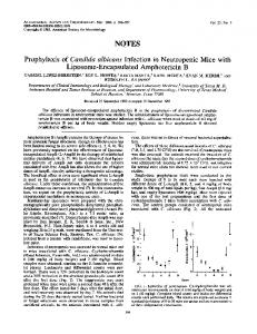
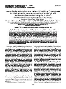
RESULTS Effects of AmB on HBV-associated DNA polymerase activity. HBV-associated DNA polymerase activity in infected plasma showed a consistent dose-response increase, as measured by incorporation of the 3H-labeled nucleotide precursors after treatment with increasing concentrations of AmB (Fig. 1). The percent increase in activity above control ranged from 11 to 34%. This effect was noted at a concentration of AmB as low as 5 yg/ml and appeared to plateau between 50 and 100 ,ug/ml. Effects of AmB on partially purified HBV. Comparison of the effects of AmB (250 ,ug/ml)-treated versus untreated control HBV particles, as determined by isopycnic banding in parallel sucrose gradients, is shown in Fig. 2. Two peaks of HBsAg activity, as measured by RIA, were demonstrated in the control gradient (Fig. 2A). The second HBsAg peak banded at a density of 1.165 g/ml, was coincident with peak HBcAg activity, and was therefore indicative of HBV particles. After treatment with AmB (Fig. 2B), peak HBcAg activity was again coincident with the second peak of HBsAg activity, as measured by RIA, but banded at a density of
828
KESSLER ET AL.
ANTIMICROB. AGENTS CHEMOTHER.
2.0.
-
P
Il.a}
I,
I
4-) ._
4
0
C. x
A
e
C 1.5
IIa I
E
F)o
4
0 CL
= O
0
0
40 50 ------------100-----------------250 Amphotericin B ug/ml FIG. 1. Effects of treatment with various concentrations of AmB on HBV-associated DNA polymerase activity (counts per minute) in pelleted HB V-infectedplasma; each point represents the mean ± one standard deviation of eight separate experiments. 0
10
20
30
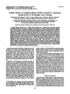
1.225 g/ml. In addition, the HBcAg activity after AmB treatment was more than twice that in the control gradient (Fig. 2B). Also noted was an 84% decrease in activity of the first peak of HBsAg as compared with the parallel control gradient. The distribution of AmB in the gradient was determined by measurement of the optical density at 360 nm, one of the absorption maxima of AmB (data not shown). AmB was found to comigrate exactly with the HBcAg activity. To determine whether there were 27nm core particles in the AmB-treated HBV gradient fraction with peak HBcAg activity, immune EM, using anti-HBcAg, was performed. Both 27-nm and 45-nm particles were observed clumped together by antibody. Routine EM of the fraction did not reveal aggregated particles. Ultrastructurally, the AmB induced several distinct changes in the partially purified HBV preparations as observed by negative-stain EM (Fig. 3). In the control preparation (Fig. 3A), normally appearing 42-nm to 45-nm HBV particles and 22-nm HBsAg particles and filaments were observed. Treatment with concentrations of 5 to 15 ,tg of AmB per ml resulted in the formation of aggregates of HBV and HBsAg particles not observed in the control preparations. These aggregates gave the impression that the material had been prepared for immune EM. Also, an increase in size of some HBV particles, from their usual 42-nm to 45-nm diameters to 55-nm to 70-nm diameters, was observed. At 25 ,ug of AmB per ml, striking changes were noted
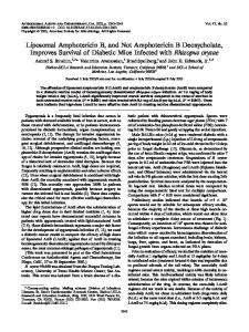
in the HBV particles, as shown in Fig. 3B. Enhanced penetration of negative stain into the outer envelope, penetration of stain into the virus with outlining of the nucleocapsid, and the actual fragmentation of the outer membrane away from the core are shown. Also noted are unusual configuations of the nucleocapsid. At concentrations above 25 jig of AmB per ml, all HBV particles appeared to be in some stage of disruption. Also observed at the higher AmB concentrations of 50, 100, and 250 yg/ml was the formation of 5-nm to 7-nm pores in some of the HBV particles (Fig. 3B, inset). To determine whether these changes were specific for AmB, similar treatment of the HBV particles with 205,ug of sodium deoxycholate per ml (the concentration present in the 250-,ig/ml AmB solution as Fungizone) was performed. This treatment did not result in any ultrastructural changes as determined by EM. Effects of AmB on HBsAg particles. In Fig. 4 are shown the results of the measurement, by RIA, of HBsAg activity remaining in the supernatants of twice-pelleted 22-nm HBsAg particles which were treated with various concentrations of AmB. The result of similar treatment with the nonionic detergent Nonidet P-40 is shown for comparison. Treatment with increasing concentrations of AmB resulted in an increase in HBsAg activity, as measured by RIA, in the supernatants. This effect was maximal at 25 ,tg of AmB per ml and was similar to treatment with 1% Nonidet P-40. No recognizable
AMPHOTERICIN B AND HEPATITIS B VIRUS
VOL. 20, 1981
829
22 20 18
[4
16 14
° S 0
.3 '30
°12 x
x_ = o
I 1O
18
2
,
6
22
5
0b
5
10
is
1S
3b
3
20 18
16 14 18 126
S
E
cyI-
oo
20 15 Fraction nunber
25
30
35
FIG. 2. Isopycnic centrifugation of HBV preparations in 30 to 65% sucrose gradients. A, Control HBV preparation treated with TNE buffer. B, HBV preparation treated with AmB (250 pg/ml). @-*, HBsAg; , density (glml). * *, HBcAg; -
-
HBsAg particles were observed by EM in the AmB- or Nonidet P-40-treated samples. Titration of these samples by reverse passive hemagglutination (Table 1) revealed decreasing titers of HBsAg as the concentration of AmB treatment increased. No AmB was detected in the supernatants by optical density measurement at 360 nm. To determine whether residual AmB in the supernatants might be interfering with the reverse passive hemagglutination assay, a 1:20 dilution of 250 ,ul of the stock 500-,ug/ml AmB solution was made with the reverse passive hemagglutination assay diluent buffer and was similarly centrifuged. The resultant supernatant was then used as diluent in a control reverse passive hemagglutination assay. No difference in titer of the HBsAg-positive control was noted when the titration was performed by using the standard diluent or the AmB-treated diluent.
To further evaluate the effects of AmB on intact HBsAg particles, rate-zonal centrifugation in parallel 10 to 30% (wt/vol) linear sucrose gradients, after treatment of '25I-labeled HBsAg particles with 250 jig of AmB per ml, was performed. In the control gradient (Fig. 5A), there was a single peak of immunologically reactive HBsAg, as measured by RIA. Three peaks of 125I radioactivity were detected in this gradient, the first two of which are due to "25I-labeled albumin which dissociates from the HBsAg particles during storage (23; unpublished data). The third peak was coincident with immunologically reactive HBsAg. After AmB treatment (Fig. 5B), there was a 65 and 68% reduction in 125I radioactivity and immunoreactive HBsAg, respectively, in similar fractions from the two gradients. The lost 125I radioactivity was recovered in the last fraction and was due to an increased
830
ANTIMICROB. AGENTS CHEMOTHER.
KESSLER ET AL.
4 FIG. 3. Negative-stain EM ofpartially purified HBVpreparations. A, Control HBVpreparation; 42-nm to 45-nm HBV particles and 22-nm HBsAg particles and filaments were present. B, HBV preparation treated with 25 pg of AmB per ml. Arrow indicates a 5-nm pore in HB V particle treated with 250 pg of AmB per ml. Original magnification, x66,000; bar = 100 nm.
1.,
N P-40
12 0 x r _
ct I
10
Treatment
8 6
:3
4 2 0
10
20 30 40 50 ------250 Amphotericin B ,ug/ml
FIG. 4. Measurement of HBsAg activity by RIA in supernatants of twice-pelleted 22-nm HBsAg particles after treatment with various concentrations of AmB. The result obtained by treatment with 1% Nonidet P-40 (NP-40) is indicated by the solid line. rate of migration of the Amb-treated '25I-labeled
HBsAg particles. No HBsAg immunoreactivity present in the fraction and could not be accounted for on the basis of a prozone effect or interference with the RIA by AmB (data not shown). Optical density measurement revealed that the Amb also comigrated with the '25I-labeled HBsAg in the last fraction. was
~~~HBsAgtitera
Control ......................... 12,800 200 Nonidet P-40, 1% .................. 400 AmB, 10 ,ug/ml ........... ........ 16 .................. AmB, 25 ,tg/ml . 32 AmB, 50 ug/ml ........... ........ 16 ................. AmB, 250 ,ug/ml a Reverse passive hemagglutination.
E C) 4J a
TABLE 1. Reciprocal titer of HBsAg by reverse passive hemagglutination in supernatants of twicepelleted HBsAg particles after treatment with various concentrations of AmB or Nonidet P-40 reciprocal Treatment
DISCUSSION
The inability to culture HBV in vitro has made the isolation of large quantities of HBV and HBsAg particles for in vitro studies dependent upon chronically infected human carriers and chimpanzees. HBV and HBsAg particles isolated from chronically infected chimpanzees have been shown to be serologically and biochemically identical to those from infected humans (21, 23). The production of HBsAg particles and HBV in chronically infected individuals is particularly useful in the study of the effects of polyene antibiotics, such as AmB, on these particles in that the effects of the drug on the intact HBV can be studied separately from the effects on the HBsAg particles.
AMPHOTERICIN B AND HEPATITIS B VIRUS
VOL. 20, 1981
831
12
A
11 10
9
8
=
3
0
x
6
5 w _ _
10c1 C
O
3
1,' I, 7 10 9oa
2
7 V.;, O X 6 '> 12 11
L
B
10
3
9' 8
.4
.-7 c -
Ej
6
5
=
.
m
0
s 5 0
4
4
3
3
2
2
se,%
1.
I_'
'1
t lm%,M
T~~~~l
- 0- 0- Al.- 2-- 2-,A-
~
----
-
Fraction number
20i
21
--
m- I 3b
FIG. 5. Rate-zonal centrifugation of radioiodinated HBsAg particles in 10 to 30%1 sucrose gradients. A, Control HBsAg particles treated with TNE buffer. B, HBsAg particles treated with 250 pg of Amb per ml. liquid fractions; -* - *, HBsAg by RIA. ,125I counts per minute in
100-[il
It has been suggested that AmB would not interact with particles in the size range of viruses owing to its preparation as a micelle suspension stabilized with deoxycholate (11). The results of the experiments in this study clearly indicate that AmB in concentrations as low as 5 [Lg/ml causes significant ultrastructural and biochemical changes in HBV particles. For example, initial experiments showed that treatment of pelleted HBV particles with increasing concentrations of AmB resulted in a corresponding increase in HBV-associated DNA polymerase ac-
tivity. It is possible that the increasing concentrations of AmB used in these experiments resulted in an increase in the activity of the DNA polymerase enzyme. However, it has been shown that AmB causes the enhanced penetration of both large and small molecules through AmBtreated fungal and animal cell membranes (5, 6, 15, 16). Therefore, it is more likely that the increase in DNA polymerase activity observed in our experiments was a result of increased penetration of nucleotide precursors through the HBV envelope to the polymerase enzyme within
832
KESSLER ET AL.
the HBV nucleocapsid. This is the first report of AmB enhancing the penetration of small molecules through the lipoprotein membrane of a virus particle. Although the effect of AmB on the chemical composition of lipid-enveloped viruses has not been analyzed, previous stqdies have shown the effects of two related polyene antibiotics, filipin and AmB methyl ester, on vesicular stomatitis virus, as measured by buoyant density determination in sucrose gradients; neither study reported any change in density (11, 14). Our results, however, showed a significant increase in density of AmB-treated HBV from 1.165 to 1.225 g/ml in sucrose gradients. The increase in density was not related to a difference in binding of AmB to HBV, as the AmB was found to comigrate in the gradient with the HBV, similar to the finding for filipin and AmB methyl ester treatments of vesicular stomatitis virus (11, 14). The increase in density can be partly attributed to the presence of free 27-nm HBV core particles, as demonstrated by immune EM. Also observed on these same grids were 42-nm HBV particles clumped to free HBV core particles. This suggests that AmB not only caused stripping of the lipid envelope away from the core, but also had an intermediate effect in which the physical properties of the HBV were changed in the absence of complete disruption. A similar spectrum of effects on biological membranes has been observed for other amphiphatic molecules
(8).
The exact mechanism of action of AmB on lipid membranes is unknown. Although the association kinetics of AmB with artificial phosphatidylcholine vesicles is independant of sterols, it has been shown that the presence of 3,8hydroxyl sterols such as cholesterol or ergosterol is necessary for AmB to cause alterations in the permeability of mammalian and fungal cell membranes (4, 7, 15, 26). The ultrastructural changes caused by AmB in both artificial and naturally occurring lipid membranes have been reported (4, 14, 26). The EM changes observed have varied from the swelling of artificial cholesterol-containing vesicles because of disorganization of the peripheral concentric lipid bilayer to the actual fragmentation of the membrane as observed with vesicular stomatitis virus (4, 14). These different effects seem dependent upon the conditions of the experiment and the type of membrane being investigated (15). The ultrastructural effects of AmB on HBV showed a similar spectrum of change dependent upon the concentration of AmB. At AmB concentrations of higher than 25 ,ug/ml, all HBV particles appeared disrupted to various degrees. The formation of pores or pits, 5 nm to 7 nm in diameter,
ANTIMICROB. AGENTS CHEMOTHER.
in the HBV membrane were also observed at high concentrations (Fig. 3B, inset). Whether these are pores through the entire thickness of the membrane or merely craters caused by the aggregation of membrane-associated particles, as seen in AmB-treated Epidermophyton floccosum, will require further study with more sensitive techniques such as freeze-etch EM (18). Although deoxycholate is also a detergent compound, treatment of HBV with deoxycholate alone had no effect on the ultrastructural appearance of the HBV particles (8). This suggests that the observed ultrastructural effects in our experiments are specific for AmB. Others have demonstrated inhibition of the activity of AmB in fungi and human erythrocytes by the addition of exogenous sterols (12). As inhibition studies were not carried out in our experiments, we cannot comment upon the molecular basis for the observed effects of AmB on HBV. AmB treatment of HBsAg particles resulted in two structural alterations. First, HBsAg particles were disrupted, resulting in a nonparticulate soluble fraction in which HBsAg immunoreactivity was detectable by RIA but not by reverse passive hemagglutination and was similar in effect to treatment with the nonionic detergent Nonidet P-40. This observation may be similar to that reported for purified influenza hemagglutinin which, in the presence of the ionic detergent sodium dodecyl sulfate, will not hemagglutinate erythrocytes. This is thought to be due to the monomeric hemagglutinin subunits being monovalent and attaching to cell receptors at one end only (13). Alternatively, the decrease in the reverse passive hemagglutination titer observed in our experiments may have been secondary to fragments of HBsAg particles which retained antigenic activity as measured by RIA but were too small to cause cross-bridging of the erythrocytes in the assay. The migration of the immunoreactive HBsAg in sucrose suggests that this HBsAg-reactive material is not composed of solubilized antigenic subunits but rather fragments of HBsAg particles not complexed to AmB. The second more striking alteration induced by AmB was the loss of specific HBsAg immunoreactivity of HBsAg particles complexed with AmB. This loss of antigenicity was suggested by the results of the HBV density gradient experiment (Fig. 2). As noted, the 84% decrease in HBsAg activity in the first HBsAg peak, after AmB treatment, was not recovered in any other fraction. This was confirmed by a similar finding after treatment of '251-labeled HBsAg 22-nm particles with AmB. As shown in Fig. 5B, the HBsAg particles which complexed to AmB and were detected in the last fraction of the sucrose
AMPHOTERICIN B AND HEPATITIS B VIRUS
VOL. 20, 1981
gradient lost HBsAg immunoreactivity. The mechanism of this structural alteration is unknown, but may be due to the rearrangement of antigenic glycoprotein subunits in the HBsAg particles by AmB-cholesterol complexes. These results suggest that the structure of HBsAg particles is similar to that of the outer lipid membrane of intact HBV. From our experiments, it is not possible to predict the effects on HBV and HBsAg particles in AmB-treated patients with chronic HBV infection. The pharmacokinetics of AmB in humans are unusual in that above a daily intravenous dose of 50 mg, it is difficult to demonstrate increases in serum levels above 2 ,ug/ml (1). This is probably due to extensive binding of the drug to sterol-containing membranes throughout the body (1, 3). Although the concentrations of AmB used in our study were higher than those obtained in the serum of patients, AmB should be evaluated either alone or in combination with other antiviral drugs in the treatment of chronic HBV infections.
7. 8. 9. 10. 11. 12.
13.
14.
15.
ACKNOWLEDGMENTS We thank J. Skelly, P. Young, and G. Trenholme for helpful comments in the preparation of this manuscript and J. Skelly for providing the purified HBsAg particles. We also thank George Tovey of the Electron Microscopy Unit, London School of Hygiene and Tropical Medicine, for technical assistance. H.A.K. is a visiting fellow from Rush-Presbyterian-St. Luke's Medical Center, Chicago, Ill., and is a recipient of the Pillsbury Trust Fellowship. The work on viral hepatitis at the London School of Hygiene and Tropical Medicine was supported by generous grants from the Medical Research Council, the Department of Health and Social Security, the Wellcome Trust, and the World Health Organization.
16.
17.
18.
LITERATURE CITED
19.
1. Bennett, J. E. 1974. Chemotherapy of systemic mycoses. N. Engl. J. Med. 290:30-32. 2. Bennett, J. E., W. E. Dismukes, R. J. Duma, G. Medoff, M. A. Sande, H. Gallis, J. Leonard, B. T. Fields, M. Bradshaw, H. Hayward, Z. A. McGee, T. R. Cote, C. G. Cobbs, J. F. Warner, and D. W. Alling. 1979. A comparison of amphotericin B alone and combined with fluorocytosine in the treatment of crytococcal meningitis. N. Engl. J. Med. 301:129-131. 3. Bindschadler, D. D., and J. E. Bennett. 1969. A pharmacologic guide to the clinical use of amphotericin B.
20.
J. Infect. Dis. 120:427-436. 4. Bittman, R., W. C. Chen, and 0. R. Anderson. 1974. Interaction of filipin III and amphotericin B with lecithin-sterol vesicles and cellular membranes. Spectral and -electron microscope studies. Biochemistry 13:
1364-1373. 5. Borden, E. C., B. W. Booth, and J. A. McBain. 1979. Enhancement of infectivity of encephalomyocarditis virus RNA by amphotericin B methyl ester. J. Gen. Virol. 42:297-303. 6. Borden, E. C., J. A. McBain, and P. H. Leonhardt. 1979. Effects of amphotericin B and its methyl ester on
21.
22. 23.
24.
25. 26.
833
the antiviral activity of polyinosinic-polycytidylic acid. Antimicrob. Agents Chemother. 16:203-209. Chen, W. C., and R. Bittman. 1977. Kinetics of association of amphotericin B with vesicles. Biochemistry 16: 4145-4149. Helenius, A., and K. Simons. 1975. Solubilization of membranes by detergents. Biochem. Biophys. Acta 415:29-79. Howard, C. R. 1978. The detection of DNA polymerase activity in the diagnosis of HBV infection. J. Med. Virol. 3:81-86. Howard, C. R., and A. J. Zuckerman. 1977. Core antigen and circulating anti-core antibody in hepatitis B infection. J. Immunol. Methods 14:291-301. Jordon, G. W., and E. C. Seet. 1978. Antiviral effects of amphotericin B methyl ester. Antimicrob. Agents Chemother. 13:199-204. Kotler-Brajtiburg, J., H. D. Price, G. Medoff, D. Schlessinger, and G. S. Kobayashi. 1974. Molecular basis for the selective toxcity of amphotericin B for yeast and filipin for animal cells. Antimicrob. Agents Chemother. 5:377-382. Laver, W. G., and R. C. Valentine. 1969. Morphology of the isolated hemagglutinin and neuraminidase subunits of influenza virus. Virology 38:105-119. Majuk, Z., R. Bittman, F. R. Landsberger, and R. W. Compans. 1977. Effects of filipin on the structure and biological activity of enveloped viruses. J. Virol. 24: 883-892. Medoff, G., and G. A. Kobayashi. 1980. The polyenes, p. 3-33. In D. Speller (ed.), Antifungal chemotherapy. John Wiley & Sons Ltd., London. Medoff, G., G. A. Kobayashi, C. N. Kwan, D. Schlessinger, and P. Venkov. 1972. Potentiation of rifampicin and 5-fluorocytosine as antifungal antibiotics by amphotericin B. Proc. Natl. Acad. Sci. U.S.A. 69:296299. Medoff, G., F. Valeriote, R. G. Lynch, D. Schlessinger, and G. A. Kobayashi. 1974. Synergistic effect of amphotericin B and 1,3-bis(2-chloroethyl)-1-nitrosourea (BCNU) against a syngenic transplantable AKR leukemia. Cancer Res. 34:974-978. Nozoura, Y., T. Kitajima, T. Sekiya, and Y. Ito. 1974. Ultrastructural alterations induced by amphotericin B in the plasma membrane of Epidermophyton floccosum as revealed by freeze-etch electron microscopy. Biochem. Biophys. Acta 367:32-38. Presant, C. A. 1980. Amphotericin B. New perspectives. Arch Intern. Med. 140:469-470. Presant, C. A., C. Klahr, and R. Santala. 1977. Amphotericin B induction of sensitivity to adriamycin, 1,3bis(2-chloroethyl)-1-nitrosourea (BCNU) plus cyclophosphamide in human neoplasia. Ann. Intern. Med. 86:47-51. Robinson, W. S. 1977. The genome of hepatitis B virus. Annu. Rev. Microbiol. 31:357-377. Skelly, J., C. R. Howard, and A. J. Zuckerman. 1978. Labeling of galactose residues in hepatitis B surface antigen glycoprotein. J. Gen. Virol. 41:447-457. Skelly, J., C. R. Howard, and A. J. Zuckerman. 1979. Analysis of hepatitis B surface antigen components solubilized with Triton X-100. J. Gen. Virol. 44:679689. Stevens, N. M., C. G. Engle, R. B. Fisher, W. Mechlinski, and C. P. Schaffher. 1975. In vitro antiherpetic activity of water-soluble amphotericin B methyl ester. Arch. Virol. 48:391-394. Takahashi, T. 1975. Physical and chemical properties of HBsAg. J. Jpn. Med. Assoc. 73:225-234. Weissman, G., and G. Sessa. 1967. The action of polyene antibiotics on phospholipid-cholesterol structures. J. Biol. Chem. 242:616-626.